Podcast
Questions and Answers
What are the structures that limit the basal surface of the pons superiorly?
What are the structures that limit the basal surface of the pons superiorly?
- Anterior Cerebral peduncles
- Middle Cerebellar peduncles
- Inferior Cerebellar peduncles
- Superior Cerebral peduncles (correct)
What is the component of the posterior or middle lobe of the cerebellum?
What is the component of the posterior or middle lobe of the cerebellum?
- Nodulus
- Uvula (correct)
- Central Lobule
- Lingula cerebelli
Which cranial nerves leave the brain on the borders of the pons at the basal surface of the cerebellum?
Which cranial nerves leave the brain on the borders of the pons at the basal surface of the cerebellum?
- Olfactory (I), Optic (II), Oculomotor (III)
- Trochlear (IV), Trigeminal (V), Abducent (VI)
- Glossopharyngeal (IX), Vagus (X), Hypoglossal (XII)
- Trigeminal (V), Abducent (VI), Facial (VII), Vestibulo-cochlear (VIII) (correct)
What forms the dorsal surface of the cerebellum?
What forms the dorsal surface of the cerebellum?
Which nuclei are found on the vermis of the cerebellum?
Which nuclei are found on the vermis of the cerebellum?
What is the component of the Floculonodular lobe of the cerebellum?
What is the component of the Floculonodular lobe of the cerebellum?
What limits the basal surface of the cerebellum inferiorly?
What limits the basal surface of the cerebellum inferiorly?
How is the dorsal surface of the cerebellum separated from the inferior part of the rhomboid fossa?
How is the dorsal surface of the cerebellum separated from the inferior part of the rhomboid fossa?
What laterally limits the dorsal surface of the cerebellum?
What laterally limits the dorsal surface of the cerebellum?
What is the pontine part of the rhomboid fossa?
What is the pontine part of the rhomboid fossa?
What are the superior limits of the medulla oblongata?
What are the superior limits of the medulla oblongata?
What does the basal surface of the medulla oblongata include?
What does the basal surface of the medulla oblongata include?
Where does the hypoglossal nerve leave the brain?
Where does the hypoglossal nerve leave the brain?
What limits the inferior part of the dorsal surface of the rhomboid fossa?
What limits the inferior part of the dorsal surface of the rhomboid fossa?
What does the vestibular area on the dorsal surface of the brain stem lead to laterally?
What does the vestibular area on the dorsal surface of the brain stem lead to laterally?
What is located on the inferolateral part of the dorsal surface of the brain stem?
What is located on the inferolateral part of the dorsal surface of the brain stem?
What is the location of the median aperture in the 4th ventricle?
What is the location of the median aperture in the 4th ventricle?
What part of the spinal cord is characterized by the vertebral levels C5-T1?
What part of the spinal cord is characterized by the vertebral levels C5-T1?
What is the inferior wall of the 4th ventricle composed of?
What is the inferior wall of the 4th ventricle composed of?
What is the structure at the floor of the rhomboid fossa?
What is the structure at the floor of the rhomboid fossa?
What is the level of the spinal cord termination?
What is the level of the spinal cord termination?
What is the range of the lumbosacral enlargement?
What is the range of the lumbosacral enlargement?
What are the lateral apertures of the 4th ventricle located in?
What are the lateral apertures of the 4th ventricle located in?
What is the composition of the superior wall of the 4th ventricle?
What is the composition of the superior wall of the 4th ventricle?
Flashcards are hidden until you start studying
Study Notes
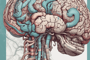
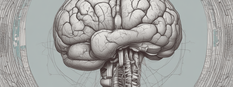
Metencephalon
- Composed of Pons and Cerebellum
Cerebellum
- Superior surface has anterior and superior notch
- Inferior surface has vallecula cerebelli, anterior and posterior edges, and hemispheres and vermis separated by median sulcus
- Divided into three lobes: Anterior lobe, Floculonodular lobe, and Posterior or middle lobe
- Anterior lobe has culmen, central lobule, and lingula cerebelli
- Floculonodular lobe has nodulus and flocculus
- Posterior or middle lobe has declive, piramid, uvula, folium vermis, and tuber
Nuclei of the Cerebellum
- Vermis has fastigial nucleus
- Hemispheres have dentate nucleus, emboliform nucleus, and globosus nucleus
Pons
- Basal surface limited superiorly by superior cerebral peduncles
- Basal surface limited inferiorly by medulla oblongata
- Continues as middle cerebellar peduncles laterally on the basal surface of the cerebellum
- Cranial nerves leaving the brain on the borders of the pons at the basal surface of the cerebellum: Trigeminal (V), abducent (VI), facial (VII), and vestibulo-cochlear (VIII)
Dorsal Surface of the Cerebellum
- Formed by the superior part of the rhomboid fossa (floor of the IVth ventricle)
- Separated from the inferior part of the rhomboid fossa by striae medullares
- Laterally limited by superior cerebellar peduncles
Rhomboid Fossa
- Pontine part includes structures such as median sulcus, medial eminences, facial colliculi, sulcus limitans, superior foveae, upper portion of the vestibular area, and locus coeruleus
Medulla Oblongata
- Basal surface has anterior median fissure, pyramids, anterior lateral sulcus, lateral funiculus, and the olive
- Hypoglossal nerve leaves the brain at the anterior lateral sulcus on the basal surface
- Superior limits: striae medullares (dorsally) and inferior border of pons with foramen caecum (ventrally)
- Inferior limits: decussation of pyramids (ventrally) and point of emergence of C1 cranial nerves (dorsally)
Rhomboid Fossa (continued)
- Inferior part of the dorsal surface limited by sulcus limitans on the lateral side
- Vagal triangle on the dorsal surface limited by funiculus separans
- Area postrema on the dorsal surface connected with its counterpart by obex
- Vestibular area on the dorsal surface leads laterally to the lateral recess with the acoustic tubercle
Structures Found on the Rhomboid Fossa
- Median sulcus
- Sulcus limitans
- Locus coeruleus
- Medial eminance
- Vagal triangle
- Hypoglossal triangle
- Funiculus separans
- Area postrema
- Superior vestibular area
4th Ventricle
- Floor formed by the rhomboid fossa
- Superior wall composed of superior cerebellar peduncles and superior medullary vellum
- Inferior wall formed by inferior medullary velum, nodulus of the vermis, and tela choroidea
- Median aperture located at the inferior wall, leads to the cerebromedullar cisterna (subarachnoid space)
- Lateral apertures located in the apex of the lateral recessus, lead to the pons cisterna (subarachnoid space)
Spinal Cord
- Termination at L1/L2 (Lumbar vertebrae 1/Lumbar vertebrae 2)
- Cervical enlargement at C5 - T1 (Cervical vertebrae 5 - Thoracic vertebrae 1)
- Lumbosacral enlargement at L1 - S3 (Lumbar vertebrae 1 - Sacral vertebrae 3)
Fissures and Sulci in the Spinal Cord
- Anterior median fissure
- Posterior medial sulcus
- Posterolateral sulcus
- Anterolateral sulcus
- Posterior intermediate sulcus
Studying That Suits You
Use AI to generate personalized quizzes and flashcards to suit your learning preferences.