Podcast
Questions and Answers
What is the primary function of the tibia in the lower limb?
What is the primary function of the tibia in the lower limb?
- To resist compressive forces of gravity (correct)
- To facilitate plantar flexion
- To provide stability to the ankle joint
- To articulate with the pelvis
What type of joint is formed by the tibia and fibula at the ankle?
What type of joint is formed by the tibia and fibula at the ankle?
- Hinge joint
- Ball-and-socket joint
- Gliding joint
- Stable joint (correct)
What is the direction of weight transmission from the ankle joint?
What is the direction of weight transmission from the ankle joint?
- Anteriorly via the tarsal bones
- Medially via the phalanges
- Posteriorly via the tarsal bones (correct)
- Laterally via the metatarsal bones
What is the primary movement of the foot and ankle?
What is the primary movement of the foot and ankle?
What is the purpose of the interosseous membrane in the lower limb?
What is the purpose of the interosseous membrane in the lower limb?
What is the shape of the foot in relation to the leg?
What is the shape of the foot in relation to the leg?
Which nerve supplies the powerful extensor of the hip?
Which nerve supplies the powerful extensor of the hip?
Which nerve supplies the muscles on the sole of the foot?
Which nerve supplies the muscles on the sole of the foot?
What is the name of the compartment that contains the flexors of the knee?
What is the name of the compartment that contains the flexors of the knee?
How many main nerves are there in the lower limb?
How many main nerves are there in the lower limb?
Which nerve supplies the anterior compartment of the thigh?
Which nerve supplies the anterior compartment of the thigh?
Which nerve divides into two branches to supply the leg?
Which nerve divides into two branches to supply the leg?
Which nerve supplies the adductor muscles of the hip?
Which nerve supplies the adductor muscles of the hip?
Which nerve supplies the lateral compartment of the leg?
Which nerve supplies the lateral compartment of the leg?
What is the main reason for the strength of the capsule?
What is the main reason for the strength of the capsule?
Which ligament has a tensile strength 20 times that of reinforced concrete?
Which ligament has a tensile strength 20 times that of reinforced concrete?
What is the shape of the iliofemoral ligament?
What is the shape of the iliofemoral ligament?
What is the function of the zona orbicularis?
What is the function of the zona orbicularis?
What is the distal attachment of the ischiofemoral ligament?
What is the distal attachment of the ischiofemoral ligament?
What is the attachment of the iliofemoral ligament proximally?
What is the attachment of the iliofemoral ligament proximally?
Which ligament arises from the pubic margin of the acetabulum?
Which ligament arises from the pubic margin of the acetabulum?
Where do all the ligaments attach distally?
Where do all the ligaments attach distally?
What type of muscles are found in the posterior compartment of the leg?
What type of muscles are found in the posterior compartment of the leg?
Which nerve is often referred to by the older name peroneal nerve?
Which nerve is often referred to by the older name peroneal nerve?
What is the function of the posterior muscles in the leg?
What is the function of the posterior muscles in the leg?
Which bone is located on the lateral side of the leg?
Which bone is located on the lateral side of the leg?
What is the purpose of the small lateral compartment in the leg?
What is the purpose of the small lateral compartment in the leg?
Which nerve is not one of the nerves of the lower limb?
Which nerve is not one of the nerves of the lower limb?
What is the significance of the nerves of the lower limb?
What is the significance of the nerves of the lower limb?
Which compartment contains the extensor muscles?
Which compartment contains the extensor muscles?
What is the main direction of blood flow to the head of the femur?
What is the main direction of blood flow to the head of the femur?
Which artery is responsible for the most significant contribution to the blood supply of the femoral head?
Which artery is responsible for the most significant contribution to the blood supply of the femoral head?
What is the term for the folds of the capsule and synovium that the circumflex arteries run along?
What is the term for the folds of the capsule and synovium that the circumflex arteries run along?
What is the consequence of the retinacular arteries being impeded due to a fracture of the femur?
What is the consequence of the retinacular arteries being impeded due to a fracture of the femur?
What type of fracture would spare the retinacular vessels?
What type of fracture would spare the retinacular vessels?
Which artery provides an additional blood supply to the femoral head, but is insufficient on its own?
Which artery provides an additional blood supply to the femoral head, but is insufficient on its own?
What is the term for the condition resulting from the death of the femoral head due to a lack of blood supply?
What is the term for the condition resulting from the death of the femoral head due to a lack of blood supply?
What is the purpose of the circumflex arteries in relation to the femoral head?
What is the purpose of the circumflex arteries in relation to the femoral head?
Flashcards are hidden until you start studying
Study Notes
Bones of the Lower Limb
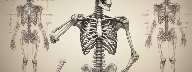
- The distal part of the limb consists of the tibia and fibula, with the tibia being the largest and weight-bearing bone.
- The tibia articulates with the knee joint, while the fibula is excluded from the knee joint but participates in the ankle joint.
- The leg bones are held together by joints at both ends and by an interosseous membrane.
- The foot is held at right angles to the leg and contains tarsal bones, metatarsal bones, and phalanges.
- In standing, weight is transmitted from the ankle joint posteriorly via the tarsal bones to the heads of the metatarsals, forming an arch between the areas of contact with the ground.
Nerves of the Lower Limb
- There are six main nerves of the lower limb:
- Superior gluteal nerve, which supplies the abductors of the hip.
- Inferior gluteal nerve, which supplies the gluteus maximus muscle.
- Femoral nerve, which supplies the anterior compartment of the thigh.
- Obturator nerve, which supplies the medial compartment of the thigh.
- Tibial nerve, which supplies the posterior muscles in the thigh and leg, and also supplies the muscles on the sole of the foot.
- Common fibular (common peroneal) nerve, which divides into superficial and deep fibular nerves.
Compartments of the Thigh
- The thigh has three compartments: anterior, medial, and posterior.
- The anterior compartment contains the extensors of the knee joint.
- The medial compartment, also known as the adductor compartment, contains the adductor muscles of the hip.
- The posterior compartment contains the flexors of the knee.
Compartments of the Leg
- The leg has three compartments: anterior, posterior, and lateral.
- The posterior compartment contains the plantar flexors of the ankle and flexors of the toes.
- The anterior compartment contains the extensor muscles.
- The lateral compartment contains muscles that aid the posterior muscles in flexion of the ankle.
Capsule Attachments
- The capsule is strong due to the presence of thickenings of the capsule, called capsular ligaments.
- There are three ligaments: iliofemoral, pubofemoral, and ischiofemoral.
- These ligaments attach proximally to the acetabulum and distally to the intertrochanteric line.
Capsular Ligaments
- The iliofemoral ligament is the strongest ligament in the human body, with a tensile strength 20 times that of reinforced concrete.
- The iliofemoral ligament has a Y-shape and attaches to the iliac margins of the acetabular labrum and extends proximally towards the anterior inferior iliac spine.
- The pubofemoral ligament arises from the pubic margin of the acetabulum and inserts onto the lesser trochanter.
- The ischiofemoral ligament arises from the ischial margin of the acetabulum and inserts largely into the iliofemoral ligament, but also onto the greater trochanter.
Zona Orbicularis
- The zona orbicularis is a circular ligament that arises from the ischiofemoral ligament and serves to pinch the capsule around the neck of the femur.
- It aids in holding the femoral head into the acetabular socket, acting like a purse-string.
Blood Supply of the Femoral Head
- The blood supply to the joint is complex and involves five different arteries and two separate anastomoses.
- The head of the femur depends on its blood being delivered from distal to proximal, mainly through the medial circumflex femoral artery.
- Branches from the circumflex arteries run along the neck of the femur, dragging the capsule and synovium into folds, and then enter nutrient foraminae to reach the femoral head.
- If there is a fracture of the femur, these vessels may be at risk of rupture, leading to avascular necrosis of the femoral head.
Studying That Suits You
Use AI to generate personalized quizzes and flashcards to suit your learning preferences.


