Podcast
Questions and Answers
What distinguishes the periosteum from the endosteum?
What distinguishes the periosteum from the endosteum?
- The periosteum contains spongy bone, whereas the endosteum contains compact bone.
- The periosteum is a hard connective tissue, whereas the endosteum is soft.
- The periosteum is a specific connective tissue present outside the bone, whereas the endosteum is the interior part of the bone. (correct)
- The periosteum is present inside the bone, whereas the endosteum is present outside the bone.
If a bone fracture occurred in the diaphysis, and a doctor mentioned issues with the bone's primary marrow source, which type of marrow would be of concern?
If a bone fracture occurred in the diaphysis, and a doctor mentioned issues with the bone's primary marrow source, which type of marrow would be of concern?
- Yellow bone marrow, due to its presence in the diaphysis or shaft. (correct)
- Neither red nor yellow bone marrow, as marrow is not found in the diaphysis.
- Red bone marrow, due to its location in the epiphysis.
- Both red and yellow bone marrow equally.
In bone remodeling, how do osteoclasts facilitate changes in bone structure and composition?
In bone remodeling, how do osteoclasts facilitate changes in bone structure and composition?
- By destroying bone through phagocytosis, allowing for the release of calcium and phosphorus. (correct)
- By transporting calcium from bone into the blood stream.
- By secreting unmineralized ground substances to build new bone.
- By transforming into osteocytes, embedding within the bone matrix.
Which characteristic distinguishes hyaline cartilage from fibrous and elastic cartilage?
Which characteristic distinguishes hyaline cartilage from fibrous and elastic cartilage?
What role do blood vessels play within bone tissue?
What role do blood vessels play within bone tissue?
Which characteristic is associated with the epiphysis of a long bone?
Which characteristic is associated with the epiphysis of a long bone?
What are the key structural components of the skeleton?
What are the key structural components of the skeleton?
What happens to red blood cells (RBCs) at the end of their lifespan?
What happens to red blood cells (RBCs) at the end of their lifespan?
Which of the following best describes the role of osteoblasts in bone tissue?
Which of the following best describes the role of osteoblasts in bone tissue?
What type of protein is elastin?
What type of protein is elastin?
Which type of cells are present inside the lacunae?
Which type of cells are present inside the lacunae?
A patient experiences a joint injury that affects their ability to move in multiple directions freely. Which type of joint is most likely involved?
A patient experiences a joint injury that affects their ability to move in multiple directions freely. Which type of joint is most likely involved?
Which of the following is the primary function of ligaments in the skeletal system?
Which of the following is the primary function of ligaments in the skeletal system?
Which of the following joints facilitates movement in one direction only?
Which of the following joints facilitates movement in one direction only?
What criteria defines synovial joints?
What criteria defines synovial joints?
Where is elastic cartilage typically found in the human body?
Where is elastic cartilage typically found in the human body?
What is the axial skeleton primarily responsible for?
What is the axial skeleton primarily responsible for?
From the options provided, what bone is part of the axial skeleton?
From the options provided, what bone is part of the axial skeleton?
Which of these is a paired bone and part of the facial skeleton?
Which of these is a paired bone and part of the facial skeleton?
What is the only moving bone of the skull?
What is the only moving bone of the skull?
How many cervical vertebrae are there in the vertebral column?
How many cervical vertebrae are there in the vertebral column?
Which of the following describes the arrangement of ribs in the rib cage?
Which of the following describes the arrangement of ribs in the rib cage?
What is the primary function of the appendicular skeleton?
What is the primary function of the appendicular skeleton?
The pectoral girdle consists of which of the following?
The pectoral girdle consists of which of the following?
How many bones make up the pelvic girdle?
How many bones make up the pelvic girdle?
A football player injures their ankle, and the doctor suspects damage to the ligaments. What type of injury is most likely?
A football player injures their ankle, and the doctor suspects damage to the ligaments. What type of injury is most likely?
Which joint type is exemplified by the pubic symphysis?
Which joint type is exemplified by the pubic symphysis?
What is the initial step in bone repair after a fracture?
What is the initial step in bone repair after a fracture?
How does soft callus formation contribute to bone repair?
How does soft callus formation contribute to bone repair?
What is the role of osteoclasts in bone remodeling after a fracture?
What is the role of osteoclasts in bone remodeling after a fracture?
How does bone typically heal after a fracture?
How does bone typically heal after a fracture?
What condition is represented when a joint's normal articulation is disrupted, leading to misalignment?
What condition is represented when a joint's normal articulation is disrupted, leading to misalignment?
Which of the following is NOT a symptom of dislocation?
Which of the following is NOT a symptom of dislocation?
According to the text what causes a sprain injury?
According to the text what causes a sprain injury?
What is the scientific name for 'muscle'?
What is the scientific name for 'muscle'?
What is the main difference between skeletal versus smooth muscle tissue.
What is the main difference between skeletal versus smooth muscle tissue.
Which muscle type contains intercalated discs?
Which muscle type contains intercalated discs?
If a particular protein is NOT arranged, what tissue would it most likely be?
If a particular protein is NOT arranged, what tissue would it most likely be?
Which tissue layer directly covers individual muscle cells or fibers?
Which tissue layer directly covers individual muscle cells or fibers?
What is the store house within the sarcoplasmic reticulum?
What is the store house within the sarcoplasmic reticulum?
Where does the myosin head specifically to bind in muscle contraction?
Where does the myosin head specifically to bind in muscle contraction?
What happens to the H-zone when a muscles contracts?
What happens to the H-zone when a muscles contracts?
Why does rigor mortis occur after death?
Why does rigor mortis occur after death?
Where does muscle action will detach from in order to relax the muscle?
Where does muscle action will detach from in order to relax the muscle?
What is known as antagonists?
What is known as antagonists?
Flashcards
Skeleton
Skeleton
A rigid framework of bones and cartilage; supports, provides movement, and locomotion.
Bone
Bone
Hard connective tissue made of different bone cells.
Periosteum
Periosteum
Specific connective tissue present outside the bone surface.
Epiphysis
Epiphysis
Signup and view all the flashcards
Diaphysis
Diaphysis
Signup and view all the flashcards
Endosteum
Endosteum
Signup and view all the flashcards
Compact bone
Compact bone
Signup and view all the flashcards
Spongy bone
Spongy bone
Signup and view all the flashcards
Red bone marrow
Red bone marrow
Signup and view all the flashcards
Yellow Bone Marrow
Yellow Bone Marrow
Signup and view all the flashcards
Blood vessels
Blood vessels
Signup and view all the flashcards
Osteoblast
Osteoblast
Signup and view all the flashcards
Osteocytes
Osteocytes
Signup and view all the flashcards
Osteoclasts
Osteoclasts
Signup and view all the flashcards
Cartilage
Cartilage
Signup and view all the flashcards
Perichondrium
Perichondrium
Signup and view all the flashcards
Perichondrium matrix
Perichondrium matrix
Signup and view all the flashcards
Lacunae
Lacunae
Signup and view all the flashcards
Hyaline cartilage
Hyaline cartilage
Signup and view all the flashcards
Fibrous cartilage
Fibrous cartilage
Signup and view all the flashcards
Elastic Cartilage
Elastic Cartilage
Signup and view all the flashcards
Axial skeleton
Axial skeleton
Signup and view all the flashcards
Head Bones
Head Bones
Signup and view all the flashcards
Hyoid Bone
Hyoid Bone
Signup and view all the flashcards
Vertebral Column
Vertebral Column
Signup and view all the flashcards
Sternum
Sternum
Signup and view all the flashcards
Pectoral girdle
Pectoral girdle
Signup and view all the flashcards
Pelvic Girdle
Pelvic Girdle
Signup and view all the flashcards
movable joints
movable joints
Signup and view all the flashcards
CARTILIGENOUS JOINTS
CARTILIGENOUS JOINTS
Signup and view all the flashcards
SYNOVIAL (JOINTS)
SYNOVIAL (JOINTS)
Signup and view all the flashcards
HINGE JOINTS
HINGE JOINTS
Signup and view all the flashcards
PIVOT JOINT
PIVOT JOINT
Signup and view all the flashcards
BALL AND SOCKET JOINTS
BALL AND SOCKET JOINTS
Signup and view all the flashcards
Slipped Disc
Slipped Disc
Signup and view all the flashcards
SPONDYLOSIS
SPONDYLOSIS
Signup and view all the flashcards
SCIATICA
SCIATICA
Signup and view all the flashcards
ARTHRITIS
ARTHRITIS
Signup and view all the flashcards
OSTEOARTHRITIS
OSTEOARTHRITIS
Signup and view all the flashcards
SIMPLE FRACTURE
SIMPLE FRACTURE
Signup and view all the flashcards
Study Notes
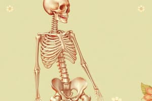
- Support and movement is achieved via the skeleton
Skeleton
- The skeleton is a rigid framework of bone and cartilage
- It provides support and is involved in movement and locomotion
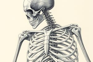
Bone (Osteo)
- Bone is hard connective tissue made up of different bone cells
- Periosteum is a specific connective tissue present outside the bone
- Both ends of the bone are the epiphysis
- The region between both epiphysis is the diaphysis, or shaft
- The interior part of the bone is called endosteum
- The peripheral part of endosteum is compact bone
- The inside of compact bone has a soft region called spongy bone
- Most of the red bone marrow is present in the epiphysis, but some is in the diaphysis
- Yellow bone marrow is present in the diaphysis or shaft
- Blood vessels are also present and transport blood to different parts of the body
- The liver destroys red blood cells after 120 days
- Every blood cell is produced in bone marrow, except platelets
Bone Cells (Osteogenic Cells)
- Bone cells include osteoblasts, osteocytes, and osteoclasts
- Osteoblasts are bone-forming cells
- Osteoblasts are uninucleated
- Osteoblasts secrete unmineralized ground substances
- Osteocytes are bone-maturing cells
- An osteocyte is a bone cell after the accumulation of matrix
- Elastin is a protein created by fibroblasts that form a chain of elastic fiber
- Osteoblasts eventually convert into osteocytes
- Osteocytes are mononucleated
- Osteocytes transport calcium from bone into blood
- Osteoclasts are bone-destructive cells and are called osteoclasts
- Osteoclasts destroys extra bone through phagocytosis
- Osteoclasts are multinucleated
- Osteoclasts secrete calcium and phosphorus into blood
- Osteoclasts participate in demineralization, which is the removal of minerals
- More osteoclasts indicates more soft bone
Cartilage (Chondrio)
- Collagen is a group of proteins found mainly in the connective tissue of skin
- Almost 30% of the body's protein is in the form of collagen
- Cartilage is connective tissue made up of chondrocytes, or cartilage cells
- All cartilage have same composition
- A specific connective tissue present outside the cartilage is perichondrium
- Inside the perichondrium matrix is collagen and elastin
- Open spaces in the matrix are known as lacunae
- Chondrocytes are present inside the lacunae
- Blood vessels are absent in cartilage; nutrients are transported via diffusion
Types of Cartilage
- Includes hyaline, fibrous, and elastic cartilage
- Hyaline cartilage is cartilage having equal concentrations of collagen and elastin
- This cartilage is present at the tip of the humerus, femur, and radius
- It is also present in the nose, larynx, and trachea
- Fibrous cartilage is cartilage having more collagen
- Elastic cartilage is cartilage having more elastin protein
- It is more flexible than the other two
- For example, it is in the ear pinna and epiglottis
Human Skeleton
- The axial and appendicular skeletons make up the human skeleton
Axial Skeleton
- The axial skeleton consists of the skull, vertebral column, and rib cage
- It makes the main axis of the human body
- The head has cranial bones, facial bones, middle ear ossicles, and the hyoid bone
- Paired bones include the temporal and parietal bones
- Unpaired bones include the frontal, occipital, sphenoid, and ethmoid bones
- Facial bones can also be paired or unpaired
- Paired facial bones: Nasal, lacrimal, zygomatic, maxilla, inferior nasal concha, palatine
- Unpaired facial bones: Mandible and vomer
- The only moving bone of the skull is the mandible, or jaw bone
- The hyoid bone is at the base of the skull
- There are 6 ear bones
- Malleus (1+1)
- Incus (1+1)
- Stapes (1+1)
- There are 33 bones in the vertebral column
- 7 Cervical
- Made up of the Atlas, and Anis
- The thoracic column contains 12 vertebrae, present in the lower vertebral column
- The lumbar vertebrae contains 5 vertebrae
- The sacral vertebrae (5) fuse to form the sacrum
- There are 4 coccygeal vertebrae
- The coccyx and sacral vertebrae makes the pelvic vertebrae
- The rib cage is made up of 24 bones and 1 sternum
- The sternum is not part of the rib cage
- The first seven pairs of ribs are true ribs
- The 8th, 9th, and 10th pairs are false ribs because they are attached to the sternum with the help of coastal cartillage
- The 11th and 12th pair are called floating ribs because they do not attach to the sternum directly or indirectly
Appendicular Skeleton
- It contains the pectoral and pelvic girdles, forelimbs, hind limbs, and feet
- The pectoral girdle contains the scapula and clavicle
- The tip of the humerus is called ball
- The arm contains the humerus (1), carpels (8), radius (1), metacarpels (5), ulna (1), and phalanges (14)
- The pelvic girdle contains the ilium, ischium, and pubis
- The three bones make up one unit
- The leg has two extra bones called the patella
- The leg contains the femur (1), tarsal/ankle (7), tibia (1), meta tarsal/sole (5), fibula (1), and phalanges (24)
Joints
- Joints are where two or more bones articulate
Fibrous Joints
- Fibrous joints are held together with the help of a fibrous band
- These joints are immovable and mainly consist of collagen
- Fibrous joints are present in the head region called sutures
- Fibrous joints are present between the radius and ulna
Cartilaginous Joints
- Cartilaginous joints are joints in which cartilage is present between bone
- These joints are slightly moveable
- These joints have either no cavity or fluid
- Joint of vertebral column present in the hip region is called pubic symphysis
- Joint present between rib and coastal cartilage + sternum
Synovial Joints
- Synovial joints contain the presence of hyaline cartilage
- These joints can move in one or more direction
- These joints have a cavity known as a synovial cavity
- A specific lubricant called synovial fluid is present in these joints
- Synovial fluid membrane is on the outside
- Tissue holds the bones with each other and is called ligament
- The synovial membrane and ligament collectively are called the capsule
Hinge Joints
- Hinge joints are joints that can move in one direction
- Examples are the elbow and knee joints
Pivot Joint
- Pivot joints provide rotational movement
- Carpals and metacarpals
- Tarsals and metatarsals
Ball and Socket Joints
- Ball and socket joints can move in all directions freely
- Examples include the pectoral and pelvic girdle joints
Disorders of the Skeleton
- Include slipped disc, disc displacement, spondylosis, sciatica, arthritis, and bone fractures
Slipped Disc (Herniation)
- Nucleus pulposus is like a material present between vertebrae
- Annulus fibrosus is the gelatinous filling inside the disc
- Annulus is a fibrous cartilage that protects the nucleus pulposus
- Both the nucleus pulposus and annulus fibrosus collectively are called the disc
Disc Displacement
- Disc displacement is a condition in which the disc slips or herniates from its original position
Reasons for Disc Displacement
- Protrusion, which is extension beyond the limit
- Lifting heavy weight
- Accidental injury
- Falling from a height Symptoms
- Failure of bending and or neck movement
- Immobility
- Extensive pain Treatment
- Bed rest
- Painkillers
- Surgery
Spondylosis
- Spondylosis is the fusion of vertebrae, particularly lumbar or cervical region
- Reasons
- Aging -Disc slip -Deterioration of the vertebra
- Falling from a height Symptoms
- Extensive pain
- Immobility
- Severe pain
Sciatica
- Sciatica is the inflammation of the sciatic nerve present in the thigh region
- Reasons
- Tumor
- Disc slip
- Wrong injection in hip region
- Hip injury Symptoms
- Radiating pain
- Tingling
- Numbness
- Temporary paralysis of leg region Treatment
- Painkillers
- Physiotherapy
- Surgery
Arthritis
- Arthritis is the inflammation of a joint Symptoms
- Immobility
- Creaking sound from joints
- Difficulty to move up and down the stairs
- Swelling
- Severe pain
Types of Arthritis
- Osteoarthritis is a disease which represent the breakdown of cartilage between the joints
- Rheumatoid is an autoimmune disease is a disease in which the synovial membranes which connect the joints degrades
- Gout is a condition of arthritis which is caused by uric acid build up
Bone Fracture
- Bone fracture is the breakdown of bone
- Types of fractures include simple, compound, and complex
- Simple fracture is closed fractures where the fractured bone remains inside the muscle
- Compound fracture is an open fracture where the fractured bone comes out of skeletal muscle
- Complex fracture is where neighboring bones damage a broken bone
Bone Repair
- Hematoma Formation: After the breakdown of bone, clotted blood will form and this clot is is known as a haemoatoma
- After bone fracture the blood capillaries will rupture and cause hemorrhage
- Deficiency of nutrients in the blood cells occurs at the fracture area
- Pain and swelling are induced
Soft Callus Formation
- Soft Callus Forms after Fibrocartiliginous Callus Formation: Soft callus is a secretion composed of porous material
- Soft callus is secreted from Forming Osteoblasts
- Area undergoes phagocytosis to clear the damaged cells
- Capillaries grow into bone and start to provide nutrient to the growing cells
- The process takes appoximately 3-4 weeks
- Callus that is formed has a porous material that clears clotted blood so bonds can be made
Bony Callus Formation
- Phase which involves replacement of cartilage by bone forms bony callus over time
- In this Cartilage gets destroyed by osteoclast
- Ends of bones will join completely and forms bony callus in the 4th to 8th week.
- Bony Callus will be stiff while still fractured
Bone Remodelling
- Phase in which remodelling of the bone structure results in fracture is called bone remodelling
- Retains its previous condition is called as bone remodelling
- Fracture bone normally glows more rapidly as compared to the original bone.
- Extra bone is degraded
- Differentiated area cannot be degraded
- Dislocation-Of joint represents the displacement of the joint is termed from its region Reasons
- Phematoid-Arthritis
- weak Liggaments -Portrusion
- Accident Injure
- Aging
- Symptoms
- Immobility
- Extensive joint
- Swelling
Treatment
- Physiotherapy
- Bandage
- Surgery
Sprain
- Breakdown of joints represents the Sprain Sprained in the following joins the Ankle the knee the wrist
- Symptoms-Immobility Severe pain Swelling redness Treatment
- Ice -Therapy
- Used pain killer
- Dressing
- Bandage
- Physiotherapy
Muscles
-
Specilized issues that contract or relax In movement or locomotion
-
Study of muscles Called MYOLOGY .Types include Skeletal Cardiac Smooth muscles
-
Skeletal musices are voluntary Stripped
-
Cardiac musces are involuntary Striped
-
Smooth muscle are involuntary Striped
-
Skeletal muscle is present bones connected or movement Location: Bone Present: heart
-
Skeletal muscle and cardlac is involed in locomotion Location: Pump blood to muscle
-
Skeletal muscle speed depends on what you are doing In nervous is voluntary
-
Cardiac muscle is regular system Location
-
Muscle .skeiatal is spingle .Cardiac thread Like Muscle smooth spinal cord,
-
Actin myosyn is are arranged
-
Cardiac: actnin mysoin nor arranged
-
Skeletal muscel is present outside the bone Location Present Outside:
-
Cardiac muscle present heart: Location present:
-
Smooth muscle is everywhere
-
EPIYSIYM protection convergin
-
Muscles
-
Muscle protection covergin of facclike.
-
Muscles made
-
Actin
-
Some Cell .Have complete mcyoin some cell .That part of muscle Call having is called zone h the mean light Thick fillament Reflect thin fillament straight passes through endoysiym That has selerical muscle which is called Sarcoremma actin
Thin Fillamanet
- Diameater has so filament
- Celi hosin some Cell is alled light aniso
Bnad actin
.Called mysin to light That line transversed.
that Cell I BAND That Cell isotropic light
Sarcomoere
- Distancel Between Cell Interlations.
- Sarcomeme has invagtional has tubule .
- Cell hhas skleletal
- Cytoplasm- contains storred Catz the Cytoplasm of skleletal Contains
- The Cytoplasm of Retlum of Catz
- Muscle IS the Cell is made up of Shape F-Tubule H.zone abnd Sarcoremm Intercations
- Sarcomeme made up Tnt Actm tropoinin troponyn
- Tripol y peptide chain
- That Poly Pepel hbinds to troinina hbinds
- That polypeptide chaion tropinny attached t the Aches.
Tnt1 + th
- Attach to inhibitation My son
- Chain has Actan sych
- ATP has 12 bondues has .Bondue motor .Brain .The cycles atn Breecing amino acds Amnion Acis-Uvera Hemoglobin
- HBY code Nnerve - Impuile, sarcorelamm Neurom
- Trany impulse Sarcorelmam T Tublules. T tubules nervous - Sarcorelum
- Calcum gates. Catons Sarco.
"
"
Actin-troponin
- Bindeing troponin .Head
- That .Thes .Bridges .the cell
Types of Munsels
- Atp miosin
- Cell conracted head
Ruget
- Afery is Stiffnes Liner cell line 1-band s and close.
- Atp has Munsels Relxsed
Of Muvers
"Atps the -Atos
-
That Cell attlachs one munsels .Attach Mumsely
-
Attaclhes to bend
-
Extenson Toes Cell-sert -Tibia joint
-
Hamstindthighmusels cell
-
Femurs -Bendind-musles Cella
- Quadrilce Cellulems -Lined 2 Tibia -Disal Cells
Mustile
"Cell Mussels cellal The end. of the text
Studying That Suits You
Use AI to generate personalized quizzes and flashcards to suit your learning preferences.