Podcast
Questions and Answers
What factors can affect the quality of a bone scan?
What factors can affect the quality of a bone scan?
Factors that impair renal function
How does Fluorine-18 sodium fluoride exchange in the bone?
How does Fluorine-18 sodium fluoride exchange in the bone?
It exchanges for a hydroxyl ion on the surface of the hydroxyapatite matrix and migrates into the crystalline matrix of bone
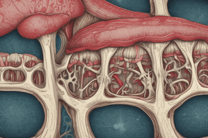
What are the two main components of bone?
What are the two main components of bone?
Inorganic mineral phase of crystals bound to protein, largely collagen
What is the main difference between technetium-99m diphosphonate and fluorine-18 sodium fluoride for bone scanning?
What is the main difference between technetium-99m diphosphonate and fluorine-18 sodium fluoride for bone scanning?
What is the purpose of voiding before imaging in bone scans?
What is the purpose of voiding before imaging in bone scans?
What is the process responsible for remodeling bone?
What is the process responsible for remodeling bone?
Where is increased activity commonly seen in older patients with arthritis in a bone scan?
Where is increased activity commonly seen in older patients with arthritis in a bone scan?
What are the two main parts of the anatomical skeleton?
What are the two main parts of the anatomical skeleton?
Which diseases may accelerate the bone turnover process?
Which diseases may accelerate the bone turnover process?
What are bone-seeking radiopharmaceuticals commonly used for skeletal imaging?
What are bone-seeking radiopharmaceuticals commonly used for skeletal imaging?
What can cause false-negative scans on a 99mTc-diphosphonate bone scan in the early stages?
What can cause false-negative scans on a 99mTc-diphosphonate bone scan in the early stages?
How is septic arthritis characterized on bone scans?
How is septic arthritis characterized on bone scans?
What imaging techniques can be used to evaluate osteomyelitis?
What imaging techniques can be used to evaluate osteomyelitis?
What is the characteristic appearance of hypertrophic pulmonary osteoarthropathy on bone scans?
What is the characteristic appearance of hypertrophic pulmonary osteoarthropathy on bone scans?
What can diffuse increased activity throughout the skeleton indicate in metabolic bone diseases?
What can diffuse increased activity throughout the skeleton indicate in metabolic bone diseases?
What imaging techniques are useful for identifying hidden hip and knee fractures?
What imaging techniques are useful for identifying hidden hip and knee fractures?
How long does the acute phase of a bone scan last after a fracture?
How long does the acute phase of a bone scan last after a fracture?
What type of fractures can be recognized by their location in consecutive ribs?
What type of fractures can be recognized by their location in consecutive ribs?
What imaging technique can provide early diagnosis and treatment options for stress injuries?
What imaging technique can provide early diagnosis and treatment options for stress injuries?
What imaging technique is more specific for identifying the causes of prosthesis failure, including infection?
What imaging technique is more specific for identifying the causes of prosthesis failure, including infection?
What imaging techniques are commonly used to differentiate normal marrow from leukocyte accumulation in cases of suspected infected joint replacements?
What imaging techniques are commonly used to differentiate normal marrow from leukocyte accumulation in cases of suspected infected joint replacements?
What is the most widely used method for accurately determining Bone Mineral Density (BMD)?
What is the most widely used method for accurately determining Bone Mineral Density (BMD)?
How is BMD expressed in terms of T-scores and Z-scores?
How is BMD expressed in terms of T-scores and Z-scores?
When should screening for osteoporosis begin in women and men without risk factors?
When should screening for osteoporosis begin in women and men without risk factors?
What tool provides a 10-year risk of hip and global fractures for assessing fracture risk?
What tool provides a 10-year risk of hip and global fractures for assessing fracture risk?
What are some common indications for bone scanning?
What are some common indications for bone scanning?
How do bone scans compare to radiography in detecting metastatic lesions?
How do bone scans compare to radiography in detecting metastatic lesions?
What is the role of radionuclide bone scans in imaging primary bone tumors?
What is the role of radionuclide bone scans in imaging primary bone tumors?
How do benign osseous neoplasms typically appear on bone scans?
How do benign osseous neoplasms typically appear on bone scans?
What factors can cause soft-tissue activity on bone scans?
What factors can cause soft-tissue activity on bone scans?
What is the typical dose range for Strontium-89 (89Sr) therapy?
What is the typical dose range for Strontium-89 (89Sr) therapy?
How is Strontium-89 (89Sr) chloride primarily excreted from the body?
How is Strontium-89 (89Sr) chloride primarily excreted from the body?
What is the typical interval between repeated doses of Strontium-89 (89Sr) therapy?
What is the typical interval between repeated doses of Strontium-89 (89Sr) therapy?
When does pain relief typically become apparent after administering Strontium-89 (89Sr) therapy?
When does pain relief typically become apparent after administering Strontium-89 (89Sr) therapy?
What are two other successful beta-emitting radiopharmaceuticals mentioned in the text?
What are two other successful beta-emitting radiopharmaceuticals mentioned in the text?
What is the physical half-life of Rhenium-186?
What is the physical half-life of Rhenium-186?
What is the soft-tissue range of Samarium-153 lexidronam (Quadramet)?
What is the soft-tissue range of Samarium-153 lexidronam (Quadramet)?
Why are alpha-emitting radionuclides like Radium-223 (223Ra) preferred over beta-emitters for treating bone metastases?
Why are alpha-emitting radionuclides like Radium-223 (223Ra) preferred over beta-emitters for treating bone metastases?
What is the primary elimination route for Radium-223 (223Ra) after administration?
What is the primary elimination route for Radium-223 (223Ra) after administration?
How many intravenous doses are included in the treatment course of Radium-223 (223Ra)?
How many intravenous doses are included in the treatment course of Radium-223 (223Ra)?
Flashcards are hidden until you start studying