Podcast
Questions and Answers
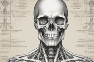
What classification does the skull belong to?
What classification does the skull belong to?
- Irregular
- Appendicular
- Axial (correct)
- Short
Which type of bone is the clavicle classified as according to developmental classification?
Which type of bone is the clavicle classified as according to developmental classification?
- Membranous (correct)
- Short
- Long
- Irregular
What type of bone is the femur classified as based on morphological classification?
What type of bone is the femur classified as based on morphological classification?
- Sesamoid
- Flat
- Short
- Long (correct)
What is the classification of the sternum?
What is the classification of the sternum?
Which type of bone is categorized as irregular among the following?
Which type of bone is categorized as irregular among the following?
What type of bone is the scapula classified as in the axial and appendicular classification?
What type of bone is the scapula classified as in the axial and appendicular classification?
In the context of structural types, which type forms the compact bone?
In the context of structural types, which type forms the compact bone?
Which of the following is classified as a sesamoid bone?
Which of the following is classified as a sesamoid bone?
What type of joint is the elbow?
What type of joint is the elbow?
Which type of joint is the pubic symphysis?
Which type of joint is the pubic symphysis?
What type of joint is the atlantoaxial joint?
What type of joint is the atlantoaxial joint?
Which muscle is primarily attached to the subclavius groove of the clavicle?
Which muscle is primarily attached to the subclavius groove of the clavicle?
What is the primary function of the biceps brachii muscle?
What is the primary function of the biceps brachii muscle?
Which of the following muscles is NOT part of the rotator cuff group?
Which of the following muscles is NOT part of the rotator cuff group?
What is the articulation point of the humerus referred to as the trochlea?
What is the articulation point of the humerus referred to as the trochlea?
Which type of muscle is skeletal muscle classified as?
Which type of muscle is skeletal muscle classified as?
What type of joint are the intercarpals associated with?
What type of joint are the intercarpals associated with?
Which nerve is related to the structures around the shoulder joint?
Which nerve is related to the structures around the shoulder joint?
Which muscle in the pectoral region is innervated by the medial pectoral nerve?
Which muscle in the pectoral region is innervated by the medial pectoral nerve?
What forms the posterior wall of the axilla?
What forms the posterior wall of the axilla?
Which artery branches into the axillary artery?
Which artery branches into the axillary artery?
Which nerve is responsible for innervating the deltoid muscle?
Which nerve is responsible for innervating the deltoid muscle?
What is the type of the elbow joint?
What is the type of the elbow joint?
Which of the following is NOT part of the contents of the cubital fossa?
Which of the following is NOT part of the contents of the cubital fossa?
Which muscle is primarily responsible for flexion of the radius at the elbow?
Which muscle is primarily responsible for flexion of the radius at the elbow?
What nerve primarily innervates the flexor carpi radialis muscle?
What nerve primarily innervates the flexor carpi radialis muscle?
Which muscle is NOT innervated by the radial nerve?
Which muscle is NOT innervated by the radial nerve?
Which muscle acts as the main shoulder flexor?
Which muscle acts as the main shoulder flexor?
Which structure separates the compartments of the forearm?
Which structure separates the compartments of the forearm?
Which of the following nerves innervates the serratus anterior muscle?
Which of the following nerves innervates the serratus anterior muscle?
What is the function of the palmaris brevis muscle?
What is the function of the palmaris brevis muscle?
What type of joint is the wrist joint?
What type of joint is the wrist joint?
Flashcards
Axial Bones
Axial Bones
Bones that make up the skull, vertebral column and ribs.
Appendicular Bones
Appendicular Bones
Bones that make up the limbs, shoulder girdle and pelvic girdle.
Cartilaginous Bones
Cartilaginous Bones
Bones that develop from a cartilage template. This template is replaced by bone cells.
Membranous Bones
Membranous Bones
Signup and view all the flashcards
Mixed Bones
Mixed Bones
Signup and view all the flashcards
Long Bones
Long Bones
Signup and view all the flashcards
Flat Bones
Flat Bones
Signup and view all the flashcards
Irregular Bones
Irregular Bones
Signup and view all the flashcards
Sutures
Sutures
Signup and view all the flashcards
Secondary Cartilaginous
Secondary Cartilaginous
Signup and view all the flashcards
Synovial Hinge
Synovial Hinge
Signup and view all the flashcards
Fibrous
Fibrous
Signup and view all the flashcards
Synovial Plane
Synovial Plane
Signup and view all the flashcards
Synovial Pivot
Synovial Pivot
Signup and view all the flashcards
Synovial Bicondylar
Synovial Bicondylar
Signup and view all the flashcards
Synovial Ellipsoid
Synovial Ellipsoid
Signup and view all the flashcards
Synovial Ball and Socket
Synovial Ball and Socket
Signup and view all the flashcards
Clavicle
Clavicle
Signup and view all the flashcards
Pectoralis major
Pectoralis major
Signup and view all the flashcards
Pectoralis minor
Pectoralis minor
Signup and view all the flashcards
Subclavius
Subclavius
Signup and view all the flashcards
Clavipectoral fascia
Clavipectoral fascia
Signup and view all the flashcards
Subscapularis
Subscapularis
Signup and view all the flashcards
Teres major
Teres major
Signup and view all the flashcards
Latissimus dorsi
Latissimus dorsi
Signup and view all the flashcards
Biceps brachii
Biceps brachii
Signup and view all the flashcards
Coracobrachialis
Coracobrachialis
Signup and view all the flashcards
Brachialis
Brachialis
Signup and view all the flashcards
Triceps brachii
Triceps brachii
Signup and view all the flashcards
Supinator
Supinator
Signup and view all the flashcards
Pronator teres
Pronator teres
Signup and view all the flashcards
Flexor carpi radialis
Flexor carpi radialis
Signup and view all the flashcards
Flexor digitorum superficialis
Flexor digitorum superficialis
Signup and view all the flashcards
Study Notes
Presentation Overview
- Presenter: Dr. Eman El Sawaf
- Department: Anatomy & Embryology
- University: Helwan University
Practical & Oral Sheet Revision
- The presentation involved practical and oral sheet revisions of anatomical topics.
Bone Classification: Regional
- Skull: Axial
- Humerus: Appendicular
- Sternum: Axial
- Vertebra: Axial
- Rib: Axial
- Scapula: Appendicular
- Hip bone: Appendicular
- Femur: Appendicular
Bone Classification: Developmental
- Scapula: Membranous
- Clavicle: Mixed
- Skull vault: Membranous
- Femur: Cartilaginous
- Mandible: Mixed
Bone Classification: Morphological
- Humerus: Long
- Scapula: Flat
- Sternum: Flat
- Clavicle: Long
- Vertebra: Irregular
- Patella: Sesamoid
- Hip bone: Irregular
- Carpals: Short
- Tarsals: Short
Bone Structural Type
- Long bone: 1- Cancellous, 2- Compact, 3- Compact
- Vertebra: 1- Compact, 2- Cancellous
Muscle Type, Movement & Nerve Supply
- Visceral: Involuntary, Autonomic
- Cardiac: Involuntary, Autonomic
- Skeletal: Voluntary, Somatic
Joint Classification
- Sutures: Fibrous
- IVD (Intervertebral disc): Secondary cartilaginous
- Elbow: Synovial hinge
- Inferior tibio-fibular joint: Fibrous
- Pubic symphysis: Secondary cartilaginous
- Intercarpals: Synovial plane
- Ankle: Synovial hinge
- Intertarsals: Synovial plane
- Shoulder: Synovial ball and socket
- Knee: Synovial bicondylar
- Wrist: Synovial ellipsoid
Bones (General Features)
- Clavicle (Superior view): Acromial end, Posterior border, Superior surface, Anterior border, Sternal end
- Clavicle (Inferior view): Subclavius groove
Clavicle (Articulations)
- Sterno-clavicular joint
- Acromio-clavicular joint
Scapula (Anterior view)
- Acromion process
- Superior angle
- Superior border
- Coracoid process
- Lateral angle (Glenoid cavity)
- Medial border
- Subscapular fossa
- Lateral border
- Inferior angle
Scapula (Posterior view)
- Acromion process
- Supraspinous fossa
- Spine
- Infraspinous fossa
Scapula (Articulations)
- Acromio-clavicular joint
- Shoulder joint
Humerus (Anterior view)
- Head
- Anatomical neck
- Surgical neck
Humerus (Anterior view)
- Greater tuberosity
- Deltoid tuberosity
- Lesser tuberosity
- Bicipital groove
Humerus (Anterior view)
- Radial fossa
- Coronoid fossa
- Lateral epicondyle
- Medial epicondyle
- Capitulum
- Trochlea
Humerus (Posterior view)
- Radial (Spiral) groove
- Medial epicondyle
- Olecranon fossa
- Trochlea
- Lateral epicondyle
Humerus (Articulations)
- Shoulder joint
- Elbow joint
Radius (Anterior view)
- Neck
- Oblique line
- Head
- Radial tuberosity
- Interosseous border
- Ulnar notch
- Styloid process
Radius (Posterior view)
- Dorsal tubercle
Radius (Articulation)
- Elbow joint
- Superior Radio Ulnar Joint
- Wrist Joint
- Inferior Radio Ulnar Joint
Ulna (Anterior view)
- Trochlear notch
- Radial notch
- Supinator fossa
- Olecranon process
- Coronoid process
- Ulnar tuberosity
- Interosseus border
- Head
- Styloid process
Ulna (Articulation)
- Elbow Joint
- Superior Radio Ulnar Joint
- Inferior Radio Ulnar Joint
Bones (Attached & Related Structures)
Identify the Attached Muscle
- Pectoralis major (Clavicular head & Sternocostal head)
- Pectoralis minor
- Subclavius
- Trapezius
- Latissimus Dorsi
- Levator scapulae
- Rhomboid minor
- Rhomboid major
- Deltoid
- Subscapularis
- Supraspinatus & Infraspinatus
- Teres Minor
- Teres Major
- Serratus anterior
- Biceps brachii
- Coracobrachialis
- Brachialis
- Triceps brachii
- Common extensor origin
- Common flexor origin
Mention the Related Structure (Arm/Bone specific)
- Axillary nerve
- Posterior circumflex humeral artery
- Radial nerve
- Profunda brachii artery
- Ulnar nerve
Identify the Following
-
Subclavius
-
Pectoralis minor
-
Pectoralis major
-
First rib
-
Superior border of scapula
-
Clavicle
-
Clavipectoral fascia
-
Bicipital groove of the humerus
-
Scapula & Subscapularis muscle
-
Teres major muscle
-
Latissimus dorsi muscle
-
Skin & fascia (axillary fascia)
-
Cords of the brachial plexus
-
Biceps & Coracobrachialis muscles
-
Lymph nodes
-
Axillary artery
-
Axillary vein
-
Brachial plexus
-
Levator scapulae
-
Rhomboid minor
-
Rhomboid major
-
Supraspinatus
-
Infraspinatus
-
Teres minor
-
Teres major
-
Subscapularis
-
Serratus anterior
-
Long head of Biceps brachii
-
Short head of Biceps brachii
-
Tendon of biceps brachii
-
Bicipital aponeurosis
-
Lateral head of Triceps brachii
-
Long head of Triceps brachii
-
Medial head of Triceps brachii
-
Cubital fossa
-
Brachial artery
-
Median nerve
-
Pronator teres
-
Flexor carpi radialis
-
Palmaris longus
-
Flexor carpi ulnaris
-
Flexor digitorum superficialis
-
Flexor digitorum profundus
-
Flexor pollicis longus
-
Pronator quadratus
-
Brachioradialis
-
Extensor carpi radialis longus
-
Extensor carpi radialis brevis
-
Extensor digitorum
-
Extensor digiti minimi
-
Extensor carpi ulnaris- Anconeus
-
Supinator
-
Abductor pollicis longus
-
Extensor pollicis brevis
-
Extensor pollicis longus
-
Extensor indicis
-
Radial artery
-
Ulnar artery
-
Axillary vein
-
Basilic vein
-
Brachial veins
-
Cephalic vein
-
Median cubital vein
-
Axillary nerve
-
Musculocutaneous nerve
-
Median nerve
-
Ulnar nerve
-
Flexor retinaculum
-
Extensor retinaculum
-
Palmar aponeurosis
-
Hypothenar muscles
-
Palmaris brevis
-
Adductor pollicis
-
Thenar muscles
-
Lumbrical muscles
-
Palmar interosseus muscles
-
Dorsal interosseus muscles
Joint section (name, type, articulating bones)
- Elbow joint: Synovial hinge (Humerus, Radius, Ulna)
- Wrist joint: Synovial ellipsoid (Radius, scaphoid, lunate, triquetrum)
Additional notes
- The presentation heavily emphasizes identification and classification of anatomical structures.
Studying That Suits You
Use AI to generate personalized quizzes and flashcards to suit your learning preferences.