Podcast
Questions and Answers
What is the primary role of skeletal muscles in body movement?
What is the primary role of skeletal muscles in body movement?
- Generating heat for the body
- Providing structural support to the bones
- Contracting and relaxing to mechanically move the body (correct)
- Converting chemical energy into electrical energy
What structure forms the postsynaptic part of the neuromuscular junction?
What structure forms the postsynaptic part of the neuromuscular junction?
- Myelin sheath
- Synaptic cleft
- Axon terminal
- Motor end plate (correct)
Which neurotransmitter is involved in the activation of skeletal muscle fibers at the neuromuscular junction?
Which neurotransmitter is involved in the activation of skeletal muscle fibers at the neuromuscular junction?
- Acetylcholine (correct)
- Serotonin
- Norepinephrine
- Dopamine
What condition results from antibodies against cholinergic receptors in the postsynaptic membrane?
What condition results from antibodies against cholinergic receptors in the postsynaptic membrane?
What is the purpose of junctional folds in the motor end plate?
What is the purpose of junctional folds in the motor end plate?
What initiates the functional activation of skeletal muscle fibers to contract?
What initiates the functional activation of skeletal muscle fibers to contract?
Which disorder is characterized by antibodies to calcium channels in the presynaptic axon terminal?
Which disorder is characterized by antibodies to calcium channels in the presynaptic axon terminal?
What role do voltage-gated sodium channels (VGSCs) play in neuromuscular transmission?
What role do voltage-gated sodium channels (VGSCs) play in neuromuscular transmission?
What is the primary role of myosin in skeletal muscle contraction?
What is the primary role of myosin in skeletal muscle contraction?
What must occur for a skeletal muscle contraction to take place?
What must occur for a skeletal muscle contraction to take place?
What process leads to the reuptake of calcium ions into the sarcoplasmic reticulum during muscle relaxation?
What process leads to the reuptake of calcium ions into the sarcoplasmic reticulum during muscle relaxation?
What is the main consequence of the body entering rigor mortis after death?
What is the main consequence of the body entering rigor mortis after death?
What happens to calcium ions when neural stimulation of muscle fibers stops?
What happens to calcium ions when neural stimulation of muscle fibers stops?
What triggers the muscle fiber repolarization during relaxation?
What triggers the muscle fiber repolarization during relaxation?
What is the function of ATP in the myosin-actin contraction cycle?
What is the function of ATP in the myosin-actin contraction cycle?
What initiates the contraction cycle in skeletal muscle fibers?
What initiates the contraction cycle in skeletal muscle fibers?
What effect does botulinum toxin have on neuromuscular function?
What effect does botulinum toxin have on neuromuscular function?
What is the role of Dihydropyridine (DHP) receptors in skeletal muscle contraction?
What is the role of Dihydropyridine (DHP) receptors in skeletal muscle contraction?
Which statement accurately describes the organization of a sarcomere?
Which statement accurately describes the organization of a sarcomere?
Which of the following best describes the sliding filament theory?
Which of the following best describes the sliding filament theory?
What is the primary source of calcium influx in skeletal muscles?
What is the primary source of calcium influx in skeletal muscles?
How many actin filaments can one myosin filament interact with in a sarcomere?
How many actin filaments can one myosin filament interact with in a sarcomere?
What characterizes the I-band in a sarcomere?
What characterizes the I-band in a sarcomere?
During repolarization of the T-tubule, what happens to the calcium release channels in the SR?
During repolarization of the T-tubule, what happens to the calcium release channels in the SR?
Flashcards
Skeletal Muscle Contraction
Skeletal Muscle Contraction
Skeletal muscles move the body by contracting and relaxing. This movement is coordinated with bones.
Neuromuscular Junction (NMJ)
Neuromuscular Junction (NMJ)
The connection point between a motor neuron and a muscle fiber where signals are passed to make the muscle contract.
Synaptic Cleft
Synaptic Cleft
The small gap between the nerve ending and muscle fiber in the NMJ.
Motor End Plate
Motor End Plate
Signup and view all the flashcards
Acetylcholine (ACh)
Acetylcholine (ACh)
Signup and view all the flashcards
Eaton-Lambert Syndrome
Eaton-Lambert Syndrome
Signup and view all the flashcards
Myasthenia Gravis
Myasthenia Gravis
Signup and view all the flashcards
Motor Neuron
Motor Neuron
Signup and view all the flashcards
Botulism
Botulism
Signup and view all the flashcards
Neuromuscular Junction
Neuromuscular Junction
Signup and view all the flashcards
Sarcomere
Sarcomere
Signup and view all the flashcards
Sliding Filament Theory
Sliding Filament Theory
Signup and view all the flashcards
Excitation-Contraction Coupling
Excitation-Contraction Coupling
Signup and view all the flashcards
Ca++2
Ca++2
Signup and view all the flashcards
Myofibrils
Myofibrils
Signup and view all the flashcards
Sarcoplasmic Reticulum (SR)
Sarcoplasmic Reticulum (SR)
Signup and view all the flashcards
Myosin
Myosin
Signup and view all the flashcards
Sarcomere contraction
Sarcomere contraction
Signup and view all the flashcards
ATP's role in muscle relaxation
ATP's role in muscle relaxation
Signup and view all the flashcards
Rigor Mortis
Rigor Mortis
Signup and view all the flashcards
Role of Calcium in Muscle Contraction
Role of Calcium in Muscle Contraction
Signup and view all the flashcards
SERCA
SERCA
Signup and view all the flashcards
Muscle relaxation triggers
Muscle relaxation triggers
Signup and view all the flashcards
Contraction cycle
Contraction cycle
Signup and view all the flashcards
Study Notes
Body Movement - Year 2, Unit 1
- Course: Body Movement
- Year: 2
- Unit: 1
- Academic Year: 2024-2025
- Instructor: Dr. Tariq Alshaibani
Learning Objectives
- Describe the neuromuscular junction (NMJ) and how action potentials in the motor neuron lead to skeletal muscle excitation and contraction.
- Detail the molecular and electrical makeup of muscle cell excitation-contraction coupling.
- Define the elements of the sarcomere that underlie striated muscle contraction.
Skeletal Muscle Contraction & Body Movement
- Skeletal muscles contract and relax to move the body (e.g., walking, running, manipulating objects).
- Skeletal muscle works with bones to create body movements.
- Muscle fibers contract, generating tension transmitted through tendons to bones, enabling movement.
Neuromuscular Control
- Stimulation of skeletal muscle fibers (motor neurons in the spinal cord/brainstem) is the only way to make them contract.
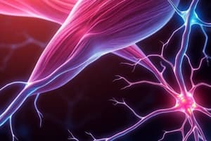
The Neuromuscular Junction (NMJ)
- The NMJ is where the axon terminal of a motor neuron meets a muscle fiber.
- The synaptic cleft is a 20-30nm gap containing acetylcholinesterase (AChE).
- The motor end plate is the postsynaptic part of the NMJ; it features junctional folds increasing the surface area of the postsynaptic membrane.
- The motor end plate contains ACh receptors and voltage-gated sodium channels.
Acetylcholine Release
- Calcium channels are localized around dense bars on the presynaptic membrane where vesicles containing ACh fuse.
- Acetylcholine (ACh) receptors are located at the top of the subneural cleft, and voltage-gated sodium channels are found in the bottom half of the subneural cleft.
- Voltage-gated sodium channels facilitate action potential generation.
Main Schema for Neuromuscular Transmission
- Action potentials travel down motor neuron axons, triggering calcium release.
- Calcium release triggers the release of acetylcholine (ACh).
- ACh binds to receptors on the muscle fiber, initiating an action potential.
- This action potential triggers calcium release from the sarcoplasmic reticulum, leading to muscle contraction.
Disorders
- Eaton-Lambert syndrome: Autoimmune disorder affecting calcium channels in the presynaptic axon terminal.
- Botulism: Toxin produced by Clostridium botulinum bacteria, inhibiting the release of acetylcholine (ACh).
- Myasthenia gravis: Autoimmune disorder affecting cholinergic receptors in the postsynaptic membrane.
Excitation-Contraction Coupling
- Describes how the action potential entering the muscle cell causes a contraction within the muscle.
Ca+2 for Excitation-Contraction Coupling
- Action potentials travel through T-tubules.
- Calcium released from the sarcoplasmic reticulum (SR).
- Calcium binds to troponin, causing tropomyosin to shift, exposing binding sites on actin.
- Myosin heads bind to actin, producing a power stroke, pulling actin filaments.
Release of Ca+2 from Sarcoplasmic Reticulum
- Dihydropyridine (DHP) receptors sense the muscle action potential in the membrane.
- DHP receptors are linked to calcium release channels (ryanodine receptors).
- Triggering calcium release from the sarcoplasmic reticulum by activating DHP receptors in the T-tubules.
Organization of Skeletal Muscle
- Describes how skeletal muscle fibers, fascicles, and bundles are organized.
Skeletal Muscle Structure- Gross to Cellular
- Muscle, tendon, and bone relationships are described, from whole muscle to individual muscle fibers.
- Myofibril structure is described.
The Myofilaments
- Arrangement of thin (actin) and thick (myosin) filaments creates the sarcomere structure that is responsible for skeletal muscle contraction.
The Sarcomere
- The sarcomere is the fundamental unit of skeletal muscle contraction.
- A band contains thick filaments; an I-band contains thin filaments.
- The structure of Z-lines, A-bands, I-bands, and M-lines is detailed for understanding skeletal muscle contraction.
The Sliding Filament Theory of Contraction
- Myosin filaments pull actin filaments closer together, shortening sarcomeres within a muscle fiber.
- Sliding of actin past myosin generates muscle tension.
Ultrastructure of Skeletal Muscle – Molecular Basis of Contraction
- Description of the structural components related to skeletal muscle contraction.
Contractile Proteins of Skeletal Muscle
- Myosin and actin filament structure are described to explain how they enable skeletal muscle contraction.
The Molecular Basis of Skeletal Muscle Contraction
- How Ca+2 levels and ATP hydrolysis lead to skeletal muscle contraction and enable myosin to detach from actin are explained.
The Contraction Cycle - Role of Ca+2 and ATP
- The process, starting with ATP hydrolysis, of how myosin heads bind to actin followed by the power stroke (myosin heads pull actin) and detachment, and repeating to enable skeletal muscle contraction is explained.
Rigor Mortis
- After death, the process of rigor mortis, where muscles stiffen, occurs because there is no more ATP to detach myosin from actin.
In order for a skeletal muscle contraction to occur: (Summary)
- There must be a neural stimulus.
- There must be Ca+2 in the cytosol of the muscle cells.
- ATP must be available for energy.
- Relaxation occurs when Ca+2 levels decrease and ATP is available.
Studying That Suits You
Use AI to generate personalized quizzes and flashcards to suit your learning preferences.