Podcast
Questions and Answers
What structural feature distinguishes arteries from veins at a microscopic level?
What structural feature distinguishes arteries from veins at a microscopic level?
- The tunica media in arteries is generally more muscular and thicker. (correct)
- Veins lack a tunica media entirely.
- The presence of a thicker tunica intima in veins.
- Arteries have valves within the tunica adventitia.
Which characteristic of veins is crucial for counteracting the effects of gravity in the lower extremities?
Which characteristic of veins is crucial for counteracting the effects of gravity in the lower extremities?
- The presence of valves that prevent backflow. (correct)
- The ability to actively contract and propel blood upwards.
- The thicker muscular walls of the tunica media.
- The smaller lumen size which increases blood velocity.
What is the primary functional difference between elastic arteries and muscular arteries?
What is the primary functional difference between elastic arteries and muscular arteries?
- Elastic arteries maintain consistent blood pressure, while muscular arteries control blood flow to organs. (correct)
- Elastic arteries primarily facilitate gas exchange, while muscular arteries transport blood away from the heart.
- Elastic arteries are involved in vasoconstriction, while muscular arteries are not.
- Elastic arteries primarily distribute blood to specific organs, while muscular arteries handle pressure from the heart.
What structural adaptation allows sinusoids, unlike capillaries, to accommodate the exchange of large molecules and cells?
What structural adaptation allows sinusoids, unlike capillaries, to accommodate the exchange of large molecules and cells?
If a patient has a thrombus (blood clot) that obstructs a main arterial supply to a region, what circulatory adaptation helps maintain blood flow to the affected tissue?
If a patient has a thrombus (blood clot) that obstructs a main arterial supply to a region, what circulatory adaptation helps maintain blood flow to the affected tissue?
Why is the tunica adventitia thicker in veins than in corresponding arteries?
Why is the tunica adventitia thicker in veins than in corresponding arteries?
How does the lymphatic system assist the cardiovascular system?
How does the lymphatic system assist the cardiovascular system?
What is the role of central lymphoid tissues such as bone marrow in the context of the cardiovascular and lymphatic systems?
What is the role of central lymphoid tissues such as bone marrow in the context of the cardiovascular and lymphatic systems?
What is the functional significance of arteriovenous anastomoses (shunts)?
What is the functional significance of arteriovenous anastomoses (shunts)?
What effect does vasoconstriction have on vascular resistance and blood flow?
What effect does vasoconstriction have on vascular resistance and blood flow?
What mechanism primarily drives venous return from the lower extremities back to the heart?
What mechanism primarily drives venous return from the lower extremities back to the heart?
What is the role of the vasa vasorum in the walls of large blood vessels?
What is the role of the vasa vasorum in the walls of large blood vessels?
What is the primary factor determining the distribution of cardiac output to different tissues?
What is the primary factor determining the distribution of cardiac output to different tissues?
How does blood viscosity affect vascular resistance?
How does blood viscosity affect vascular resistance?
End arteries are characterized by which of the following properties?
End arteries are characterized by which of the following properties?
Why is knowledge about end arteries clinically significant?
Why is knowledge about end arteries clinically significant?
How do the respiratory movements aid in venous return?
How do the respiratory movements aid in venous return?
Where are superficial veins located, and what is their function?
Where are superficial veins located, and what is their function?
What function do valves serve in deep veins?
What function do valves serve in deep veins?
Which vascular structure connects arterioles directly to venules, bypassing capillaries?
Which vascular structure connects arterioles directly to venules, bypassing capillaries?
What is the composition of the tunica intima of a blood vessel?
What is the composition of the tunica intima of a blood vessel?
Which of the features is characteristic of veins but not of arteries?
Which of the features is characteristic of veins but not of arteries?
Which vessels are primarily responsible for regulating blood flow into capillary beds?
Which vessels are primarily responsible for regulating blood flow into capillary beds?
What is the function of the lymphatic system in relation to the cardiovascular system?
What is the function of the lymphatic system in relation to the cardiovascular system?
If a patient's blood pressure is recorded as 120/80 mmHg, what does the '80' signify?
If a patient's blood pressure is recorded as 120/80 mmHg, what does the '80' signify?
What effect does an increase in blood vessel length have on vascular resistance, all other factors being equal?
What effect does an increase in blood vessel length have on vascular resistance, all other factors being equal?
Which type of blood vessel is best suited for substance exchange between blood and tissues?
Which type of blood vessel is best suited for substance exchange between blood and tissues?
What would be the effect of removing the spleen?
What would be the effect of removing the spleen?
Which scenario best describes 'collateral supply' in the circulatory system?
Which scenario best describes 'collateral supply' in the circulatory system?
What is the significance of the difference in pressure between a venule (16 mmHg) and the right ventricle (0 mmHg) in venous return?
What is the significance of the difference in pressure between a venule (16 mmHg) and the right ventricle (0 mmHg) in venous return?
What is the role of the thoracic duct in the lymphatic system?
What is the role of the thoracic duct in the lymphatic system?
Why are valves absent in the venae cavae, hepatic, renal, uterine, ovarian, cerebral, spinal, and pulmonary veins?
Why are valves absent in the venae cavae, hepatic, renal, uterine, ovarian, cerebral, spinal, and pulmonary veins?
Venules drain capillaries and converge to form which of the following blood vessels?
Venules drain capillaries and converge to form which of the following blood vessels?
According to the Hemodynamics model, what blood vessels impact blood flow?
According to the Hemodynamics model, what blood vessels impact blood flow?
Arteries are characterized as having which of the following features?
Arteries are characterized as having which of the following features?
Flashcards
What is the heart?
What is the heart?
A four-chambered muscular organ that pumps blood throughout the body.
What are arteries?
What are arteries?
Large blood vessels carrying oxygenated blood away from the heart.
What are arterioles?
What are arterioles?
Minute branches of arteries, visible to the naked eye.
What are capillaries?
What are capillaries?
Signup and view all the flashcards
What are veins?
What are veins?
Signup and view all the flashcards
What are venules?
What are venules?
Signup and view all the flashcards
What are sinusoids?
What are sinusoids?
Signup and view all the flashcards
What is the tunica intima?
What is the tunica intima?
Signup and view all the flashcards
What is the tunica media?
What is the tunica media?
Signup and view all the flashcards
What is the tunica adventitia/externa?
What is the tunica adventitia/externa?
Signup and view all the flashcards
What is endothelium?
What is endothelium?
Signup and view all the flashcards
What is arterial anastomosis?
What is arterial anastomosis?
Signup and view all the flashcards
What is collateral supply?
What is collateral supply?
Signup and view all the flashcards
What are end arteries?
What are end arteries?
Signup and view all the flashcards
What is Arterio-venous anastomosis (shunt)?
What is Arterio-venous anastomosis (shunt)?
Signup and view all the flashcards
What is vasa vasorum?
What is vasa vasorum?
Signup and view all the flashcards
What is a lymphatic system?
What is a lymphatic system?
Signup and view all the flashcards
What is lymph?
What is lymph?
Signup and view all the flashcards
What is central lymphoid tissue?
What is central lymphoid tissue?
Signup and view all the flashcards
What are peripheral lymphoid organs?
What are peripheral lymphoid organs?
Signup and view all the flashcards
What is blood flow?
What is blood flow?
Signup and view all the flashcards
What is cardiac output (CO)?
What is cardiac output (CO)?
Signup and view all the flashcards
What is systolic blood pressure?
What is systolic blood pressure?
Signup and view all the flashcards
What is diastolic blood pressure?
What is diastolic blood pressure?
Signup and view all the flashcards
What is vascular resistance?
What is vascular resistance?
Signup and view all the flashcards
What is venous return?
What is venous return?
Signup and view all the flashcards
What is the skeletal muscle pump?
What is the skeletal muscle pump?
Signup and view all the flashcards
Study Notes
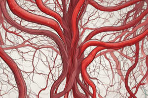
Classification of Blood Vessels
- The blood vascular system consists of the heart, arteries, veins, and capillaries.
- The heart is a four-chambered muscular organ which pumps the blood.
- Each half of the heart has an atrium for receiving blood and a ventricle for pumping blood.
Arteries
- Arteries are large blood vessels that carry oxygenated blood away from the heart.
- Characteristically, arteries are thick-walled, have a smaller lumen, and do not have valves.
- Arteries are usually accompanied by veins and nerves, forming a neurovascular bundle.
- Arterioles are minute branches of arteries visible to the naked eye.
Types of Arteries
- Large sized arteries (Elastic arteries) include the pulmonary trunk, aorta, and their branches like brachiocephalic, subclavian, and common carotid artery
- Medium sized arteries (Muscular arteries) include the radial, popliteal, and temporal arteries.
- Smaller arteries are called arterioles.
Veins
- Veins are blood vessels that carry deoxygenated blood back to the heart.
- Veins have thin walls, a larger lumen, and valves to maintain unidirectional blood flow against gravity.
- Valves are absent in the venae cavae, hepatic, renal, uterine, ovarian, cerebral, spinal, pulmonary, and umbilical veins.
- Venules are smaller veins.
Types of Veins
- Large-sized veins include the vena cava and portal vein with their tributaries.
- Medium-sized and small-sized veins, also known as venules, are other types of veins.
Capillaries and Sinusoids
- Capillaries are networks of microscopic vessels connecting arterioles and venules, facilitating nutrient exchange with tissues.
- Sinusoids are large, irregular vascular spaces surrounded by organ parenchyma.
- Sinusoids differ from capillaries by having a wider and irregular lumen and thinner, possibly incomplete walls.
- Sinusoids exist in the liver, spleen, and bone marrow.
Microscopic Structure of Arteries and Veins
- Both arteries and veins have three coats or tunics, which include the tunica intima (innermost), tunica media (middle), and tunica adventitia (outermost), along with an endothelial layer.
Endothelium
- The endothelium is a special type of epithelium lining the interior surface of blood and lymphatic vessels.
- It forms an interface between circulating blood or lymph in the lumen and the vessel wall.
- The endothelium is a simple squamous type of epithelium.
Microscopic Structure of Arteries: Tunica Intima
- The tunica intima, the innermost layer, is composed of the endothelium (simple squamous epithelium), basal lamina, subendothelial connective tissue, and internal elastic lamina made of elastic material.
Microscopic Structure of Arteries: Tunica Media
- The tunica media surrounds the tunica intima.
- It consists of elastic fibers and smooth muscles.
Microscopic Structure of Arteries: Tunica Adventitia/Externa
- The tunica adventitia/externa, the outermost layer, is composed mainly of connective tissue.
- It is separated from the tunica media by the external elastic lamina.
Microscopic Structure of Veins
- Veins also have tunics similar to that of arteries.
- The three layers of veins are not well defined.
Microscopic Structure of A Large (Elastic) artery
- Tunica intima consists of all 4 components
- Tunica media is the thickest of the three layers, contains a high proportion of elastic fibers, and has layers of smooth muscle cells between elastic lamellae
- Tunica adventitia is made up of connective tissues, is relatively thinner, and contains collagen fibers
Microscopic Structure of A Medium Sized (Muscular) Artery
- Tunica intima contains all 4 components
- Internal elastic lamina is more clearly visible
- Tunica media contains high proportion of smooth muscle cells
- Tunica adventitia is made of connective tissue, and is thicker than that of elastic artery
Microscopic Structure of a Large Vein
- Tunica intima consists of endothelial cells, subendothelial connective tissues, and few smooth muscle cells
- Tunica media consists of smooth muscle cells, collagen fibers and fibroblasts, and is thinner than that of in arteries
- Tunica adventitia is thicker, consists of smooth muscle cells, collagen, elastic fibers & fibroblasts
Microscopic Structure of a Medium Sized Vein
- Tunica intima consists of little or no sub endothelial connective tissue
- Tunica media consists of few layers of smooth muscle cells and collagen fibres and elastic fibers
- Tunica adventitia is thicker than the media, and consists of collagen and elastic fibers
Differences Between Artery and Vein
- Arteries have a smaller overall diameter and smaller lumen.
- Artery walls are thicker and do not collapse after death, and there is more rapid blood flow.
- Arteries carry blood with a high oxygen content, and their tunica intima is relatively thicker.
- Arteries have well-developed internal and external elastic laminae.
- The tunica media in arteries is muscular and considerably thicker.
- The tunica adventitia in arteries is about half the thickness of the media and has a high elastin content, and they have no valves.
- Veins have a large overall diameter and larger lumen.
- Vein walls are thinner and collapse after death, with slower blood flow, and have a low oxygen content.
- The tunica intima in veins is relatively thinner.
- Veins have less developed internal and external elastic laminae, and their tunica media is generally a thin muscular layer.
- The tunica adventitia is the thickest coat of wall and is composed chiefly of collagen fibers, and they have valves.
Arterial Supply and Venous Drainage
- Arterial anastomosis is the communication between arteries or their branches.
- Collateral supply includes circulation through the anastomosis and supply of blood through a network of minor vessels joining adjacent vessels when a major vessel is obstructed.
- End arteries do not anastomose with neighbors, such as the central artery of the retina, central branches of cerebral arteries, and arteries of the spleen and kidney.
Arterio-Venous Anastomosis (Shunt)
- It is the communication between an artery and a vein
- When the organ is active these shunts are closed and the blood circulates through the capillaries
- When the organ is at rest, the blood bypasses the capillary bed and is shunted back through the arterio-vnous anastomosis
- The shunt vessels may be straight or coiled, possess a thick muscular coat, and are under the influence of the sympathetic system
- Shunts are found in simple structures found on the skin of the nose, lips, erectile tissue of the sexual organ, and thyroid gland.
Deep and Superficial Veins; Venous Valves
- Deep veins are located deep within the body, often near arteries with the same name, such as the femoral vein with the femoral artery.
- Superficial veins are closer to the body surface.
- Venous valves maintain unidirectional blood flow against gravity.
- Due to low venous pressure (7 mmHg), valves are important for venous return, and they prevent backflow.
Vasa Vasorum
- Vasa vasorum is the network of small blood vessels supplying large blood vessels.
Lymphoid Tissues
- Lymphoid tissues offer a drainage system accessory to the venous system.
- Lymphatics remove larger particles, including proteins and particulate matter.
- The lymphatics act as a 'drainage system of coarse type’.
- Lymph is the tissue fluid flowing in the lymphatic system.
Lymphoid Tissues in the Body
- Central lymphoid tissue includes bone marrow and the thymus.
- Peripheral lymphoid organs are lymph nodes and the spleen.
- Circulating lymphocytes, and lymphatic follicles (nodules)
- The bone marrow aids in B-lymphocyte differentiation into antibody-synthesizing plasma cells.
- The thymus facilitates T-lymphocyte differentiation.
- Lymph nodes filter the lymph and remove particulate matter via phagocytic action, and produce lymphocytes, and Plasma cells produce antibodies
- The spleen filters blood by removing worn-out RBCs, WBCs, and platelets.
Major Lymphatic Ducts
- Filtered lymph passes through larger lymphatics.
- It is collected into two large trunks: the thoracic duct and the right lymphatic duct.
Hemodynamics
- Blood flow equals the volume of blood flowing through a tissue in a given period (mL/min)
- Total blood flow is the cardiac output (CO)
- CO is the volume of blood circulating through systemic (or pulmonary) blood vessels/minute
- Can be calculated by the formula : CO = heart rate (HR) x stroke volume (SV)
- The distribution of CO relies on pressure differences driving blood flow from higher to lower pressure and resistance to blood flow, as a higher resistance leads to smaller blood flow.
Blood pressure
- Contraction of the Ventricles generates the blood pressure
- The systolic blood pressure (BP) is the highest pressure attained during systole
- The diastolic blood pressure (DBP is the lowest arterial pressure during diastole
- The pressure falls progressively with distance from the left ventricle
- The blood pressure also depends on the total volume of blood.
Vascular resistance
- Opposition to blood flow due to friction between blood and walls of blood vessels
- Size of lumen – vasoconstriction makes the lumen smaller, causing greater resistance
- Blood viscosity - ratio of RBCs to plasma and protein concentration, higher viscosity means higher resistance
- Total blood vessel length - resistance is directly proportional to the length of the vessel since 400 miles of additional blood vessels are required for each 2.2lb. of fat
Venous return
- Volume of returning blood flow to the heart throughout the systemic veins
- Occurs due to pressure generated via constriction of the left ventricle
- A small amount of pressure the Venule (16 mmHg) to the right ventricle (0 mmHg) that is a sufficient amount
Studying That Suits You
Use AI to generate personalized quizzes and flashcards to suit your learning preferences.