Podcast
Questions and Answers
Following a blood draw and subsequent spin in a centrifuge, which layer contains leukocytes and platelets?
Following a blood draw and subsequent spin in a centrifuge, which layer contains leukocytes and platelets?
- Formed elements
- Buffy coat (correct)
- Plasma
- Hematocrit
Which of the following is a primary function of blood?
Which of the following is a primary function of blood?
- Filtering metabolic waste for excretion
- Producing digestive enzymes
- Synthesizing hormones
- Transporting oxygen and carbon dioxide (correct)
If a patient's blood sample shows a higher than normal viscosity, which component of the blood is most likely affected?
If a patient's blood sample shows a higher than normal viscosity, which component of the blood is most likely affected?
- Higher water content
- Lower blood volume (correct)
- Elevated erythrocyte count
- Increased plasma protein
What role does albumin play in maintaining fluid balance within the bloodstream?
What role does albumin play in maintaining fluid balance within the bloodstream?
In erythrocytes, what is the role of hemoglobin?
In erythrocytes, what is the role of hemoglobin?
Which characteristic of erythrocytes is most important for their function of gas exchange?
Which characteristic of erythrocytes is most important for their function of gas exchange?
What is the primary event that triggers the start of erythropoiesis?
What is the primary event that triggers the start of erythropoiesis?
In the process of erythropoiesis, what stimulates erythrocyte CFUs to differentiate into proerythroblasts?
In the process of erythropoiesis, what stimulates erythrocyte CFUs to differentiate into proerythroblasts?
What is the role of the spleen in erythrocyte regulation?
What is the role of the spleen in erythrocyte regulation?
In erythrocyte destruction, what is heme converted into initially?
In erythrocyte destruction, what is heme converted into initially?
Which primary cause is most likely in a patient diagnosed with iron deficiency anemia?
Which primary cause is most likely in a patient diagnosed with iron deficiency anemia?
What is the function of neutrophils?
What is the function of neutrophils?
What is the role of eosinophils in the immune response?
What is the role of eosinophils in the immune response?
How do lymphocytes recognize specific antigens?
How do lymphocytes recognize specific antigens?
What cells become macrophages once they exit the capillaries and enter the tissues?
What cells become macrophages once they exit the capillaries and enter the tissues?
What is the function of platelets?
What is the function of platelets?
What characteristic do platelets lack that is found in most whole cells?
What characteristic do platelets lack that is found in most whole cells?
Which event is directly facilitated by platelet aggregation?
Which event is directly facilitated by platelet aggregation?
What is the role of fibrin in the coagulation process?
What is the role of fibrin in the coagulation process?
What happens after a blood vessel is injured that triggers the start of the hemostasis process?
What happens after a blood vessel is injured that triggers the start of the hemostasis process?
Which of the following formed elements is responsible for transporting oxygen and carbon dioxide?
Which of the following formed elements is responsible for transporting oxygen and carbon dioxide?
What is the primary component of plasma?
What is the primary component of plasma?
Which of the following is a function of blood?
Which of the following is a function of blood?
During blood clot formation, what role do platelets play?
During blood clot formation, what role do platelets play?
What stimulates erythrocyte CFUs to differentiate into proerythroblasts?
What stimulates erythrocyte CFUs to differentiate into proerythroblasts?
What is the role of hematopoietic stem cells (HSCs) in erythropoiesis?
What is the role of hematopoietic stem cells (HSCs) in erythropoiesis?
How does carbon dioxide affect hemoglobin?
How does carbon dioxide affect hemoglobin?
What is a reticulocyte?
What is a reticulocyte?
Which of the following initiates the intrinsic pathway of coagulation?
Which of the following initiates the intrinsic pathway of coagulation?
In the common pathway of coagulation, what is the role of thrombin?
In the common pathway of coagulation, what is the role of thrombin?
What is the role of erythropoietin (EPO) in the regulation of erythropoiesis?
What is the role of erythropoietin (EPO) in the regulation of erythropoiesis?
Which of the following is a common symptom of anemia?
Which of the following is a common symptom of anemia?
What is the fate of bilirubin following erythrocyte destruction?
What is the fate of bilirubin following erythrocyte destruction?
How do neutrophils contribute to the inflammatory response?
How do neutrophils contribute to the inflammatory response?
What is the role of von Willebrand factor (vWF) in hemostasis?
What is the role of von Willebrand factor (vWF) in hemostasis?
What is the outcome of agglutination during a blood transfusion reaction?
What is the outcome of agglutination during a blood transfusion reaction?
Which blood type is considered the universal donor?
Which blood type is considered the universal donor?
Which of the following best describes the process of fibrinolysis?
Which of the following best describes the process of fibrinolysis?
What triggers the release of erythropoietin (EPO)?
What triggers the release of erythropoietin (EPO)?
How do damaged subendothelial cells initiate the extrinsic pathway of blood coagulation?
How do damaged subendothelial cells initiate the extrinsic pathway of blood coagulation?
Which of the following mechanisms helps maintain blood pH within the narrow range of 7.35–7.45?
Which of the following mechanisms helps maintain blood pH within the narrow range of 7.35–7.45?
What is the primary significance of the biconcave shape of erythrocytes?
What is the primary significance of the biconcave shape of erythrocytes?
What is the role of transferrin following erythrocyte destruction?
What is the role of transferrin following erythrocyte destruction?
In the kidneys, erythropoietin (EPO) is produced in response to decreasing oxygen levels in the blood. What effect does erythropoietin have?
In the kidneys, erythropoietin (EPO) is produced in response to decreasing oxygen levels in the blood. What effect does erythropoietin have?
How does vitamin B12 deficiency lead to anemia?
How does vitamin B12 deficiency lead to anemia?
During an infection, injured cells release chemicals that attract neutrophils out of the bloodstream. What is this process called?
During an infection, injured cells release chemicals that attract neutrophils out of the bloodstream. What is this process called?
What role do B lymphocytes play in the immune response?
What role do B lymphocytes play in the immune response?
How are platelets formed from megakaryocytes?
How are platelets formed from megakaryocytes?
Following damage to a blood vessel, what is the role of von Willebrand factor (vWF) in platelet plug formation?
Following damage to a blood vessel, what is the role of von Willebrand factor (vWF) in platelet plug formation?
Which event directly activates Factor X to Xa in the intrinsic pathway of coagulation?
Which event directly activates Factor X to Xa in the intrinsic pathway of coagulation?
What is the direct role of thrombin in the common pathway of coagulation?
What is the direct role of thrombin in the common pathway of coagulation?
How does antithrombin III (AT-III) prevent excessive blood clotting?
How does antithrombin III (AT-III) prevent excessive blood clotting?
What is the underlying cause of bleeding disorders such as hemophilia?
What is the underlying cause of bleeding disorders such as hemophilia?
Why is O- blood considered the 'universal donor'?
Why is O- blood considered the 'universal donor'?
The ABO blood group is determined by presence or absence of A and B antigens. What antigens are present on someone with type AB blood?
The ABO blood group is determined by presence or absence of A and B antigens. What antigens are present on someone with type AB blood?
Which of the following blood components transport lipids and allows these molecules to use the blood as a transportation system?
Which of the following blood components transport lipids and allows these molecules to use the blood as a transportation system?
What is the source of the bilirubin that results from erythrocyte destruction?
What is the source of the bilirubin that results from erythrocyte destruction?
Which condition could result from bacterial infections, diseases of the immune system or liver, and lead poisoning?
Which condition could result from bacterial infections, diseases of the immune system or liver, and lead poisoning?
Which of the following best describes the role of monocytes?
Which of the following best describes the role of monocytes?
How are erythrocytes recycled for reuse?
How are erythrocytes recycled for reuse?
Flashcards
Blood
Blood
Fluid connective tissue composing about 8% of total body weight.
Plasma
Plasma
The liquid extracellular matrix of blood.
Formed elements of blood
Formed elements of blood
Erythrocytes, leukocytes, and platelets suspended in plasma.
Erythrocytes (RBCs)
Erythrocytes (RBCs)
Signup and view all the flashcards
Leukocytes (WBCs)
Leukocytes (WBCs)
Signup and view all the flashcards
Platelets
Platelets
Signup and view all the flashcards
Exchanging Gases
Exchanging Gases
Signup and view all the flashcards
Distributing Solutes
Distributing Solutes
Signup and view all the flashcards
Performing immune functions
Performing immune functions
Signup and view all the flashcards
Sealing Damaged Vessels
Sealing Damaged Vessels
Signup and view all the flashcards
Plasma
Plasma
Signup and view all the flashcards
Erythropoiesis
Erythropoiesis
Signup and view all the flashcards
Erythropoietin (EPO)
Erythropoietin (EPO)
Signup and view all the flashcards
Erythrocyte Death
Erythrocyte Death
Signup and view all the flashcards
Anemia
Anemia
Signup and view all the flashcards
Neutrophils
Neutrophils
Signup and view all the flashcards
Hemostasis
Hemostasis
Signup and view all the flashcards
Platelet plug formation
Platelet plug formation
Signup and view all the flashcards
Fibrin
Fibrin
Signup and view all the flashcards
Clot retraction
Clot retraction
Signup and view all the flashcards
Hematocrit
Hematocrit
Signup and view all the flashcards
Maintaining body temperature
Maintaining body temperature
Signup and view all the flashcards
Preserving acid-base homeostasis
Preserving acid-base homeostasis
Signup and view all the flashcards
Stabilizing blood pressure
Stabilizing blood pressure
Signup and view all the flashcards
Albumin
Albumin
Signup and view all the flashcards
Immune proteins
Immune proteins
Signup and view all the flashcards
Transport proteins
Transport proteins
Signup and view all the flashcards
Clotting proteins
Clotting proteins
Signup and view all the flashcards
Hematopoiesis
Hematopoiesis
Signup and view all the flashcards
Oxyhemoglobin (HbO2)
Oxyhemoglobin (HbO2)
Signup and view all the flashcards
Carbaminohemoglobin
Carbaminohemoglobin
Signup and view all the flashcards
Biliverdin
Biliverdin
Signup and view all the flashcards
Transferrin
Transferrin
Signup and view all the flashcards
Granulocytes
Granulocytes
Signup and view all the flashcards
Coagulation
Coagulation
Signup and view all the flashcards
Intrinsic pathway
Intrinsic pathway
Signup and view all the flashcards
Extrinsic pathway
Extrinsic pathway
Signup and view all the flashcards
Fibrinolysis
Fibrinolysis
Signup and view all the flashcards
Hemophilia
Hemophilia
Signup and view all the flashcards
Thromboembolism
Thromboembolism
Signup and view all the flashcards
Study Notes
Overview of Blood
- Blood is a fluid connective tissue constituting about 8% of total body weight
- Blood circulates continuously through blood vessels.
- Blood volume is ~5 liters
Components of Blood
- Blood consists of plasma and formed elements
Plasma
- Plasma is the liquid extracellular matrix of blood
- Plasma constitutes ~55% of total blood volume
- Plasma is pale yellow
- Plasma is ~90% water
- Water in plasma determines blood viscosity
- Less water leads to greater blood viscosity and sluggish flow
Formed Elements
- The formed elements are cells and cell fragments suspended in plasma.
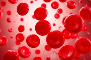
- Erythrocytes (Red blood cells or RBCs) carries oxygen
- Leukocytes (White blood cells or WBCs) are part of the immune system
- Platelets are cell fragments involved in hemostasis (blood clotting)
Blood sample layers after centrifugation (separation)
- Spinning a blood sample rapidly in a centrifuge forms three distinct layers
- Top layer: Plasma (~55% of total blood volume)
- Middle layer: Buffy coat (Leukocytes and platelets, ~1% of total blood volume)
- Bottom layer: Erythrocytes (~44% of total blood volume)
- Hematocrit is the percentage of blood volume composed of erythrocytes
Blood functions
- Exchanging gases: Transports oxygen and carbon dioxide
- Distributing solutes: Transports ions, nutrients, hormones, and wastes and also regulates ion concentrations in tissues
- Performing immune functions: Transports leukocytes and immune system proteins
- Maintaining body temperature: Carries away heat generated by chemical reactions
- Clot formation: Seals damaged vessels with blood clots using platelets and proteins, preventing blood loss.
- Preserving acid-base homeostasis: Maintains a stable blood pH in a narrow range (7.35–7.45) via buffering systems
- Stabilizing blood pressure: Blood volume is a major factor
Plasma Proteins
- Plasma contains plasma proteins including Albumin, Immune proteins, Transport proteins, and Clotting proteins
- Plasma proteins form a colloid that makes up about 9% of plasma volume
- Albumin is a large protein synthesized in the liver
- Albumin is responsible for blood's colloid osmotic pressure, drawing water into the blood via osmosis.
- Immune proteins (γ-globulins or antibodies) are produced by leukocytes
- Transport proteins bind to lipid-based molecules, allowing them to be transported in the water-based plasma
- Clotting proteins, along with platelets, stop bleeding from injured vessels by forming blood clots
Plasma miscellaneous
- The remaining 1% of plasma volume contains small molecules dissolved in the water to form a solution
- These molecules are readily exchanged between blood and interstitial fluid in most capillary beds
Erythrocyte (RBC) Structure
- Enables the transport of oxygen and carbon dioxide
- Typical erythrocyte (RBC) is biconcave disc-shaped
- Concave on both sides
- The biconcave shape increases cell surface area for gas exchange
- Mature RBCs are anucleate and lack typical cellular organelles to create space
Hemoglobin (Hb)
- This creates room for enzymes and nearly 1 billion oxygen-binding hemoglobin (Hb) proteins in the cytosol
- Hemoglobin is a large protein
- Hemoglobin has four polypeptide subunits: two alpha (α) and two beta (β) chains
- Each polypeptide chain binds to a heme group containing iron
- Iron binds to oxygen in areas of high concentration, such as the lungs, forming oxyhemoglobin (HbO2)
- This creates a red-colored molecule.
- Hemoglobin releases oxygen in tissues where oxygen concentration is low, such as the systemic capillary beds
- Carbaminohemoglobin: Hemoglobin binds to carbon dioxide when oxygen levels are low which accounts for ~23% of CO2 transport in blood
Erythrocyte Life Span
- Erythrocytes have relatively short lifespan (100–120 days)
- Cells incurs damage due to harsh environment it exists in
- The cells lack the means for repair
- Body has a mechanism for continuously producing new erythrocytes to counter this
Hematopoiesis
- The process in red bone marrow
- Formed elements in blood are produced by hematopoietic stem cells (HSCs)
Erythropoiesis (Red Blood Cell Formation)
- Erythropoiesis is the process that produces erythrocytes from hematopoietic stem cells (HSCs)
- This entire process takes ~5–7 days.
- Erythropoiesis begins when HSCs differentiate into erythrocyte colony-forming units (CFUs) that form only one cell type
- Erythropoietin (EPO) hormone, secreted by kidneys, causes erythrocyte CFUs to differentiate into proerythroblasts
- Proerythroblasts develop into erythroblasts
- Erythroblasts synthesize hemoglobin and other proteins.
- Nucleus shrinks in the erythroblast, is ejected, and becomes a reticulocyte
- Reticulocytes enter bloodstream
- After ejected from bone marrow their remaining organelles exit through pores in sinusoidal capillaries
Erythropoiesis Regulation
- Erythropoietin regulates the hematocrit within a normal range through a negative feedback loop.
- Stimulus: Blood oxygen levels fall below normal.
- Receptor: Kidney cells detect falling oxygen levels.
- Control center: Kidneys increase erythropoietin production and release into the bloodstream.
- Effector/Response: Erythrocyte production increases.
- Homeostasis: Blood oxygen levels rise to normal.
Erythrocyte Death
- Erythrocytes become less flexible and get trapped in spleen sinusoids
- Spleen macrophages digest erythrocytes
- Hemoglobin breaks down into amino acids, iron ions, and bilirubin.
- Heme converts to biliverdin and then to bilirubin.
- Iron ions and amino acids are recycled to make new hemoglobin in red bone marrow.
- Iron ions are transported back to red bone marrow in the bloodstream via the protein transferrin.
- Bilirubin is sent to the liver for excretion.
Anemia
- A condition with decreased oxygen-carrying capacity of blood.
- Three primary causes:
- Decreased hemoglobin
- Decreased hematocrit
- Abnormal hemoglobin
- Variety of events/conditions is associated with anemia.
- Symptoms includes pallor, fatigue, weakness, and shortness of breath.
- Elevated reticulocytes in blood.
- Severe anemia can elevate heart rate.
Decreased Hemoglobin (Anemia)
- Iron deficiency anemia is the most common which is caused by:
- Inadequate dietary iron intake
- Reduced intestinal absorption of iron
- Slow blood loss
- Vitamin B6 deficiency, malnutrition, poisoning with drugs or heavy metals like lead, and pregnancy can all decrease hemoglobin levels
Decreased Hematocrit (Anemia)
- The number of erythrocytes in the blood can be reduced by several factors which lowers hematocrit
- Blood loss from:
- Acute injury
- Stomach ulcers
- Pernicious anemia results from vitamin B12 deficiency
- Erythrocyte destruction can be caused by:
- Bacterial infections
- Diseases of the immune system or liver
- Lead poisoning
- Hemolytic anemia
- Aplastic anemia is caused by:
- Certain medications
- Exposure to ionizing radiation It can also inhibit or stop the production of erythrocytes in the red bone marrow
Abnormal Hemoglobin (Anemia)
- The most common cause of abnormal hemoglobin is sickle-cell disease
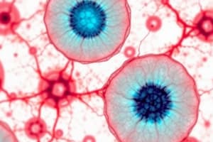
Leukocytes (White Blood Cells/WBCs)
- Larger than erythrocytes with a prominent nucleus
- Use bloodstream for transportation
- Do not generally perform their functions within the blood.
- Adhere to blood vessel walls, then squeeze between endothelial cells to enter surrounding tissue
Leukocytes categories
- Granulocytes have cytoplasmic granules released when activated
- Agranulocytes lack visible granules.
Granulocytes Characteristics
- Unusual nuclei consisting of multiple connected lobes
- Contain general lysosomal granules along with specialized granules
- Neutrophils, Eosinophils, and Basophils are three categories based on the color the granules appear (dark purple/red), after staining
Neutrophils
- Neutrophils are the most common leukocyte
- Cytoplasmic granules absorb both dyes
- Cytoplasm is light lilac
- Active phagocytes that ingest and destroy bacterial cells
- Often called polymorphonucleocytes (polys/PMNs)
- Have uniquely shaped nuclei with three to five lobes
- Chemicals released by injured cells attracts neutrophils (chemotaxis)
- Neutrophils exit bloodstream and release granules in the damaged tissue.
- Granules directly kill bacterial cells and attract more neutrophils and leukocytes, enhancing inflammation
Eosinophils
- Eosinophils have bilobed nucleus
- Appear red due to uptake of eosin dye
- Ingest foreign molecules
- Respond to infections with parasitic worms and allergic reactions
- Their granules contain enzymes and toxins that enhance those activities
Basophils
- Basophils are the least common leukocytes
- Have an S-shaped nucleus
- Appear dark purple due to uptake of methylene blue dye
- Mediate inflammation
Agranulocytes
- Includes Lymphocytes and Monocytes
- Lack visible cytoplasmic granules.
- Contain lysosomes, like granulocytes
Lymphocytes
- Lymphocytes are the second most common leukocyte in blood
- Large, spherical nuclei and light blue rim of cytoplasm when stained
- Two basic types of lymphocytes that have similar appearances but have different functions.
- Both cell types are activated by cellular markers (antigens)
- B lymphocytes (B cells) produce antibodies
- The antibodies bind to and remove antigens from tissues.
- Each population of B lymphocytes secretes antibody that binds only to a specific unique antigen
- T lymphocytes (T cells) do not produce antibodies
- Have membrane-bound receptors for individual antigens.
- Activate other immune system components.
- Directly destroy abnormal body cells, e.g., cancer cells or virally infected cells
- Antibodies and T cell receptor cells both bind to only one unique antigen based on their structure
Monocytes
- The largest cells in the leukocyte family
- Have large U-shaped nuclei surrounded by light blue or purple stained cytoplasm.
- The monocytes circulate briefly in the blood
- The monocytes exit the capillaries and enter tissues
- In the tissues, monocytes mature into macrophages
- Macrophages are phagocytic cells that ingest:
- Dead & dying cells
- Bacteria
- Antigens
- Other cellular debris
- Macrophages activate other components of the immune system by displaying phagocytosed antigens to other leukocytes
Leukopoiesis
- In bone marrow, hematopoietic stem cells form new leukocytes
- HSCs divide and split into two cell lines: myeloid and lymphoid cells line
- The myeloid cell line produces most of the formed elements (erythrocytes and platelets)
- The myeloid cell line differentiates into blast cells committed to becoming monocytes called monoblasts
- Granulocytes are derived from myleoblasts that differentiate into precursor cells called promyelocytes
- Band cells or stab cells are the final stage before mature granulocytes migrate to bloodstream
- The lymphoid cell line produces lymphoblasts
- These are committed to becoming B and T lymphocytes
- B and T lymphocytes mature in different locations
- B lymphocytes mature in bone marrow
- T lymphocytes migrate from the bone marrow to thymus gland to the mediastinum
Platelets
- Small cell fragments surrounded by a plasma membrane
- The smallest of the formed elements, involved in hemostasis (stops blood loss from injured vessels)
- Platelets lack nuclei and other organelles, but contain several types of granules
- Granules contain clotting factors, enzymes, mitochondria, and glycogen deposits
- Platelets contain cytoskeletal elements (microtubules, actin, and myosin filaments)
- Platelet lifespan is 7-10 days
- These are removed from circulation by the liver and spleen
Platelet Formation
- Platelet formation begins when HSCs differentiate into megakaryoblasts from the myeloid cell line
- Megakaryoblasts develop into megakaryocytes that undergo repeated cycles of mitosis without cytokinesis.
- Megakaryocytes are massive
- Mature megakaryocytes send cytoplasmic extension through clefts in bone marrow sinusoids, stimulated by hormones like thrombopoietin
- Pieces break off into the bloodstream, forming thousands of platelets
Hemostasis
- Hemostasis involves five events that form a gelatinous blood clot, plugging broken vessel.
- Mechanisms limit significant blood loss
Hemostasis - Vascular Spasm
- Begins immediately when a blood vessel is injured
- Blood leaks into extracellular fluid
- Vasoconstriction and increased tissue pressure decrease the blood vessel diameter
- Minimizes blood loss via reducing blood pressure and blood flow locally
Hemostasis - Platelet Plug Formation
- Platelets form a plug/patch that adheres only to injury site to reduce blood loss.
- Injured endothelial cells release von Willebrand factor (vWF)
- vWF is a glycoprotein that binds to receptors on platelet plasma membranes
- Exposed collagen plus von Willebrand factor causes platelets to become sticky
- Platelets adhere to each other and surrounding vessel walls.
- Binding of vWF and collagen to platelets triggers platelet activation
- Granule contents attract and activate nearby platelets, causing them to clump together or aggregate
- Platelet aggregation forms the platelet plug and seals the injured vessel temporarily
Hemostasis - Coagulation
- Forms molecular glue that binds platelets, endothelial cells, and other formed elements
- Coagulation occurs through a cascade of events including:
- Intrinsic pathway
- Extrinsic pathway
- The intrinsic and extrinsic pathways converge at a common pathway activating fibrin
Fibrin
- Fibrin is a threadlike protein
- Fibrin converts a soft, liquid platelet plug into a substantial solid mass
- Fibrinogen is an inactive form that is found circulating in plasma and platelets
- Fibrinogen is converted into fibrin
- Fibrin conversion is a series of reactions at the surface of platelets and/or damaged endothelial cells called the coagulation cascade.
- Depends on clotting factors
- Clotting factors are mostly inactive enzymes synthesized in liver and circulate in the blood
- Clotting factors are labeled with Roman numerals in the order of their discovery
- Clotting factors II, VII, IX, and X depend on vitamin K for synthesis
Intrinsic/Contact Activation Pathway
- Named because all involved clotting factors are found in the blood, Pathway activation occurs
- Following sequence of the events
- Inactive protein clotting factor XII comes in contact with exposed collagen fibers
- Exposed collagen fibers activate XII to XIIa
- Factors XI and IX become activated
- XIIa is an enzyme that activates XI
- XIa subsequently activates IX
- In a cascade of events, each factor activated activates another in the pathway
- Factors IXa and VIIIa along w/ calcium ions form an enzyme complex
- This complex activates factor X to Xa to complete the pathway
Extrinsic Tissue Factor Pathway
- Named because it is initiated by a factor outside of the blood.
- Damaged subendothelial cells display a protein called tissue factor.
- tissue factor activates factor from VII to VIIa.
- factor VIIa, tissue factor, and calcium ions form an enzyme complex
- the enzyme complex activates factor X to Xa.
Common Pathway
- Both the intrinsic and extrinsic pathways form factor Xa.
- Factors Xa and Va along with calcium ions form prothrombin activator
- Activator converts; prothrombin into the active form (thrombin)
- Thrombin turns fibrinogen into fibrin
- Fibrin glues the platelet plug together
Hemostasis - Clot retraction
- Actin and myosin fibers in involved platelets contract, bringing edges of wounded vessels closer
- Serum is forced out of the clot
- Serum consists of plasma but without the clotting proteins
Hemostasis - Thrombolysis
- The process that begins after injury has healed and blood clotting is no longer necessary
- Fibrinolysis is the first step in thrombolysis and it breaks down the fibrin glue
Regulation of clotting
- Requires strict regulation
- Endothelial cells regulate the first and second stages of clot formation by producing and secreting chemicals
- Endothelial cells and hepatocytes produce anticoagulants to inhibit coagulation:
- Antithrombin III (AT-III) protein binds and inhibits activity of factor Xa and thrombin.
- Antithrombin also prevents the activation of new thrombin.
- Heparin sulfate: polysaccharide that enhances antithrombin activity
- Protein C catalyzes reactions that degrade clotting factors Va and VIIIa
- Protein C is activated by protein S
Disorders of Clotting
- A disorder is a condition in which clotting is not regulated correctly and has drastic consequences for maintaining homeostasis
- Bleeding disorders
- Hypercoagulable conditions
- Bleeding disorders can increase blood loss from even minor cuts.
- A clotting protein deficiency prevents the blood from clotting (hemophilia).
Hypercoagulable Conditions
- Results in inappropriate clot formation (thrombosis).
- The clot/thrombus is dangerous, obstructing blood flow through a vessel.
- Thromboembolism: When a thrombus breaks off and occludes smaller vessels downstream.
- Thrombi commonly form in deep veins of the legs causing deep vein thrombosis (DVT).
- Pulmonary embolism: dangerous complication of DVTs in which, emboli break off thrombi in legs and lodge in small blood vessels in lungs
Blood transfusions
- Blood taken from a donor is given to recipient
- Blood transfusions were not always the common treatment modality they are now
- Discovery of surface markers/antigens biological molecules and cells (including erythrocytes) lead to safer blood transfusions
- The Antigen is recognized as foreign and the immune system trys to remove them
Blood groups
- Antigens on erythrocytes give rise to this
- Blood groups are genetically determined carbohydrate chains
- Two groups of the 30 different antigens found on erythrocytes are particularly useful for clinical use: (1) ABO blood group (2) Rh blood group
Blood Typing (ABO)
- Features two antigens: A and B antigens
- Giving rise to four ABO types: a. In Type A, only the A antigen is present on erythrocytes. b. In Type B, only the B antigen is present on erythrocytes. c. In Type AB, both the A and B antigens are present on erythrocytes. d. In Type O, neither A nor B antigens are present on erythrocytes. There is no O antigen.
Rh Blood Group
- Rh antigen was first discovered in rhesus monkeys.
- Individuals with Rh on their erythrocytes are Rh-positive (Rh+).
- Those without Rh antigen are Rh-negative (Rh-).
- The ABO and Rh blood groups combined to create eight common blood types.
- Type O+ is the most common blood type in the U.S. populations while AB- is the least.
Blood Typing in Lab
- Blood typing uses antibodies to bind to individual antigens on erythrocytes
- When antibodies (agglutinins) bind to surface antigens
- The surface antigens clump together or agglutinate.
- Agglutination promotes erythrocytes destruction (hemolysis)
- A blood sample is treated with three separate antibodies in order to determine blood type
- If agglutination occurs in response to an antibody, antigens are present
- That specific antigen is absent when no agglutination occurs
- The three antibodies used are: a. Anti-A antibodies bind and agglutinate A antigens b. Anti-B antibodies bind and agglutinate B antigens c. Anti-Rh antibodies bind and agglutinate Rh antigens
Blood Transfusions:
- Antigens and antibodies are the basis for blood matching
- Blood taken from a donor screened for compatibility prior to administration to a recipient
- A match occurs if donor blood type is compatible with recipient blood type
Transfusion Reaction
- During reaction, recipient antibodies bind to donor antigens
- Antibodies cause agglutination, subsequently destroying erythrocytes
- It can possibly lead to kidney failure and death.
Universal Donor
- Universal Donor is has blood type O- because their erythrocytes do not have A, B, or Rh surface antigens
- This blood type can be given if no blood matching is available
- Universal Recipient has blood type AB+
- This individual with blood type does not make antibodies against the A, B, or Rh antigens
- Matching is still the safest practice
Studying That Suits You
Use AI to generate personalized quizzes and flashcards to suit your learning preferences.