Podcast
Questions and Answers
What is a nevus flammeus commonly known as?
What is a nevus flammeus commonly known as?
- Mole
- Birthmark (correct)
- Skin tag
- Lentigo
Which of the following is NOT classified as a congenital or acquired lesion?
Which of the following is NOT classified as a congenital or acquired lesion?
- Nevus flammeus
- Skin cancer (correct)
- Hereditary hemorrhagic telangiectasia
- Spider telangiectasias
Hereditary hemorrhagic telangiectasia is associated with which of the following?
Hereditary hemorrhagic telangiectasia is associated with which of the following?
- Autoimmune response
- Genetic predisposition (correct)
- Infectious disease
- Environmental factors
Spider telangiectasias are often seen in association with which of the following conditions?
Spider telangiectasias are often seen in association with which of the following conditions?
When do hemangiomas typically become noticeable after birth?
When do hemangiomas typically become noticeable after birth?
Which statement is true regarding these lesions mentioned?
Which statement is true regarding these lesions mentioned?
How long do hemangiomas generally grow in size before they begin to shrink?
How long do hemangiomas generally grow in size before they begin to shrink?
What is the term used to describe the process through which hemangiomas shrink over time?
What is the term used to describe the process through which hemangiomas shrink over time?
What timeframe is typically involved in the involution of hemangiomas?
What timeframe is typically involved in the involution of hemangiomas?
Which statement about hemangiomas is accurate?
Which statement about hemangiomas is accurate?
What characteristic is commonly found in benign neoplasms such as hemangiomas?
What characteristic is commonly found in benign neoplasms such as hemangiomas?
Which type of cells line the vascular channels in benign neoplasms?
Which type of cells line the vascular channels in benign neoplasms?
Benign neoplasms like hemangiomas usually exhibit which of the following features?
Benign neoplasms like hemangiomas usually exhibit which of the following features?
Which statement about the structure of benign neoplasms is false?
Which statement about the structure of benign neoplasms is false?
What defines a hemangioma within the category of benign neoplasms?
What defines a hemangioma within the category of benign neoplasms?
What is the primary virus associated with all Kaposi's Sarcoma (KS) lesions?
What is the primary virus associated with all Kaposi's Sarcoma (KS) lesions?
How is Kaposi's Sarcoma (KS) primarily transmitted?
How is Kaposi's Sarcoma (KS) primarily transmitted?
What role does local inflammation play in the development of Kaposi's Sarcoma?
What role does local inflammation play in the development of Kaposi's Sarcoma?
Which of the following contributes to the pathogenesis of Kaposi's Sarcoma?
Which of the following contributes to the pathogenesis of Kaposi's Sarcoma?
Which statement about Kaposi's Sarcoma is true?
Which statement about Kaposi's Sarcoma is true?
What is a common gross appearance of Kaposi sarcoma?
What is a common gross appearance of Kaposi sarcoma?
What do the microscopic features of Kaposi sarcoma predominantly show?
What do the microscopic features of Kaposi sarcoma predominantly show?
Which of the following statements is false regarding the morphology of Kaposi sarcoma?
Which of the following statements is false regarding the morphology of Kaposi sarcoma?
Which feature is NOT typically seen in the microscopic examination of Kaposi sarcoma?
Which feature is NOT typically seen in the microscopic examination of Kaposi sarcoma?
What is the significance of slitlike vascular spaces in Kaposi sarcoma?
What is the significance of slitlike vascular spaces in Kaposi sarcoma?
What is a characteristic feature of the lesion described as angiosarcoma?
What is a characteristic feature of the lesion described as angiosarcoma?
Which of the following visual characteristics is associated with angiosarcoma?
Which of the following visual characteristics is associated with angiosarcoma?
What microscopic feature is notable in angiosarcoma?
What microscopic feature is notable in angiosarcoma?
What aspect of the tissue is affected in the case of angiosarcoma?
What aspect of the tissue is affected in the case of angiosarcoma?
Where is the cystic lump associated with angiosarcoma located?
Where is the cystic lump associated with angiosarcoma located?
Flashcards
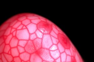
Nevus flammeus
Nevus flammeus
A type of birthmark characterized by a flat, red, and irregular-shaped mark caused by dilated blood vessels.
Spider telangiectasias
Spider telangiectasias
Small, spider-shaped blood vessels that appear as a central red dot with radiating branches.
Hereditary hemorrhagic telangiectasia (HHT)
Hereditary hemorrhagic telangiectasia (HHT)
A genetic disorder causing abnormal blood vessels in the skin and mucous membranes; leads to frequent nosebleeds and other bleeding episodes.
Congenital or Acquired Lesions
Congenital or Acquired Lesions
Signup and view all the flashcards
Vascular Lesions
Vascular Lesions
Signup and view all the flashcards
Hemangioma
Hemangioma
Signup and view all the flashcards
Benign Neoplasm
Benign Neoplasm
Signup and view all the flashcards
Vascular Channels
Vascular Channels
Signup and view all the flashcards
Monolayer of Endothelial Cells
Monolayer of Endothelial Cells
Signup and view all the flashcards
Lymph
Lymph
Signup and view all the flashcards
Involution
Involution
Signup and view all the flashcards
Peak size
Peak size
Signup and view all the flashcards
Noticeable after birth
Noticeable after birth
Signup and view all the flashcards
Hemangioma Shrinking Time
Hemangioma Shrinking Time
Signup and view all the flashcards
Angiosarcoma
Angiosarcoma
Signup and view all the flashcards
Cystic lump
Cystic lump
Signup and view all the flashcards
Soft and spongy lesion
Soft and spongy lesion
Signup and view all the flashcards
Slit-like vessels
Slit-like vessels
Signup and view all the flashcards
Dissection of dermal collagen
Dissection of dermal collagen
Signup and view all the flashcards
Kaposi's Sarcoma (KS)
Kaposi's Sarcoma (KS)
Signup and view all the flashcards
Microscopic features of Kaposi's Sarcoma
Microscopic features of Kaposi's Sarcoma
Signup and view all the flashcards
Spindle-shaped cells in KS
Spindle-shaped cells in KS
Signup and view all the flashcards
Slit-like vascular spaces in KS
Slit-like vascular spaces in KS
Signup and view all the flashcards
Diagnostic value of KS microscopic features
Diagnostic value of KS microscopic features
Signup and view all the flashcards
What causes all KS lesions?
What causes all KS lesions?
Signup and view all the flashcards
How is KSHV transmitted?
How is KSHV transmitted?
Signup and view all the flashcards
What are somatic mutations in relation to KS?
What are somatic mutations in relation to KS?
Signup and view all the flashcards
How does inflammation affect KS progression?
How does inflammation affect KS progression?
Signup and view all the flashcards
What cell type is most affected in KS?
What cell type is most affected in KS?
Signup and view all the flashcards
Study Notes
Cardiovascular System Pathology (3rd Year Medical Students)
- Topic: Pathology of the Cardiovascular System
- Target Audience: 3rd-year medical students
- Course: Histopathology
- Institution: University of Science & Technology
Veins and Lymphatics
- Veins: The pathology presentation covers varicose veins of the extremities.
- Varicose Veins: These are abnormally dilated, tortuous veins caused by increased intraluminal pressure and weakened supporting walls.
- Prevalence: Varicose veins affect up to 20% of men and a third of women.
- Risk Factors: Obesity, compression of the inferior vena cava (e.g., by a gravid uterus during pregnancy), and familial tendency are risk factors.
Learning Objectives
- Defining varicose veins
- Defining lymphangitis and lymphedema
- Classifying vascular tumors
- Discussing benign, borderline, and malignant vascular tumors considering etiology, pathogenesis, and morphology
Varicose Veins of the Extremities
- Cause: Increased intraluminal pressure and weakened supporting vessel walls lead to varicose veins.
- Superficial Veins: Frequently involved veins, affecting up to 20% of men and a third of women.
Risk Factors
- Obesity
- Compression of the inferior vena cava (e.g., gravid uterus during pregnancy)
- Familial tendency
Morphology (Varicose Veins)
- Wall Thinning: The walls of varicose veins are often thinner, especially at points of maximum dilation.
- Smooth Muscle Hypertrophy: Smooth muscle in the vein walls might undergo hypertrophy.
- Intimal Fibrosis: Fibrosis of the intimal layer can occur.
- Elastic Tissue Degeneration: Weakening of the elastic tissue within the vessel walls.
- Medial Calcifications: Spotty calcifications within the medial layer of the vein.
- Phlebosclerosis: A term encompassing elastic tissue degeneration and spotty medial calcifications.
- Intraluminal Thrombosis: Blood clots often form within the veins due to stasis.
- Venous Valve Deformities: Rolling and shortening of venous valves are common occurrences.
Clinical Features of Varicose Veins
- Lower Extremity Stasis and Thrombosis: Stagnant blood flow and blood clots can occur in the lower extremities.
- Congestion and Edema: Fluid buildup (edema) and congestion in the affected area.
- Ischemia and Ulceration: Compromised blood supply (ischemia) and potential skin ulceration.
- Varicose Ulcers: Skin sores developing directly as a consequence of varicose vein pathology.
Sites of Varicose Vein Formation
- Esophageal Varices: Dilated veins in the esophagus resulting from portal hypertension caused by liver cirrhosis, portal vein obstruction, or hepatic vein thrombosis.
- Hemorrhoids: Varicose veins in the rectum or anus. They can be uncomfortable, lead to bleeding, thrombosis, or painful ulceration.
Lymphangitis and Lymphedema
- Uncommon Primary Disorders: Primary abnormalities of lymph vessels are unusual.
- Common Secondary Processes: Conditions developing due to inflammation or malignancy such as trauma, infection, or malignancy.
- Causes of Secondary Obstructive Lymphedema: Neoplasms, surgeries, radiation fibrosis, filariasis (e.g., elephantiasis), and post-inflammatory processes.
- Lymphangitis Characteristics: Inflammation of lymphatic vessels often appearing as red, painful, subcutaneous streaks and swollen lymph nodes. Infection, possibly bacteremia (bacteria in the blood), and sepsis pose serious risks.
- Lymphedema Characteristics: Buildup of lymph fluid causing swelling (edema). Causes include congenital defects, obstruction, or inflammatory processes. It often results in chronic edema, fibrosis, a "peau d'orange" appearance (skin texture irregularities), and skin ulcers. Possible complications include chylous ascites, chylothorax, or chylopericardium.
Vascular Neoplasms
- Classification: Benign (developmental or acquired); Intermediate grade; Malignant.
- Benign Lesions/Tumors: Vascular ectasias (nevus flammeus, spider telangiectasia, hemorrhagic telangiectasia), hemangioma, lymphangioma, and glomus tumors.
- Intermediate Grade Lesions/Tumors: Kaposi sarcoma.
- Malignant Lesions/Tumors: Angiosarcoma and hemangioendothelioma.
Benign Developmental Lesions
- Telangiectasia: Permanent dilation of small blood vessels (capillaries, venules, arterioles) in the skin or mucous membranes forming discete red lesions. These are not true neoplasms. Examples include nevus flammeus (birthmarks), spider telangiectasia, and hereditary hemorrhagic telangiectasia.
Benign Neoplasms: Hemangioma
- Definition: A common tumor in infancy and childhood that may be localized or extensive, consisting of blood vessels lined by endothelial cells.
- Types: Capillary hemangioma, cavernous hemangioma, and pyogenic granuloma. This encompasses juvenile hemangiomas.
Hemangioma Details
- Benign growths of blood vessels, appearing as birthmarks.
- Usually grow in size for 6-12 months, then shrink.
- Mostly do not need treatment.
- Some can cause problems like ulceration, blocking organs or cosmetic concerns
Lymphangioma Variants
- Simple capillary lymphangioma: Commonly called cystic hygroma.
- Cavernous lymphangioma: lymphatic counterpart of hemangioma. Characterized by cystic spaces lined by endothelial cells with lymphoid aggregates. Spaces usually contain pale fluid.
Kaposi Sarcoma (KS)
- Cause: A vascular neoplasm linked to Kaposi's sarcoma-associated herpesvirus (KSHV). Highly prevalent in AIDS patients.
- Types: Classic, endemic African, transplantation-associated, and AIDS-associated.
- Pathogenesis: KSHV infection, somatic mutations, and local inflammation contribute to cellular proliferation and endothelial cell progression. This disease is transmitted (sexually or non-sexually).
Morphology (Kaposi Sarcoma)
- GROSS: Patches, raised plaques or nodular lesions. These can range from smooth to bumpy skin surfaces.
- MICROSCOPIC: Sheets of spindle-shaped cells and slit-like vascular spaces.
Hemangioendothelioma:
- Tumor cells are plump and cuboidal, not forming well-defined vascular channels.
- Sometimes mistaken for metastatic epithelioid or melanomas.
Malignant Tumor (Angiosarcoma)
- Cellular Density: Highly cellular, displaying cytological atypia.
- Vascular Organization: Fail to form well-organized vessels.
- Differentiation: Range from highly differentiated to poorly differentiated forms (anaplastic).
- Patient Population: Most common in older adults and arises in various locations in the body.
Morphology (Angiosarcoma)
- Gross: Large lesion, fleshy red-tan to gray-white with necrosis and hemorrhage.
- Microscopic: Shows various degrees of atypical endothelial cells, ranging from plump to spindle-shaped cells; may not form discernible vascular channels.
- Diagnostic Markers: Can use immunohistochemistry (staining) to spot endothelial cell markers (like CD34, CD31, Von Willebrand factor) in poorly differentiated lesions.
- Aggressive Properties: Aggressive local spread, and can metastasize.
- Examples: Cystic lump in the ear, a soft, spongy lesion with purple streaks.
Studying That Suits You
Use AI to generate personalized quizzes and flashcards to suit your learning preferences.