Podcast
Questions and Answers
What heart rhythm is characterized by a regular rhythm and a rate between 60-100 bpm?
What heart rhythm is characterized by a regular rhythm and a rate between 60-100 bpm?
- Sinus Tachycardia
- Normal Sinus Rhythm (correct)
- Sinus Bradycardia
- Atrial Fibrillation
Which heart rhythm is characterized by a slow heart rate of less than 60 bpm?
Which heart rhythm is characterized by a slow heart rate of less than 60 bpm?
- Sinus Bradycardia (correct)
- Atrial Fibrillation
- Sinus Tachycardia
- Normal Sinus Rhythm
Which of the following rhythms presents with irregularly irregular rhythm and no distinct P waves?
Which of the following rhythms presents with irregularly irregular rhythm and no distinct P waves?
- Sinus Bradycardia
- Normal Sinus Rhythm
- Sinus Tachycardia
- Atrial Fibrillation (correct)
What heart rhythm is often associated with stress, fever, or exertion?
What heart rhythm is often associated with stress, fever, or exertion?
Which heart rhythm requires immediate defibrillation?
Which heart rhythm requires immediate defibrillation?
Which of the following best describes asystole?
Which of the following best describes asystole?
What can sustained ventricular tachycardia (VT) lead to?
What can sustained ventricular tachycardia (VT) lead to?
What type of waves are characteristic of atrial flutter?
What type of waves are characteristic of atrial flutter?
An EEG records brainwave activity, and what other activity?
An EEG records brainwave activity, and what other activity?
Why is it important to differentiate between cardiac syncope and epileptic seizures?
Why is it important to differentiate between cardiac syncope and epileptic seizures?
Bradycardia or asystole may show what on EEG?
Bradycardia or asystole may show what on EEG?
In cardiac arrest, what may an EEG become after prolonged hypoxia?
In cardiac arrest, what may an EEG become after prolonged hypoxia?
Monitoring ECG during EEG is especially important in patients with which condition?
Monitoring ECG during EEG is especially important in patients with which condition?
In convulsive syncope, what may be seen on EEG?
In convulsive syncope, what may be seen on EEG?
Which arrhythmia is often associated with SUDEP (Sudden Unexpected Death in Epilepsy)?
Which arrhythmia is often associated with SUDEP (Sudden Unexpected Death in Epilepsy)?
Cardiac rhythms can sometimes mimic what condition on an EEG?
Cardiac rhythms can sometimes mimic what condition on an EEG?
What cardiac change is common during seizures?
What cardiac change is common during seizures?
What should be checked on the ECG trace, when the EEG shows slowing or flattening?
What should be checked on the ECG trace, when the EEG shows slowing or flattening?
Understanding cardiac rhythms helps in preventing what type of misdiagnosis?
Understanding cardiac rhythms helps in preventing what type of misdiagnosis?
What type of care should be provided by collaborating with cardiology?
What type of care should be provided by collaborating with cardiology?
In a patient experiencing convulsive syncope, what EEG finding would be most indicative of the condition rather than an epileptic seizure?
In a patient experiencing convulsive syncope, what EEG finding would be most indicative of the condition rather than an epileptic seizure?
Why is it critical to monitor ECG during EEG in patients presenting with atypical 'seizure-like' spells?
Why is it critical to monitor ECG during EEG in patients presenting with atypical 'seizure-like' spells?
During an EEG recording, if the EEG trace shows diffuse slowing and intermittent flattening, what immediate action should be taken?
During an EEG recording, if the EEG trace shows diffuse slowing and intermittent flattening, what immediate action should be taken?
What EEG change might be expected in a patient experiencing bradycardia or asystole?
What EEG change might be expected in a patient experiencing bradycardia or asystole?
In the context of SUDEP, what is the primary significance of cardiac arrhythmias?
In the context of SUDEP, what is the primary significance of cardiac arrhythmias?
How does sinus tachycardia typically manifest on an EEG, and what condition alters this presentation?
How does sinus tachycardia typically manifest on an EEG, and what condition alters this presentation?
What EEG changes are expected in cases of cardiac arrest, ventricular fibrillation, or asystole?
What EEG changes are expected in cases of cardiac arrest, ventricular fibrillation, or asystole?
Considering the potential for misdiagnosis, which condition could cardiac syncope most closely mimic based on EEG findings?
Considering the potential for misdiagnosis, which condition could cardiac syncope most closely mimic based on EEG findings?
In which clinical scenario is cardiac monitoring during EEG most critical, necessitating immediate evaluation and potential intervention?
In which clinical scenario is cardiac monitoring during EEG most critical, necessitating immediate evaluation and potential intervention?
What specific aspect of EEG interpretation is vital for both EEG technologists and neurologists to prevent misdiagnosis involving cardiac events?
What specific aspect of EEG interpretation is vital for both EEG technologists and neurologists to prevent misdiagnosis involving cardiac events?
What underlying physiological principle directly links cardiac function to EEG patterns, especially in conditions like bradycardia or asystole?
What underlying physiological principle directly links cardiac function to EEG patterns, especially in conditions like bradycardia or asystole?
In cases of suspected ictal bradycardia/asystole, prompt detection is crucial to prevent which potential outcome?
In cases of suspected ictal bradycardia/asystole, prompt detection is crucial to prevent which potential outcome?
What type of collaborative approach is most beneficial in providing comprehensive patient care when cardiac and neurological symptoms overlap?
What type of collaborative approach is most beneficial in providing comprehensive patient care when cardiac and neurological symptoms overlap?
What is the MOST likely cause of a flatline EEG in the setting of Cardiac Arrest/VF/Asystole?
What is the MOST likely cause of a flatline EEG in the setting of Cardiac Arrest/VF/Asystole?
Why are modern EEG systems equipped with simultaneous single-lead ECG recording?
Why are modern EEG systems equipped with simultaneous single-lead ECG recording?
Which of the following is a characteristic feature of Atrial Flutter on ECG that distinguishes it from Atrial Fibrillation?
Which of the following is a characteristic feature of Atrial Flutter on ECG that distinguishes it from Atrial Fibrillation?
Which cardiac rhythm is most likely to lead to cardiac arrest if sustained, according to provided information?
Which cardiac rhythm is most likely to lead to cardiac arrest if sustained, according to provided information?
A patient's ECG shows a chaotic rhythm with no pulse. What immediate intervention is required?
A patient's ECG shows a chaotic rhythm with no pulse. What immediate intervention is required?
What is the key distinguishing feature observed on an ECG for Normal Sinus Rhythm?
What is the key distinguishing feature observed on an ECG for Normal Sinus Rhythm?
Which patient demographic is most likely to exhibit Sinus Bradycardia?
Which patient demographic is most likely to exhibit Sinus Bradycardia?
Flashcards
Normal Sinus Rhythm
Normal Sinus Rhythm
Regular rhythm, rate 60-100 bpm, P waves present before each QRS complex.
Sinus Bradycardia
Sinus Bradycardia
Slow heart rate (<60 bpm), regular rhythm; often seen in athletes or during sleep.
Sinus Tachycardia
Sinus Tachycardia
Fast heart rate (>100 bpm), regular rhythm; often due to stress, fever, or exertion.
Atrial Fibrillation
Atrial Fibrillation
Signup and view all the flashcards
Atrial Flutter
Atrial Flutter
Signup and view all the flashcards
Ventricular Tachycardia (VT)
Ventricular Tachycardia (VT)
Signup and view all the flashcards
Ventricular Fibrillation (VF)
Ventricular Fibrillation (VF)
Signup and view all the flashcards
Asystole
Asystole
Signup and view all the flashcards
Heart Blocks
Heart Blocks
Signup and view all the flashcards
Bradycardia/Asystole EEG Effects
Bradycardia/Asystole EEG Effects
Signup and view all the flashcards
Cardiac Arrest/VF/Asystole EEG Effects
Cardiac Arrest/VF/Asystole EEG Effects
Signup and view all the flashcards
Convulsive Syncope EEG
Convulsive Syncope EEG
Signup and view all the flashcards
SUDEP
SUDEP
Signup and view all the flashcards
Differentiating Syncope and Seizures
Differentiating Syncope and Seizures
Signup and view all the flashcards
EEG Slowing: Check ECG
EEG Slowing: Check ECG
Signup and view all the flashcards
Sinus Tachycardia EEG
Sinus Tachycardia EEG
Signup and view all the flashcards
Study Notes
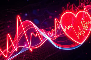
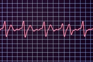
Basic Cardiac Rhythms
- Normal Sinus Rhythm has a regular rhythm, a rate of 60-100 bpm, and P waves present before each QRS complex
- Sinus Bradycardia is a slow heart rate of less than 60 bpm with a regular rhythm, often seen in athletes or during sleep
- Sinus Tachycardia is a fast heart rate of more than 100 bpm with a regular rhythm, often due to stress, fever, or exertion
- Atrial Fibrillation presents as an irregularly irregular rhythm with no distinct P waves and carries a risk of stroke
- Atrial Flutter is characterized by sawtooth flutter waves that can be regular or irregular and can reduce cardiac output
- Ventricular Tachycardia (VT) involves fast, wide QRS complexes that may lead to cardiac arrest if sustained
- Ventricular Fibrillation (VF) is a chaotic rhythm with no pulse, requiring immediate defibrillation
- Asystole is a flatline with no electrical activity or cardiac output
- Heart Blocks, including 1st, 2nd, and 3rd degree, involve delays or complete block in AV conduction
EEG and Cardiac Rhythms
- Modern EEG systems record a single-lead ECG simultaneously with brainwave activity
- Cardiac rhythms can mimic seizures on EEG, such as syncope from arrhythmias
- Epileptic seizures can cause cardiac changes like tachycardia or, rarely, life-threatening bradycardia or asystole
- Differentiation between cardiac syncope and epileptic seizures is essential for proper diagnosis
EEG Patterns and Cardiac Rhythms
- Sinus Tachycardia often shows no change in EEG unless seizure activity is present
- Bradycardia/Asystole may show slowing or flattening of the EEG background due to hypoperfusion
- Cardiac Arrest/VF/Asystole can cause the EEG to become isoelectric (flat) after prolonged hypoxia
- Convulsive Syncope presents with brief, generalized slowing or myoclonic jerks on EEG, but not epileptic spikes
- SUDEP often involves terminal cardiac arrhythmia following a seizure
Clinical Relevance
- Always monitor ECG during EEG, especially in patients with known heart disease, atypical "seizure-like" spells, or suspected ictal asystole or seizure-related arrhythmia
- Identifying artifacts versus real changes is important
- When EEG shows slowing or flattening, check the ECG trace for cardiac causes
Summary
- Understanding basic cardiac rhythms aids in avoiding misdiagnosis, such as mistaking cardiac syncope for epilepsy
- Detecting dangerous conditions like ictal bradycardia/asystole is crucial
- Collaborate with cardiology for holistic patient care
Studying That Suits You
Use AI to generate personalized quizzes and flashcards to suit your learning preferences.