Podcast
Questions and Answers
During knee flexion, which internal ligament is primarily responsible for preventing posterior displacement of the tibia?
During knee flexion, which internal ligament is primarily responsible for preventing posterior displacement of the tibia?
- Medial Collateral Ligament (MCL)
- Lateral Collateral Ligament (LCL)
- Anterior Cruciate Ligament (ACL)
- Posterior Cruciate Ligament (PCL) (correct)
A patient presents with excessive hyperextension of the knee. Which abnormal knee alignment is most likely the cause?
A patient presents with excessive hyperextension of the knee. Which abnormal knee alignment is most likely the cause?
- Genu Valgum
- Plica Syndrome
- Genu Recurvatum (correct)
- Genu Varum
Which of the following best describes the primary function of the menisci within the knee joint?
Which of the following best describes the primary function of the menisci within the knee joint?
- To facilitate medial and lateral rotation of the tibia.
- To stabilize the femur on the tibia and distribute forces within the joint. (correct)
- To directly connect the quadriceps to the tibia.
- To prevent valgus and varus stresses on the knee.
A soccer player sustains an injury to their knee after a valgus force was applied. Which ligament is most likely to be injured?
A soccer player sustains an injury to their knee after a valgus force was applied. Which ligament is most likely to be injured?
What type of synovial joint is the tibiofemoral joint and what movements are possible?
What type of synovial joint is the tibiofemoral joint and what movements are possible?
Which factor has the LEAST influence on the range of motion at a joint?
Which factor has the LEAST influence on the range of motion at a joint?
Which characteristic is LEAST typical of diarthrosis joints?
Which characteristic is LEAST typical of diarthrosis joints?
Which of the following joint types allows for the HIGHEST degree of movement around multiple axes?
Which of the following joint types allows for the HIGHEST degree of movement around multiple axes?
A gymnast performing a cartwheel primarily utilizes which type of synovial joint in their wrists?
A gymnast performing a cartwheel primarily utilizes which type of synovial joint in their wrists?
Which of the following BEST describes the primary difference between a sprain and a strain?
Which of the following BEST describes the primary difference between a sprain and a strain?
Which of the following BEST describes the role of bursae in relation to joints?
Which of the following BEST describes the role of bursae in relation to joints?
Which of the following features is present in synovial joints but NOT in cartilaginous joints?
Which of the following features is present in synovial joints but NOT in cartilaginous joints?
Synostosis is classified as what type of joint, based on its movement capabilities?
Synostosis is classified as what type of joint, based on its movement capabilities?
Flashcards
Tibiofemoral Joint
Tibiofemoral Joint
A double condyloid synovial joint connecting the femur and tibia.
Patellofemoral Joint
Patellofemoral Joint
A gliding synovial joint located between the patella and femur.
Medial Collateral Ligament (MCL)
Medial Collateral Ligament (MCL)
Resists forces that push the knee inward (toward midline).
Anterior Cruciate Ligament (ACL)
Anterior Cruciate Ligament (ACL)
Signup and view all the flashcards
Genu Recurvatum
Genu Recurvatum
Signup and view all the flashcards
Arthrology
Arthrology
Signup and view all the flashcards
Synarthrosis
Synarthrosis
Signup and view all the flashcards
Amphiarthrosis
Amphiarthrosis
Signup and view all the flashcards
Diarthrosis
Diarthrosis
Signup and view all the flashcards
Bony Fusion (Synostosis)
Bony Fusion (Synostosis)
Signup and view all the flashcards
Fibrous Joints
Fibrous Joints
Signup and view all the flashcards
Bursitis
Bursitis
Signup and view all the flashcards
Arthritis
Arthritis
Signup and view all the flashcards
Study Notes
- Arthrology is the study of joints, which are vital for movement and stability.
- The range of motion at a joint is determined by bony structure, ligaments/connective tissues, and muscle tightness.
Functional Joint Classification
- Synarthrosis: Immovable joints such as bony fusions, cartilaginous, and fibrous joints.
- Amphiarthrosis: Slightly movable joints like fibrous (syndesmosis) and cartilaginous (symphysis) joints.
- Diarthrosis: Freely movable joints or Synovial Joints, are the most common type (e.g., knee, elbow).
Structural Joint Classification
- Bony Fusion (Synostosis): Direct bone connections, exemplified by skull sutures in adults.
- Fibrous Joints: Held together by connective tissue (e.g., sutures, syndesmoses, gomphoses).
- Cartilaginous Joints: Held together by cartilage e.g., synchondrosis, symphysis.
- Synovial Joints: Freely movable, featuring a joint capsule, articular cartilage, joint cavity with synovial fluid, synovial membrane, and ligaments (extrinsic, intrinsic, capsular), fibrocartilage pads & fat pads.
Types of Synovial Joints
- Gliding Joint: e.g., intercarpal joints
- Hinge Joint: e.g., elbow, knee
- Pivot Joint: e.g., atlas-axis joint in the neck
- Ellipsoidal (Condyloid) Joint: e.g., wrist
- Saddle Joint: e.g., thumb joint
- Ball and Socket Joint: e.g., shoulder, hip
Common Joint Disorders
- Bursitis: Inflammation of bursae.
- Arthritis: Inflammation and degeneration of joints.
- Osteoarthritis: Wear and tear of hyaline cartilage.
- Rheumatoid Arthritis: Autoimmune disorder attacking synovial joints.
- Synovitis: Inflammation of synovial membrane.
- Joint Dislocation (Luxation): Complete misalignment of joint.
- Subluxation: Partial joint dislocation.
- Sprains and Strains: Ligament or tendon injuries.
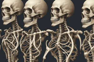
Knee Joint (Genu) Anatomy
- The knee joint is the body's largest synovial joint.
- Bones include the Femur, Tibia, and Patella
Knee Articulations
- Tibiofemoral Joint: A double condyloid synovial joint between the femur and tibia.
- Patellofemoral Joint: A gliding synovial joint between the patella and femur.
Knee Movements
- Sagittal Plane: Flexion, Extension, Hyperextension
- Transverse Plane: Medial and Lateral Rotation
- Frontal Plane: Abduction (Valgus), Adduction (Varus)
External Knee Ligaments
- Medial Collateral Ligament (MCL): Resists valgus forces.
- Lateral Collateral Ligament (LCL): Resists varus forces.
- Patellar Tendon: Connects quadriceps to tibia.
- Patellar Retinaculum: Provides medial and lateral knee support.
Internal Knee Ligaments
- Anterior Cruciate Ligament (ACL): Prevents anterior displacement of tibia and posterior displacement of femur and is tight in extension.
- Posterior Cruciate Ligament (PCL): Prevents posterior displacement of tibia and anterior displacement of femur and is tight in flexion.
Other Knee Structures
- Menisci (Medial & Lateral): Stabilize femur on tibia and spread out forces within the joint; outer 1/3 is vascularized.
- Plica: Permanent fold of synovial membrane.
Abnormal Knee Alignments
- Genu Recurvatum: Excessive hyperextension in the sagittal plane.
Frontal Plane Issues
- Genu Valgum (Knock-Knees): Medial tibiofemoral angle >195°.
- Genu Varum (Bowlegs): Medial tibiofemoral angle <180°.
Studying That Suits You
Use AI to generate personalized quizzes and flashcards to suit your learning preferences.