Podcast
Questions and Answers
Which of the following describes the correct sequence of layers from the outside to the inside of an anther wall?
Which of the following describes the correct sequence of layers from the outside to the inside of an anther wall?
- Epidermis, Middle layer, Endothecium, Tapetum
- Epidermis, Endothecium, Middle layer, Tapetum (correct)
- Endothecium, Epidermis, Tapetum, Middle layer
- Epidermis, Tapetum, Endothecium, Middle layer
What is the function of the tapetum layer within the anther?
What is the function of the tapetum layer within the anther?
- To provide nutrition to the developing microspores (correct)
- To facilitate the dehiscence of the anther
- To protect the anther from environmental stress
- To conduct water and nutrients to the anther
Which of the following features is characteristic of endothecium cells?
Which of the following features is characteristic of endothecium cells?
- Cells with callose bands along the radial walls (correct)
- Single-celled thick layer below the epidermis
- Cells with dense cytoplasm and prominent nuclei
- Thin-walled cells lacking any thickening
What is the role of the callase enzyme in microsporogenesis?
What is the role of the callase enzyme in microsporogenesis?
Which of the following best describes a 'bithecous' anther?
Which of the following best describes a 'bithecous' anther?
How does the tapetum contribute to the formation of the pollen wall?
How does the tapetum contribute to the formation of the pollen wall?
What is the main function of the Ubisch bodies (orbicules) during pollen development?
What is the main function of the Ubisch bodies (orbicules) during pollen development?
Which of the following statements is true regarding the middle layer in the anther wall?
Which of the following statements is true regarding the middle layer in the anther wall?
What is the role of the connective tissue in the stamen?
What is the role of the connective tissue in the stamen?
In plants with glandular tapetum, what is the fate of the pro-ubisch bodies?
In plants with glandular tapetum, what is the fate of the pro-ubisch bodies?
Which process is directly associated with the formation of microspores?
Which process is directly associated with the formation of microspores?
How does the hygroscopic nature of the endothecium contribute to anther dehiscence?
How does the hygroscopic nature of the endothecium contribute to anther dehiscence?
What happens to the middle layer in plants with ameboid tapetum?
What happens to the middle layer in plants with ameboid tapetum?
Which of the following is the correct definition of microsporogenesis?
Which of the following is the correct definition of microsporogenesis?
What distinguishes archesporial cells from the surrounding cells in the developing anther?
What distinguishes archesporial cells from the surrounding cells in the developing anther?
In the context of pollen development, what is a 'tetrad'?
In the context of pollen development, what is a 'tetrad'?
Which layer of the anther is single-celled thick and continuous and forms the outermost protective layer?
Which layer of the anther is single-celled thick and continuous and forms the outermost protective layer?
What is the function of pollenkitt, which is formed by the tapetum?
What is the function of pollenkitt, which is formed by the tapetum?
How does the filament contribute to the structure and function of the stamen?
How does the filament contribute to the structure and function of the stamen?
What is tryphine, and where is it produced?
What is tryphine, and where is it produced?
Flashcards
Androecium
Androecium
Male reproductive part of a flower; its unit is a stamen.
Stamen
Stamen
Also known as microsporophyll, it has three parts: anther, connective and filament.
Anther
Anther
The upper fertile part of stamen; a broader, knob-like, bi-lobed structure.
Connective
Connective
Signup and view all the flashcards
Filament
Filament
Signup and view all the flashcards
Anther Function
Anther Function
Signup and view all the flashcards
Bithecous Anther
Bithecous Anther
Signup and view all the flashcards
Pollen Sacs (Microsporangia)
Pollen Sacs (Microsporangia)
Signup and view all the flashcards
Epidermis (Anther)
Epidermis (Anther)
Signup and view all the flashcards
Endothecium
Endothecium
Signup and view all the flashcards
Stomium
Stomium
Signup and view all the flashcards
Middle Layer (Anther)
Middle Layer (Anther)
Signup and view all the flashcards
Tapetum
Tapetum
Signup and view all the flashcards
Ameboid Tapetum
Ameboid Tapetum
Signup and view all the flashcards
Glandular Tapetum
Glandular Tapetum
Signup and view all the flashcards
Ubisch Bodies
Ubisch Bodies
Signup and view all the flashcards
Microsporogenesis
Microsporogenesis
Signup and view all the flashcards
Meristematic Homogeneous Cells
Meristematic Homogeneous Cells
Signup and view all the flashcards
Vascular Tissue Cells (Anther)
Vascular Tissue Cells (Anther)
Signup and view all the flashcards
Microspore Tetrad
Microspore Tetrad
Signup and view all the flashcards
Study Notes
Structure and Development of Anther & Microsporogenesis
- A bisexual or perfect flower contains both male (stamens) and female (pistil) parts.
- In contrast, a unisexual flower contains either a pistil or stamens, but not both.
Stamen
- It's the male reproductive part of a flower; its unit is the stamen and collectively known as androecium.
- Stamen is also referred to as microsporophyll.
- The number and length of stamens can vary among different species.
Parts of a Typical Stamen
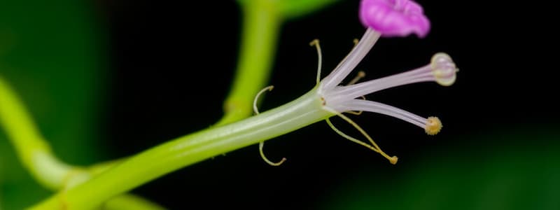
- Anther: The upper fertile part, broader and knob-like, is a bi-lobed structure.
- Connective: The median sterile part is known as the connective, which joins the lobes.
- Each lobe has two pollen sacs in a mature anther.
- Filament: A long, thin structure that joins the stamen to the thalamus or petal of the flower.
Anther
- Anther is the part of a stamen that contains the pollen.
- The anther's function is to produce and provide the pollen required for reproduction.
Structure of Anther
- A normal bithecous or dithecous anther consists of two anther lobes connected by a connective.
- The lobes contain four elongated cavities or pollen sacs (microsporangia), where pollen grains are produced.
- A normal or complete anther has four microsporangia called Tetrasporangiate.
- Some plants have a single lobe with two microsporangia, known as monothecous, such as Hibiscus and Moringa.
- Rarely, an anther has only one lobe with a single microsporangium, called unisporangiate, like Arceuthiobium.
Development of Anther
- Anther development originates from the eusporangiate type, arising from multiple archesporial cells.
- A cross-section (T.S.) of the anther reveals parietal layers (wall layers) and pollen chambers, or microsporangia, containing sporogenous tissue or archesporial cells.
Parietal Layers (Wall Layers) of Anther
- From the outside to the inner side, the anther wall consists of the following layers:
Epidermis
- The outermost layer is single-celled and continuous but not archesporial in origin.
- It forms the outermost protective layer.
Endothecium
- Present below the epidermis; it's a single-celled thick layer.
- During maturation, the cells undergo changes in different walls.
- The outer wall remains thin, while inner and radial walls thicken due to α-cellulose fibers.
- Callose bands are present along the radial walls, but are absent in some places called stomium.
- Dehiscence occurs only from these places.
- It becomes hygroscopic due to fibrous thickening, aiding dehiscence.
Middle Layer
- Consists of parenchymatous cells.
- This layer is one to three cells thick and stores food.
- It's ephemeral or short-lived in nature and absent in mature anthers.
- In Holoptelia plants, it's 3 to 4 cells thick, while it's absent in Najadaceae & Lemnaceae families.
Tapetum
- It's the innermost, single layer of cells with dense cytoplasm and prominent nuclei.
- Provides nutrition to developing microspores.
- Tapetum cells are initially diploid but become polyploid due to endomitosis.
- Tapetum absorbs food from the middle layer, providing nutrition to microspore mother cells or microspores.
- Tapetum cells secrete hormones and enzymes and disappear in the mature anther, but can be multilayered in Nicodia and Costum plants.
Types of Tapetum
Amoeboid / Invasive tapetum / Periplasmodial tapetum
- This type is found in primitive Angiosperms.
- It absorbs all foods from the middle layer, which immediately degenerates.
- Tapetal cells convert absorbed food into special food granules called protoplast bodies.
- The innermost layer dissolves, releasing protoplast bodies into the microsporangium cavity.
- Protoplast bodies become known as periplasmodium, surrounding and nourishing the microspore mother cells.
Glandular / Secretary tapetum
- It is more developed and does not degenerate quickly.
- It absorbs nutrients from the middle layer and secretes them into the microsporangia (pollen sacs).
- Found in most flowering plants.
- It secretes enzymes and hormones, forming special granules called Pro ubisch bodies in the cytoplasm before degeneration.
- Pro ubisch bodies transfer between the cell wall and cell membrane of tapetal cells and are surrounded by sporopollenin.
- These are now called Ubisch bodies/orbicules, and they release into pollen sacs when tapetum degenerates.
- Sporopollenin participates in the formation of other coverings (exine) of pollen grains.
- The tapetum helps in the transfer and storage of food, formation of sporopollenin, pollen kitt materials, and tryphine.
Functions of Tapetum
- Secretion of callase enzyme (β-1,3- glucanase) to dissolve the callosic wall of the tetrad and release them.
- Secretion of polysaccharides into the locules during the free microspore stage, which are absorbed by the microspores.
- The tapetal cells secrete a sporopollenin precursor, though its role in sporopollenin synthesis is unclear.
- In secretory tapetum, sporopollenin is deposited as orbicules on the inner phase of the tapetal cells.
- Formation of Ubisch bodies, pollenkitt, and tryphine, where later two are deposited on the pollen surface to bind pollen grains together for efficient insect pollination.
- Ultrastructural studies show that a special population of plastids synthesizes these substances in the tapetal cells.
- Pollen wall is formed during the post-meiotic period.
Microsporogenesis
- Primary sporogenous cells give rise to microspore mother cells.
- Each microspore mother cell undergoes reduction division to produce four microspores or pollens.
- The process of microspore or pollen formation is microsporogenesis.
- A young anther consists of a mass of meristematic homogenous cells covered by a single layer of epidermis.
- It becomes four-lobed, and each lobe forms a microsporangium with microspores or pollen grains.
- Vascular tissue is formed in the middle region, and simultaneously, four cells below the epidermis in vertical rows become large with visible nuclei and dense cytoplasm.
- These are called archesporial cells.
- Archesporial cells divide periclinally to form primary parietal cells below the epidermis and primary sporogenous cells towards the center.
- Both cells undergo further division to form the anther's complete structure, excluding the epidermis.
- Primary parietal cells undergo further periclinal and anticlinal divisions to form a series of 3-5 layers, forming the anther walls.
- The outermost layer just below the epidermis formed by the primary parietal cells is called the endothecium or fibrous layer, followed by a 1-3 celled middle layer and the innermost layer called tapetum.
- Tapetal cells play a significant role during meiotic cell division in microsporogenous cells and pollen development.
- The primary sporogenous cells divide twice or more by mitotic division to form sporogenous cells, which later differentiate into microspore mother cells during pollen sac wall formation.
- Each microspore mother cell divides to form four haploid microspores or pollen grains by meiotic division or reduction division.
- During, this period, spherical bodies are formed inside the tapetal cells which are known as Ubisch-body.
- Ubisch body is made up of a complex substance called sporopollenin, a polymer of carotenoids.
- The tapetum layer degenerates after formation with Ubisch bodies participating in the formation of the exine of microspores inside pollen sacs.
- Then, thick-walled microspores are called pollen grains.
- At the initial stage, all four microspores are attached together with the help of a callose layer, forming something called a tetrad.
- The callose layer dissolves by callase enzyme, which the tapetum secretes.
Studying That Suits You
Use AI to generate personalized quizzes and flashcards to suit your learning preferences.


