Podcast
Questions and Answers
What is the key anatomical feature defining the anterolateral abdominal wall?
What is the key anatomical feature defining the anterolateral abdominal wall?
- A rigid bony structure providing fixed support.
- A flat, inflexible surface primarily composed of adipose tissue.
- A rigid cartilaginous structure providing fixed support.
- A curved, flexible hexagonal area composed of skin, muscle and connective tissue. (correct)
Which of the following is the MOST accurate description of the number of layers of superficial fascia in the anterior abdominal wall in adults?
Which of the following is the MOST accurate description of the number of layers of superficial fascia in the anterior abdominal wall in adults?
- Four layers, each with distinct fibrous and adipose components.
- Two layers: Camper's fascia and Scarpa's fascia.
- Three layers: Camper's fascia, Scarpa's fascia, and a deep fatty layer. (correct)
- A single, undifferentiated layer.
Which statement BEST describes the clinical significance of the differential metabolic activity observed between the superficial and deep fatty layers of the abdominal wall?
Which statement BEST describes the clinical significance of the differential metabolic activity observed between the superficial and deep fatty layers of the abdominal wall?
- It determines the extent of pubic hair migration in postpubertal individuals.
- It is clinically insignificant, as both layers respond similarly to dietary changes.
- It informs liposuction techniques to preferentially remove the deep fatty layer, minimizing skin contour irregularities. (correct)
- It dictates the distribution of superficial blood vessels, influencing flap viability.
During a laparoscopic inguinal hernia repair, a surgeon identifies a thickened band of tissue incorrectly labeled as the inguinal ligament. Which anatomical structure is the surgeon MOST likely visualizing?
During a laparoscopic inguinal hernia repair, a surgeon identifies a thickened band of tissue incorrectly labeled as the inguinal ligament. Which anatomical structure is the surgeon MOST likely visualizing?
What is the most crucial implication of the varying amounts of fat in the extraperitoneal connective tissue during hernia repair?
What is the most crucial implication of the varying amounts of fat in the extraperitoneal connective tissue during hernia repair?
When planning incisions for abdominal surgery, what is the MOST crucial anatomical consideration regarding the blood supply of the anterior abdominal wall?
When planning incisions for abdominal surgery, what is the MOST crucial anatomical consideration regarding the blood supply of the anterior abdominal wall?
A surgeon encounters significant bleeding during an incision extending alongside the xiphoid process. Which anatomical structures are MOST likely compromised?
A surgeon encounters significant bleeding during an incision extending alongside the xiphoid process. Which anatomical structures are MOST likely compromised?
What is the primary clinical significance of the inferior epigastric artery (IEA) lying posterior to the spermatic cord, separated by the transversalis fascia?
What is the primary clinical significance of the inferior epigastric artery (IEA) lying posterior to the spermatic cord, separated by the transversalis fascia?
Which of the following BEST describes the vascular supply distribution in Zone 2 of the anterior abdominal wall?
Which of the following BEST describes the vascular supply distribution in Zone 2 of the anterior abdominal wall?
A surgeon performing a myocutaneous flap harvests the mid to lower rectus abdominis based on the superior epigastric artery. Why is preliminary ligation of the inferior epigastric artery often performed?
A surgeon performing a myocutaneous flap harvests the mid to lower rectus abdominis based on the superior epigastric artery. Why is preliminary ligation of the inferior epigastric artery often performed?
In portal hypertension, why do small tributaries connect the inferior epigastric vein with the umbilical vein?
In portal hypertension, why do small tributaries connect the inferior epigastric vein with the umbilical vein?
During anterior abdominal wall reconstruction, why is knowledge of the course and location of the tenth and eleventh posterior intercostal, subcostal, and lumbar arteries so important?
During anterior abdominal wall reconstruction, why is knowledge of the course and location of the tenth and eleventh posterior intercostal, subcostal, and lumbar arteries so important?
What clinical signs might suggest that a patient has an enlarged lymph node in the umbilical region?
What clinical signs might suggest that a patient has an enlarged lymph node in the umbilical region?
Why can significant cutaneous anesthesia and muscle denervation only be seen if at least two sequential nerves are surgically sectioned?
Why can significant cutaneous anesthesia and muscle denervation only be seen if at least two sequential nerves are surgically sectioned?
What is the MOST accurate purpose of the transversus abdominis plane (TAP) blockade?
What is the MOST accurate purpose of the transversus abdominis plane (TAP) blockade?
Which of the following statements accurately describes the function(s) of the anterolateral abdominal muscles?
Which of the following statements accurately describes the function(s) of the anterolateral abdominal muscles?
Which anatomical feature explains why denervation from segmental nerve injury results in subtle and clinically difficult to detect muscle tone differences in the anterolateral abdominal wall?
Which anatomical feature explains why denervation from segmental nerve injury results in subtle and clinically difficult to detect muscle tone differences in the anterolateral abdominal wall?
How is it possible for the abdominal muscles to maintain tone and assist pressure regulation?
How is it possible for the abdominal muscles to maintain tone and assist pressure regulation?
There is variation in the amount and number of fibrous bands or tendinous intersections, how does this present?
There is variation in the amount and number of fibrous bands or tendinous intersections, how does this present?
In the context of rectus sheath anatomy, what is the clinical significance of transitioning from the posterior to the anterior layer?
In the context of rectus sheath anatomy, what is the clinical significance of transitioning from the posterior to the anterior layer?
While preparing a surgical incision, what is the importance of identifying decussating fibres?
While preparing a surgical incision, what is the importance of identifying decussating fibres?
How can a hernia occur during linea alba/rectus abdominis thinning?
How can a hernia occur during linea alba/rectus abdominis thinning?
If only have can be considered as an anomaly - what can never be considered an anomaly?
If only have can be considered as an anomaly - what can never be considered an anomaly?
When does it become important to start recognizing and taking the Inguinal ligament?
When does it become important to start recognizing and taking the Inguinal ligament?
If the body had missing section/areas, where would be the expected?
If the body had missing section/areas, where would be the expected?
Name a surgery, that when combined released the oblique aponeurosis from insertion can cause issues.
Name a surgery, that when combined released the oblique aponeurosis from insertion can cause issues.
What would be the result of not providing proper care on those muscles?
What would be the result of not providing proper care on those muscles?
Without the usage of more muscles, or external support, what would be the result on smaller pressure changes.
Without the usage of more muscles, or external support, what would be the result on smaller pressure changes.
With regards to inguinal, what area relates most strongly with it?
With regards to inguinal, what area relates most strongly with it?
What is commonly required (and must be taken in account) when a case of strangulation occurs?
What is commonly required (and must be taken in account) when a case of strangulation occurs?
From all options provided, that can result with or without birth, what can only be attributed with time? (or acquired through age)
From all options provided, that can result with or without birth, what can only be attributed with time? (or acquired through age)
During surgical treatment, there are often re-approximations needed to make a proper alignment, but what must be considered?
During surgical treatment, there are often re-approximations needed to make a proper alignment, but what must be considered?
Is it ever possible to have tissue expansion aided by component separation operations, still cause problems? Is it just a simple case, with easy operation.
Is it ever possible to have tissue expansion aided by component separation operations, still cause problems? Is it just a simple case, with easy operation.
What is the risk with surgical incision procedures, and it is important to have knowledgable surgical team, especially with hernias caused because of birth?
What is the risk with surgical incision procedures, and it is important to have knowledgable surgical team, especially with hernias caused because of birth?
Direct hernias most often arise from weakness of that specific tissue in the area, can some times be confused or similar - what is a key difference?
Direct hernias most often arise from weakness of that specific tissue in the area, can some times be confused or similar - what is a key difference?
A femoral will cause a certain area to be wider, to aid which other procedures? (especially during what period of time being considered
A femoral will cause a certain area to be wider, to aid which other procedures? (especially during what period of time being considered
Can it be easily dismissed and said to be easy to point one from the other with a single touch?
Can it be easily dismissed and said to be easy to point one from the other with a single touch?
With surgical intervention what type of operation will be done, and what will be considered?
With surgical intervention what type of operation will be done, and what will be considered?
What is the main goal of procedures performed in this situation?
What is the main goal of procedures performed in this situation?
What is the MOST critical anatomical consideration when managing surgical approaches to Spigelian hernias?
What is the MOST critical anatomical consideration when managing surgical approaches to Spigelian hernias?
During a surgical procedure to address portal hypertension, which vascular anastomosis is MOST likely targeted for manipulation or assessment?
During a surgical procedure to address portal hypertension, which vascular anastomosis is MOST likely targeted for manipulation or assessment?
When performing extensive liposuction of the anterior abdominal wall, what anatomical layer should be preferentially targeted and why?
When performing extensive liposuction of the anterior abdominal wall, what anatomical layer should be preferentially targeted and why?
What anatomical structure is critical for a surgeon to identify during laparoscopic inguinal hernia repair to avoid mistaking it for the inguinal ligament?
What anatomical structure is critical for a surgeon to identify during laparoscopic inguinal hernia repair to avoid mistaking it for the inguinal ligament?
During the creation of a transverse rectus abdominis myocutaneous (TRAM) flap, what is the primary rationale for ligating the inferior epigastric artery when basing the flap on the superior epigastric artery?
During the creation of a transverse rectus abdominis myocutaneous (TRAM) flap, what is the primary rationale for ligating the inferior epigastric artery when basing the flap on the superior epigastric artery?
In the context of anterior abdominal wall reconstruction, why is it crucial to understand the innervation patterns of the anterolateral abdominal muscles?
In the context of anterior abdominal wall reconstruction, why is it crucial to understand the innervation patterns of the anterolateral abdominal muscles?
Why might division of the lacunar ligament be necessary during a femoral hernia repair, and what critical anatomical structure must be carefully protected?
Why might division of the lacunar ligament be necessary during a femoral hernia repair, and what critical anatomical structure must be carefully protected?
What is the rationale behind limiting surgical dissection to the region above the arcuate line when performing an extraperitoneal hernia repair?
What is the rationale behind limiting surgical dissection to the region above the arcuate line when performing an extraperitoneal hernia repair?
How does the transversus abdominis plane (TAP) blockade achieve its analgesic effect, and what anatomical structures are directly targeted?
How does the transversus abdominis plane (TAP) blockade achieve its analgesic effect, and what anatomical structures are directly targeted?
Why are perforating vessels important considerations in creating large lipocutaneous flaps on the abdominal wall, and what is the potential consequence of their ligation?
Why are perforating vessels important considerations in creating large lipocutaneous flaps on the abdominal wall, and what is the potential consequence of their ligation?
During surgical incisions of the anterior abdominal wall, why is it important to identify the decussating fibers at the linea alba?
During surgical incisions of the anterior abdominal wall, why is it important to identify the decussating fibers at the linea alba?
What anatomical feature is responsible for the 'shutter effect' that provides resistance to increases in intra-abdominal pressure and protects against hernia formation?
What anatomical feature is responsible for the 'shutter effect' that provides resistance to increases in intra-abdominal pressure and protects against hernia formation?
During a surgical repair in the inguinal region, inadvertent injury to the iliohypogastric nerve may lead to what potential complication?
During a surgical repair in the inguinal region, inadvertent injury to the iliohypogastric nerve may lead to what potential complication?
Why does the rectus abdominis muscle remain functional despite variations in the number and extent of tendinous intersections?
Why does the rectus abdominis muscle remain functional despite variations in the number and extent of tendinous intersections?
While closing the abdominal wall the surgeon is re-approximating the tissue - what is the most important factor for the alignment?
While closing the abdominal wall the surgeon is re-approximating the tissue - what is the most important factor for the alignment?
Where does the Cremaster consist of loosely arranged muscle fasciculi lying?
Where does the Cremaster consist of loosely arranged muscle fasciculi lying?
Superiorly, rectus abdominis is attached by three slips of muscle to the fifth, sixth and seventh costal cartilages - what about the 'most medial fibres'?
Superiorly, rectus abdominis is attached by three slips of muscle to the fifth, sixth and seventh costal cartilages - what about the 'most medial fibres'?
During creation of Transverse Rectus Abdominis Myocutaneous Flap - what needs to be taken account for?
During creation of Transverse Rectus Abdominis Myocutaneous Flap - what needs to be taken account for?
On the topic of surgical treatment, with regards to general incision creation, what is the result of not knowing abdominal wall anatomy?
On the topic of surgical treatment, with regards to general incision creation, what is the result of not knowing abdominal wall anatomy?
Ischemic bowel can be resulted with damage - what steps must be accounted for? (and why do they appear)
Ischemic bowel can be resulted with damage - what steps must be accounted for? (and why do they appear)
After trauma (in this situation direct trauma to abdominal section) what must be accounted for when looking blood?
After trauma (in this situation direct trauma to abdominal section) what must be accounted for when looking blood?
What is the MOST significant risk associated with surgical ligation of periumbilical perforator vessels during the creation of large lipocutaneous flaps?
What is the MOST significant risk associated with surgical ligation of periumbilical perforator vessels during the creation of large lipocutaneous flaps?
During component separation for ventral hernia repair, what is the primary risk associated with raising large subcutaneous flaps from the midline incision?
During component separation for ventral hernia repair, what is the primary risk associated with raising large subcutaneous flaps from the midline incision?
In the creation of a TRAM flap based on the superior epigastric artery, what is the MOST important reason to perform preliminary ligation of the inferior epigastric artery?
In the creation of a TRAM flap based on the superior epigastric artery, what is the MOST important reason to perform preliminary ligation of the inferior epigastric artery?
What is the key surgical implication of the inferior epigastric vessels ascending obliquely along the medial margin of the deep inguinal ring?
What is the key surgical implication of the inferior epigastric vessels ascending obliquely along the medial margin of the deep inguinal ring?
During an extraperitoneal hernia repair, what anatomical characteristic makes separation of the peritoneum and transversalis fascia difficult, increasing the risk of peritoneal rents?
During an extraperitoneal hernia repair, what anatomical characteristic makes separation of the peritoneum and transversalis fascia difficult, increasing the risk of peritoneal rents?
How does the 'shutter effect' contribute to abdominal wall stability and resistance to hernia formation?
How does the 'shutter effect' contribute to abdominal wall stability and resistance to hernia formation?
In anterior abdominal wall reconstruction, why is an understanding of the course of intercostal, subcostal, and lumbar arteries CRUCIAL when creating myofascial flaps?
In anterior abdominal wall reconstruction, why is an understanding of the course of intercostal, subcostal, and lumbar arteries CRUCIAL when creating myofascial flaps?
What is true when the abdominal pressure is required to increase?
What is true when the abdominal pressure is required to increase?
In individuals where the lineal area starts thinning, with relation to abdominis, what becomes of them?
In individuals where the lineal area starts thinning, with relation to abdominis, what becomes of them?
From all the list options that are described below, why might hernia occur?
From all the list options that are described below, why might hernia occur?
What anatomical factor is critical to consider when placing laparoscopic ports for a procedure to repair a Spigelian hernia?
What anatomical factor is critical to consider when placing laparoscopic ports for a procedure to repair a Spigelian hernia?
What is the primary reason for preferring liposuction in the deep fatty layer (rather than in the superficial) within the abdominal wall?
What is the primary reason for preferring liposuction in the deep fatty layer (rather than in the superficial) within the abdominal wall?
If in doubt in which tissue the pain radiates to - after dissection, in which specific location can the intercostal nerve be located to get a better reading?
If in doubt in which tissue the pain radiates to - after dissection, in which specific location can the intercostal nerve be located to get a better reading?
On certain people, the lineal alba presents itself relatively visible - what is required in those people?
On certain people, the lineal alba presents itself relatively visible - what is required in those people?
Despite what kind of action done by the cremaster, what cannot occur?
Despite what kind of action done by the cremaster, what cannot occur?
During repairs, with components separations what occurs, that can be an effect by lack of care?
During repairs, with components separations what occurs, that can be an effect by lack of care?
Due to the characteristics of the lineal alba being composed of decussations, what element is related to make it up?
Due to the characteristics of the lineal alba being composed of decussations, what element is related to make it up?
When hernias become from direct origin, what will cause in a anatomical fashion?
When hernias become from direct origin, what will cause in a anatomical fashion?
As a way to defend any individual, what kind of maneuver will occur to protect from injury?
As a way to defend any individual, what kind of maneuver will occur to protect from injury?
What is the key surgical anatomical consideration while facing a femoral hernia?
What is the key surgical anatomical consideration while facing a femoral hernia?
What occurs in multiple people (regarding the hernias) prior and after with the spermatic cord?
What occurs in multiple people (regarding the hernias) prior and after with the spermatic cord?
During laparoscopic inguinal repair, with which fiber-based band does need extra consideration?
During laparoscopic inguinal repair, with which fiber-based band does need extra consideration?
What is common factor with the different types of arteries?
What is common factor with the different types of arteries?
During any event where there is division (for what ever reason) in a segmental nerve, what is observed/detectable with most frequency?
During any event where there is division (for what ever reason) in a segmental nerve, what is observed/detectable with most frequency?
Where is the 'triangle' often seen when looking at or repairing the abdominal wall?
Where is the 'triangle' often seen when looking at or repairing the abdominal wall?
Which area is innervated more consistently the most?
Which area is innervated more consistently the most?
In cases of extreme umbilical defects, is expected for what structure to be the covering the defect?
In cases of extreme umbilical defects, is expected for what structure to be the covering the defect?
If the belly has extreme lack or absent sections of muscle, and those missing sides have an effect, what is it?
If the belly has extreme lack or absent sections of muscle, and those missing sides have an effect, what is it?
Which of the listed is the main goal while trying to fix a big issue in the abdominal section?
Which of the listed is the main goal while trying to fix a big issue in the abdominal section?
The anterior abdominal wall's superior border is defined by the intercostal muscles and the xiphisternal junction.
The anterior abdominal wall's superior border is defined by the intercostal muscles and the xiphisternal junction.
The membranous layer (Scarpa's fascia) is located superficial to the superficial fatty layer (Camper's fascia) within the superficial fascia of the abdominal wall.
The membranous layer (Scarpa's fascia) is located superficial to the superficial fatty layer (Camper's fascia) within the superficial fascia of the abdominal wall.
The superficial fatty layer is consistent in thickness across the anterior abdominal wall due to a uniform distribution of adipose tissue.
The superficial fatty layer is consistent in thickness across the anterior abdominal wall due to a uniform distribution of adipose tissue.
The transversalis fascia is a continuous sheet that does not attach to the iliac crest or the posterior margin of the inguinal ligament.
The transversalis fascia is a continuous sheet that does not attach to the iliac crest or the posterior margin of the inguinal ligament.
The iliopubic tract is often correctly identified as the inguinal ligament during laparoscopic inguinal hernia repair.
The iliopubic tract is often correctly identified as the inguinal ligament during laparoscopic inguinal hernia repair.
The inferior epigastric vessels are contained within an envelope of fat in the extraperitoneal connective tissue, serving as an anatomical landmark during hernia repairs.
The inferior epigastric vessels are contained within an envelope of fat in the extraperitoneal connective tissue, serving as an anatomical landmark during hernia repairs.
Liposuction preferentially removes the superficial fatty layer to avoid skin dimpling and contour irregularities.
Liposuction preferentially removes the superficial fatty layer to avoid skin dimpling and contour irregularities.
Vascular branches supply the rectus abdominis and penetrate the anterior layer of the rectus sheath to supply the abdominal skin and subcutaneous fat.
Vascular branches supply the rectus abdominis and penetrate the anterior layer of the rectus sheath to supply the abdominal skin and subcutaneous fat.
The superior epigastric artery anastomoses with the hepatic artery via the falciform ligament.
The superior epigastric artery anastomoses with the hepatic artery via the falciform ligament.
The musculophrenic artery arises from the external iliac artery.
The musculophrenic artery arises from the external iliac artery.
The inferior epigastric artery runs posterior to the spermatic cord and is separated from it by the transversalis fascia.
The inferior epigastric artery runs posterior to the spermatic cord and is separated from it by the transversalis fascia.
The integrity of the periumbilical perforator vessels is not a factor when creating lipocutaneous flaps on the abdominal wall.
The integrity of the periumbilical perforator vessels is not a factor when creating lipocutaneous flaps on the abdominal wall.
The ductus deferens or round ligament hooks around the superior epigastric artery at the deep inguinal ring.
The ductus deferens or round ligament hooks around the superior epigastric artery at the deep inguinal ring.
The inferior epigastric vessels form the medial border of the inguinal triangle, also known as Hesselbach's triangle.
The inferior epigastric vessels form the medial border of the inguinal triangle, also known as Hesselbach's triangle.
The caput medusae is caused by portal venous blood draining into the systemic circulation via the superior epigastric veins.
The caput medusae is caused by portal venous blood draining into the systemic circulation via the superior epigastric veins.
Transection of the nerves within the neurovascular plane during abdominal wall reconstruction will not cause any segmental denervation of portions of the rectus abdominis and the lateral anterior abdominal wall musculature.
Transection of the nerves within the neurovascular plane during abdominal wall reconstruction will not cause any segmental denervation of portions of the rectus abdominis and the lateral anterior abdominal wall musculature.
Rectus abdominis contribute to flexion of the trunk and the maintenance of abdominal wall tone required during straining.
Rectus abdominis contribute to flexion of the trunk and the maintenance of abdominal wall tone required during straining.
The arcuate line marks the inferior end of the anterior layer of the rectus sheath
The arcuate line marks the inferior end of the anterior layer of the rectus sheath
The Linea Alba consists of superficial fibres attached to the pubic symphysis, and deeper fibres that form a lamella attached behind the rectus abdominis to the posterior surface of both pubic crests.
The Linea Alba consists of superficial fibres attached to the pubic symphysis, and deeper fibres that form a lamella attached behind the rectus abdominis to the posterior surface of both pubic crests.
The actions of the External abdominal oblique include contributing to the maintenance of abdominal tone, increasing intra-abdominal pressure, and lateral flexion of the trunk against resistance.
The actions of the External abdominal oblique include contributing to the maintenance of abdominal tone, increasing intra-abdominal pressure, and lateral flexion of the trunk against resistance.
Match the layers of superficial fascia in the abdominal wall with their descriptions:
Match the layers of superficial fascia in the abdominal wall with their descriptions:
Match the following arteries with their origin and area of supply:
Match the following arteries with their origin and area of supply:
Match the type of hernia with its description:
Match the type of hernia with its description:
Match the layers forming the rectus sheath with their composition:
Match the layers forming the rectus sheath with their composition:
Match each action to the primary muscle(s) responsible for it:
Match each action to the primary muscle(s) responsible for it:
Match the following anatomical structures/concepts with their accurate description:
Match the following anatomical structures/concepts with their accurate description:
Match the location described with the possible type of herniation:
Match the location described with the possible type of herniation:
Match the following nerves with the region supplied:
Match the following nerves with the region supplied:
Match the surgical zone with the vessel that supplies that region:
Match the surgical zone with the vessel that supplies that region:
Match the layer with its relevant anatomical structure:
Match the layer with its relevant anatomical structure:
Flashcards
Anterior abdominal wall
Anterior abdominal wall
Hexagonal area defined by costal arches, mid-axillary line, iliac crests, inguinal ligament, and pubic symphysis.
Superficial fascia
Superficial fascia
Layer between dermis and muscles, divided into superficial fatty (Camper's) and membranous (Scarpa's) layers.
Superficial fatty layer
Superficial fatty layer
Layer containing fat, partitioned by fibrous septa connecting dermis and deeper membranous layer.
Membranous layer
Membranous layer
Signup and view all the flashcards
Transversalis fascia
Transversalis fascia
Signup and view all the flashcards
Extraperitoneal connective tissue
Extraperitoneal connective tissue
Signup and view all the flashcards
Superior epigastric artery
Superior epigastric artery
Signup and view all the flashcards
Inferior epigastric artery
Inferior epigastric artery
Signup and view all the flashcards
Zone 2
Zone 2
Signup and view all the flashcards
TAP blockade
TAP blockade
Signup and view all the flashcards
Muscles during pressure increase
Muscles during pressure increase
Signup and view all the flashcards
Tendinous intersections
Tendinous intersections
Signup and view all the flashcards
Rectus sheath
Rectus sheath
Signup and view all the flashcards
Linea alba
Linea alba
Signup and view all the flashcards
Umbilicus
Umbilicus
Signup and view all the flashcards
Umbilical hernia
Umbilical hernia
Signup and view all the flashcards
Pyramidalis
Pyramidalis
Signup and view all the flashcards
External abdominal oblique
External abdominal oblique
Signup and view all the flashcards
Inguinal ligament
Inguinal ligament
Signup and view all the flashcards
Internal abdominal oblique
Internal abdominal oblique
Signup and view all the flashcards
Transversus abdominis
Transversus abdominis
Signup and view all the flashcards
Conjoint aponeurosis
Conjoint aponeurosis
Signup and view all the flashcards
Inguinal canal
Inguinal canal
Signup and view all the flashcards
Superficial inguinal ring
Superficial inguinal ring
Signup and view all the flashcards
Deep inguinal ring
Deep inguinal ring
Signup and view all the flashcards
Cremaster
Cremaster
Signup and view all the flashcards
Myopectineal orifice
Myopectineal orifice
Signup and view all the flashcards
mycopectineal orifice
mycopectineal orifice
Signup and view all the flashcards
Direct and Indirect Inguinal Hernia
Direct and Indirect Inguinal Hernia
Signup and view all the flashcards
Femoral hernia
Femoral hernia
Signup and view all the flashcards
incisional hernia
incisional hernia
Signup and view all the flashcards
Epigastric hernia
Epigastric hernia
Signup and view all the flashcards
Compartment Separation
Compartment Separation
Signup and view all the flashcards
Spigelian hernia
Spigelian hernia
Signup and view all the flashcards
Parastomal hernia
Parastomal hernia
Signup and view all the flashcards
Vascular supply
Vascular supply
Signup and view all the flashcards
Vascular branches
Vascular branches
Signup and view all the flashcards
Posterior arteries
Posterior arteries
Signup and view all the flashcards
Segmental nerves
Segmental nerves
Signup and view all the flashcards
Anterolateral abdominal wall muscles
Anterolateral abdominal wall muscles
Signup and view all the flashcards
External oblique action
External oblique action
Signup and view all the flashcards
Internal Oblique
Internal Oblique
Signup and view all the flashcards
Periumbilical Perforator Vessels
Periumbilical Perforator Vessels
Signup and view all the flashcards
Positive Intra-abdominal Pressure
Positive Intra-abdominal Pressure
Signup and view all the flashcards
Transversus Abdominis Plane Block
Transversus Abdominis Plane Block
Signup and view all the flashcards
Indirect Inguinal Hernia
Indirect Inguinal Hernia
Signup and view all the flashcards
Inferior Epigastric Artery Origin
Inferior Epigastric Artery Origin
Signup and view all the flashcards
Paraspinal Region Vessels
Paraspinal Region Vessels
Signup and view all the flashcards
Intercostal Nerves Pathway
Intercostal Nerves Pathway
Signup and view all the flashcards
Subcostal Nerve Function
Subcostal Nerve Function
Signup and view all the flashcards
Transversus Abdominis Plane (TAP) Blockade
Transversus Abdominis Plane (TAP) Blockade
Signup and view all the flashcards
Component separation
Component separation
Signup and view all the flashcards
Femoral Hernia Cause
Femoral Hernia Cause
Signup and view all the flashcards
Spigelian location
Spigelian location
Signup and view all the flashcards
Anterior abdominal wall integument
Anterior abdominal wall integument
Signup and view all the flashcards
Deep fatty layer
Deep fatty layer
Signup and view all the flashcards
Male membranous layer
Male membranous layer
Signup and view all the flashcards
Rectus abdominis attachment point
Rectus abdominis attachment point
Signup and view all the flashcards
Anastomoses Vessel
Anastomoses Vessel
Signup and view all the flashcards
Different TAP block Approaches
Different TAP block Approaches
Signup and view all the flashcards
Study Notes
Anterior Abdominal Wall Boundaries and Functions
- Superior boundary is the costal arches (margins) and xiphisternal junction.
- Lateral boundary is the mid-axillary line.
- Inferior boundary is the imaginary line along the iliac crests, inguinal ligament, and pubic symphysis.
- The anterior abdominal wall is continuous with the posterior abdominal wall, forming a flexible sheet of skin, muscle and connective tissue.
- The anterior abdominal wall is Contiguous with the respiratory diaphragm, the bony and myofascial structures of the thorax.
- Tissues form the myopectineal orifice and the inguinal canal.
- Maintains abdominal shape and aids physiological functions.
- Hernia repair is a common surgical operation related to abdominal wall dysfunction.
Skin and Soft Tissue Layers
- Includes skin, soft tissues, lymphatic/vascular structures, and segmental nerves.
- The skin is non-specialized and may be hirsute depending on sex and ancestry.
- All postpubertal individuals have some extension of the pubic hair on the abdominal wall, especially in males, where the hair can extend to the umbilicus in a triangular pattern.
- Subcutaneous fat varies in thickness based on sex and caloric intake.
Superficial Fascia Layers
- Consists of three layers: superficial fatty (Camper's), membranous (Scarpa's), and deep fatty layer.
- Layers are well-defined in children; the membranous layer remains distinct in adults.
- Contains adipose tissue, blood vessels, lymphatics, and nerves, especially in the inguinal region, superficial inguinal nodes.
Superficial Fatty Layer
- Contains a variable amount of fat partitioned by fibrous septa connecting the dermis with the deeper membranous layer.
- Inferiorly it is continuous with the superficial fascia of the thigh.
- Medially, it is continuous with the linea alba.
- Extends over external genitalia in males, becomes thin and contains dartos muscle in the scrotum
- Extends into the labia majora and perineum in females.
Membranous Layer
- Made of connective tissue and elastic fibers
- Thickness varies, becoming thinner proximally on the abdomen (Lancerotto et al 2011)
- Thickness histologically is between 0.5-1mm but it appears thicker on computed tomography (Chopra et al 2011, Lancerotto et al 2011)
- Loosely connected to external abdominal oblique aponeurosis and rectus sheath by oblique fibrous septa
- Continuous with superficial fascia of the trunk
- Adherent to linea alba and pubic symphysis
- Fuses with iliac crest, extends superficial to the inguinal ligament, and fuses with the fascia lata at the inguinal crease
- In males it extends onto the dorsum of the penis, forming the fundiform ligament, and onto the scrotum where it becomes continuous with the membranous layer of the superficial fascia of the perineum (Colles’ fascia)
- In females it continues into the labia majora and is continuous with the superficial fascia of the perineum
- In males, the testis can frequently be retracted out of the scrotum into the loose areolar tissue between the membranous layer of superficial fascia over the inguinal canal and the aponeurosis of external abdominal oblique
Deep Fatty Layer
- Thickness more variable than the superficial layer, thin/absent at bony prominences and linea alba.
- Adipocytes show different metabolic activities vs superficial layer (Chopra et al 2011)
- Liposuction preferentially removes this layer to avoid skin irregularities (Markman and Barton 1987)
Transversalis Fascia
- Thin connective tissue between transversus abdominis and extraperitoneal fat.
- Part of the general fascial layer between peritoneum and abdominal wall.
- Fuses posteriorly with the anterior layer of the thoracolumbar fascia; forms a continuous sheet anteriorly.
- Superiorly blends with the fascia covering the inferior surface of the respiratory diaphragm
- Inferiorly is continuous with the iliac and pelvic parietal fasciae.
- Attached to the iliac crest between the origins of transversus abdominis and iliacus and to the posterior margin of the inguinal ligament (iliopubic tract).
- The iliopubic tract consists of transverse fibers that fan out laterally towards the anterior superior iliac spine to blend with the iliopsoas fascia and run medially to the pubic bone
- The iliopubic tract is a landmark during laparoscopic inguinal hernia repair, though often incorrectly described as the inguinal ligament (Teoh et al 1999)
- Medial to the femoral sheath the transversalis fascia is fused to the pubis behind the conjoint aponeurosis (conjoint ‘tendon’)
- An inferior extension of the fascia forms the anterior part of the femoral sheath
- A further thickening of the transversalis fascia, the interfoveolar ligament, runs inferior to the inguinal ligament at the medial margin of the deep inguinal ring; it may contain muscle fibres.
- Continues as the internal spermatic fascia over structures passing through the deep inguinal ring.
Extraperitoneal Connective Tissue
- Lies between the peritoneum and the fasciae lining the abdominal/pelvic cavities.
- Contains a variable amount of fat, often found in congenital hernias
- Adipose tissue is most common content of small congenital umbilical and epigastric hernias
- Abundant on the posterior abdominal wall around the kidneys.
- The inferior epigastric vessels contained within the fat pad as they travel from the external iliac vessels to rectus abdominis
- Fat pad serves as an anatomical landmark during various types of abdominal hernia repairs.
- Fat abundance allows for peritoneal flap creation during hernia surgery.
- Minimal fat makes separation of peritoneum and transversalis fascia difficult.
Vascular Supply: Superior Epigastric Artery/Veins
- The superior epigastric artery is a terminal branch of the internal thoracic artery.
- Arises level with the sixth costal cartilage, descends between the costal and sternal parts of diaphragm.
- They pass anterior to distal transversus thoracis and proximal transversus abdominis.
- Anastomoses with inferior epigastric arteries in the rectus abdominis (Rozen et al 2008).
- Vascular branches supply rectus abdominis and perforate the anterior rectus sheath
- Vessels penetrate rectus abdominis near the umbilicus, referred to as periumbilical perforator vessels.
- Proximal branch passes anterior to the xiphoid process, anastomosing with the contralateral branch.
- Gives small branches to the anterior diaphragm and falciform ligament (on the right).
Vascular Supply: Inferior Epigastric Artery/Veins
- Inferior epigastric artery originates from the external iliac artery proximal to the inguinal ligament.
- Vessels curve forwards in extraperitoneal tissue; ascends obliquely near the junction.
- Lies posterior to the spermatic cord, separated by the transversalis fascia.
- It pierces transversalis fascia and enters posterior layer of the rectus sheath anterior to the arcuate line.
- Its accompanying veins, usually two, unite to form a single vein that drains into the external iliac vein (Rozen et al 2009).
Blood Supply: Surgical Zones
- Surgical zones have been defined to better understand this vascular distribution.
- Zone 1 (central proximal abdomen) gets blood from the superior epigastric artery (superiorly) and inferior epigastric artery (inferiorly).
- Periumbilical perforator vessels arise from both epigastric arteries to supply overlying tissues.
- Zone 2 (hypogastric region) receives blood supply from superficial/deep branches of inferior epigastric artery (medially).
- Superficial circumflex iliac artery (from femoral artery) provides lateral blood supply.
- Zone 3 (proximal to arcuate line, lateral to semilunar line) gets blood from deep circumflex iliac artery (inferiorly).
- Musculophrenic artery (branch of internal thoracic artery) supplies zone 3 superiorly.
Inferior Epigastric Artery Branches
- Branches anastomose with the superior epigastric artery within rectus abdominis (Rozen et al 2008).
- Inferolaterally, it anastomoses with the deep circumflex iliac artery.
- Gives off the cremasteric artery, a pubic branch, and muscular/cutaneous branches.
- In the male, cremasteric artery supplies cremaster, other cord coverings, and anastomoses with the testicular artery.
- Is small in females
- A pubic branch anastomoses with a pubic branch of the obturator artery.
- Sometimes, the obturator artery is replaced with the larger inferior epigastric artery, and lies close to the femoral ring (Pai et al 2009).
- The muscular branches supply abdominal muscles and peritoneum, and Anastomose with the circumflex iliac and lumbar arteries.
- This Contributes to periumbilical perforator vessels.
Blood Supply: Additional Info
- Mapped for large lipocutaneous flaps
- Ligation of these can lead to flap necrosis (Rozen et al 2008)
- May arise from femoral artery and ascend anterior to femoral vein
- Superior and inferior epigastric arteries provide collateral blood flow between the internal thoracic artery.
- The small tributaries of the inferior epigastric vein anastomose with branches of the umbilical vein in the falciform ligament.
- In portal hypertension, leads to caput medusae and Back pressure to the skin and pattern of dilated, serpiginous superficial veins radiating out from the umbilicus
Intercostal/Subcostal/Lumbar Arteries
- Pierce transversus abdominis to enter neurovascular plane between transversus abdominis and internal abdominal oblique.
- Give muscular branches to obliques
- Before anastomosing with lateral branches of superior and inferior epigastric arteries at rectus sheath.
Anterior Abdominal Muscles
- Rectus abdominis, external/internal obliques, transversus abdominis, and pyramidalis contribute to functions involving pressure.
- Muscle tone provides support for abdominal viscera and retains normal contour.
- Contraction increases intra-abdominal pressure, mainly using respiratory diaphragm muscles.
Nerves
- Damage to nerves that innervate rectus abdominis can cause segmental denervation of portions of rectus abdominis.
- Transversus abdominis plane (TAP) blockade is a regional anesthetic technique for abdominal surgery.
Lymphatic Drainage
- Highest concentration in the dermis (Friedman et al 2015)
- Vessels from lumbar and gluteal pass with with the superficial circumflex iliac vessels
- Lymphatic vessels from the skin beyond the umbilicus is passed via the superficial epigastric vessels
- Vessels from the lumbar region drain to axillary/parasternal nodes
- The vessels can drain to the lateral aortic nodes.
- Deeper Lymphatic vessels accompany the arteries.
Segmental Nerves
- Thoracic spinal nerves innervate the abdominal wall.
- Seventh to twelfth thoracic ventral rami continue anteriorly into the abdominal wall.
- All these segmental nerves run anteriorly within a thin layer of fascia in the neurovascular plane between transversus abdominis and internal abdominal oblique.
- Supply the skin of the lateral and anterior abdominal walls
- Ninth intercostal nerve supplies the skin proximal to the umbilicus.
- Tenth intercostal nerve supplies the skin that consistently includes the umbilicus.
- Eleventh intercostal nerve supplies the skin distal to the umbilicus.
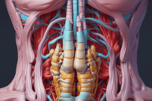
Rectus Abdominis
- Paired, long muscle extending along the anterior abdominal wall on either side of the linea alba
- Interrupted by fibrous bands (tendinous intersections) (Rai et al 2018)
- Muscle arises from the anterior border of the sixth and seventh ribs and the xiphoid process
- Supplied by superior and inferior epigastric arteries as well as the distal thee posterior Intercostal arteries
- Receives innervation from thoracic spinal nerves
- It acts in flexion of the trunk
Rectus Sheath
- Encloses the rectus abdominis
- The anterior layer extends the length of the muscle and fuses with the periosteum
- Distal to the arcuate line, the three aponeuroses from EO, IO and TA form the anterior layer with transversalis fascia on the posterior layer
- Functions as attachments for the EAO, IAO and TA muscles
External Abdominal Oblique
- Largest muscle and lies most superficially
- Curves and attaches to the ribs muscles are the serratus anterior/latissimus Dorsi and attaches to iliac crest
- Fibres diverge to attach distally
- Receives blood supply from the distal Posterior Intercostal and subcostal arteries proximally, and the deep circumflex iliac artery distally. -
- There are additional smaller contributions (Schlenz et al 1999).
- The Innervation comes form the Spinal Nerves to maintain abdominal tone, increasing intra-abdominal pressure, and lateral flexion of the trunk against resistance from the Spinal Nerves
Internal Abdominal Oblique
- Lies deeper than external abdominal oblique and thinner than EAO
- Runs from iliopectineal arch, a thickened band of the fascia (Acland 2008)
- It is attached to the last ribs and merges with internal intercostal muscles function of which is Innervation and lateral flexion of the trunk against resistance
Transversus Abdominis
- It is attached to the inner top of the anterior part of crest, the throacolumbar fascia, and the costal cartilages
- Ends that curve downwards through the fibres to have the body
- Receives blood from the superior Epigastric arteries and lumbar arteries in order to maintain the core
Conjoint Aponeurosis
- Aponeurosis in form with the muscles attached as a Conjunction
- Runs through to act
Hernias of the Anterior Abdominal Wall (Overview)
- Anterior abdominal wall hernias can occur in inguinal, femoral, umbilical, epigastric and Spigelian regions.
- These types of hernia occur where the layers of the abdominal wall are intrinsically weak
- 60% are on the right, 25% are on the left and only 15% are both
Myopectineal Orifice
- Infermedial part of anterior abdominal wall
- Encompasses the inguinal ring, and inguinal triangle, and portal that transmits femoral parts
- Is a major target, but if it is arranged poorly can result in conditions such as hernias
Indirect Inguinal
- A result of development as internal rings, direct hernias, or femoral hernias in combination with the aponeuroses
- Results in the abnormal parts
Direct Inguinal Hernias
- Arise as direct hernias within the tissue and enlarge or become larger than normal and can become a risk
Femoral Structures/Concerns of Health
- Hernias start to come from the potential of the areas of attachment causing concerns
Umbilical Abnormalities
- Occurs in the ring of the umbilical causing hernias and weaknesses
- Occurs after the return of parts of the fetus in the womb
Spigelian Hernia
- Spigelican is a hole for the fat and peritoneal sac and causes major health defects
Incisional Concerns
- Occurs with a mass that leads to failure and infection which increases major health complications
The Parastomal Area
- When there is a disruption the health leads from a hernia in the gut and can be bad for you
- Pyramidalis contributes to tensing the distal linea alba but is of doubtful physiological significance.
Studying That Suits You
Use AI to generate personalized quizzes and flashcards to suit your learning preferences.