Podcast
Questions and Answers
Besides reduced oxygen-carrying capacity, what is another significant physiological consequence of anemia?
Besides reduced oxygen-carrying capacity, what is another significant physiological consequence of anemia?
- Decreased neutrophil production
- Increased intravascular volume
- Tissue hypoxia (correct)
- Elevated platelet counts
What is the primary concern in the immediate management of acute blood loss?
What is the primary concern in the immediate management of acute blood loss?
- Restoring intravascular volume (correct)
- Administering iron supplements
- Preventing platelet aggregation
- Monitoring erythrocyte count
How does anemia of chronic disease (ACD) primarily differ from iron deficiency anemia in terms of iron availability?
How does anemia of chronic disease (ACD) primarily differ from iron deficiency anemia in terms of iron availability?
- ACD involves increased iron utilization, whereas iron deficiency anemia involves decreased iron stores.
- ACD is characterized by impaired iron utilization despite sufficient iron stores, while iron deficiency anemia involves low iron stores. (correct)
- ACD primarily affects the production of platelets, while iron deficiency anemia specifically affects hemoglobin synthesis.
- ACD results from increased erythropoiesis; iron deficiency anemia results from decreased erythropoiesis.
In hemolytic anemia, which of the following processes leads to the premature destruction of erythrocytes?
In hemolytic anemia, which of the following processes leads to the premature destruction of erythrocytes?
What genetic anomaly is most frequently linked to the development of polycythemia vera (PV)?
What genetic anomaly is most frequently linked to the development of polycythemia vera (PV)?
What condition is indicated by a platelet count of 100,000/mm³?
What condition is indicated by a platelet count of 100,000/mm³?
What is the underlying mechanism of heparin-induced thrombocytopenia (HIT)?
What is the underlying mechanism of heparin-induced thrombocytopenia (HIT)?
How do hepatic cells contribute to hemostasis?
How do hepatic cells contribute to hemostasis?
What is the primary pathological event in disseminated intravascular coagulation (DIC)?
What is the primary pathological event in disseminated intravascular coagulation (DIC)?
What is the primary characteristic of thrombophilia that increases the risk of thromboembolic events?
What is the primary characteristic of thrombophilia that increases the risk of thromboembolic events?
What is the primary distinction between acquired and inherited causes of aplastic anemia?
What is the primary distinction between acquired and inherited causes of aplastic anemia?
Which mechanism explains how penicillin can induce hemolytic anemia?
Which mechanism explains how penicillin can induce hemolytic anemia?
How does anemia of chronic disease (ACD) typically affect iron metabolism?
How does anemia of chronic disease (ACD) typically affect iron metabolism?
Which of the following is a congenital cause of hemolytic anemia?
Which of the following is a congenital cause of hemolytic anemia?
What clinical manifestation is particularly associated with polycythemia vera due to increased red blood cell mass?
What clinical manifestation is particularly associated with polycythemia vera due to increased red blood cell mass?
How is hereditary hemochromatosis (HH) typically treated to reduce the risk of organ damage?
How is hereditary hemochromatosis (HH) typically treated to reduce the risk of organ damage?
What characterizes heparin-induced thrombocytopenia (HIT)?
What characterizes heparin-induced thrombocytopenia (HIT)?
Why is plasma exchange a critical treatment component for thrombotic thrombocytopenic purpura (TTP)?
Why is plasma exchange a critical treatment component for thrombotic thrombocytopenic purpura (TTP)?
What is the primary goal of treatment for essential thrombocythemia (ET)?
What is the primary goal of treatment for essential thrombocythemia (ET)?
Which condition directly impairs the synthesis of coagulation factors in the liver?
Which condition directly impairs the synthesis of coagulation factors in the liver?
What is the underlying cause of disseminated intravascular coagulation (DIC)?
What is the underlying cause of disseminated intravascular coagulation (DIC)?
What is the significance of Factor V Leiden in thromboembolic disease?
What is the significance of Factor V Leiden in thromboembolic disease?
What is the primary mechanism by which acute blood loss leads to anemia?
What is the primary mechanism by which acute blood loss leads to anemia?
Which of the following nutritional deficiencies leads to megaloblastic anemia?
Which of the following nutritional deficiencies leads to megaloblastic anemia?
What is a common symptom specific to pernicious anemia due to vitamin B12 deficiency?
What is a common symptom specific to pernicious anemia due to vitamin B12 deficiency?
Which of the following is a typical manifestation of advanced iron deficiency anemia?
Which of the following is a typical manifestation of advanced iron deficiency anemia?
What is the underlying cause of anemia in anemia of chronic disease (ACD)?
What is the underlying cause of anemia in anemia of chronic disease (ACD)?
What is the primary characteristic of aplastic anemia?
What is the primary characteristic of aplastic anemia?
What is the typical response of leukocyte counts to stressors like infection or intense exercise?
What is the typical response of leukocyte counts to stressors like infection or intense exercise?
In the context of infection management, what does a 'shift to the left' indicate?
In the context of infection management, what does a 'shift to the left' indicate?
What cell type is primarily affected in infectious mononucleosis?
What cell type is primarily affected in infectious mononucleosis?
How are leukemias classified?
How are leukemias classified?
What is a significant risk factor associated with the development of leukemia?
What is a significant risk factor associated with the development of leukemia?
What distinguishes Hodgkin lymphoma from non-Hodgkin lymphoma?
What distinguishes Hodgkin lymphoma from non-Hodgkin lymphoma?
What is a common initial sign or symptom of Hodgkin lymphoma?
What is a common initial sign or symptom of Hodgkin lymphoma?
Why do individuals with multiple myeloma experience recurrent infections?
Why do individuals with multiple myeloma experience recurrent infections?
What is the effect of an overactive spleen on blood cells?
What is the effect of an overactive spleen on blood cells?
What hematological change is unique to children compared to adults?
What hematological change is unique to children compared to adults?
What condition develops from Rh incompatibility in mother and fetus?
What condition develops from Rh incompatibility in mother and fetus?
What primary compensatory mechanism does the body employ to mitigate tissue hypoxia resulting from anemia?
What primary compensatory mechanism does the body employ to mitigate tissue hypoxia resulting from anemia?
In acute blood loss, which physiological change occurs within the first 24 hours as the body begins to compensate?
In acute blood loss, which physiological change occurs within the first 24 hours as the body begins to compensate?
How does the pathophysiology of anemia of chronic disease (ACD) directly contribute to impaired iron utilization?
How does the pathophysiology of anemia of chronic disease (ACD) directly contribute to impaired iron utilization?
Which of the following mechanisms primarily accounts for the development of hemolytic anemia in drug-induced cases?
Which of the following mechanisms primarily accounts for the development of hemolytic anemia in drug-induced cases?
In polycythemia vera (PV), what is the most direct consequence of increased blood viscosity on cardiovascular function?
In polycythemia vera (PV), what is the most direct consequence of increased blood viscosity on cardiovascular function?
What is the central pathophysiological process in heparin-induced thrombocytopenia (HIT) that triggers the thrombotic events?
What is the central pathophysiological process in heparin-induced thrombocytopenia (HIT) that triggers the thrombotic events?
How does severe liver disease directly impair hemostasis, apart from affecting the production of coagulation factors?
How does severe liver disease directly impair hemostasis, apart from affecting the production of coagulation factors?
What is the most immediate life-threatening consequence of disseminated intravascular coagulation (DIC) that requires prompt intervention?
What is the most immediate life-threatening consequence of disseminated intravascular coagulation (DIC) that requires prompt intervention?
How does Factor V Leiden mutation lead to an increased risk of thromboembolic events at the molecular level?
How does Factor V Leiden mutation lead to an increased risk of thromboembolic events at the molecular level?
In aplastic anemia, the reduction in hematopoietic stem cells leads most directly to which of the following conditions?
In aplastic anemia, the reduction in hematopoietic stem cells leads most directly to which of the following conditions?
If a patient develops hemolytic anemia after starting penicillin, what immunologic mechanism is most likely responsible for erythrocyte destruction?
If a patient develops hemolytic anemia after starting penicillin, what immunologic mechanism is most likely responsible for erythrocyte destruction?
How does anemia of chronic disease (ACD) affect serum iron levels and total iron stores compared to iron deficiency anemia?
How does anemia of chronic disease (ACD) affect serum iron levels and total iron stores compared to iron deficiency anemia?
What intrinsic erythrocyte defect is the primary cause of hemolysis in hereditary spherocytosis?
What intrinsic erythrocyte defect is the primary cause of hemolysis in hereditary spherocytosis?
In hereditary hemochromatosis (HH), what is the direct physiological effect of the HFE gene mutation on iron metabolism?
In hereditary hemochromatosis (HH), what is the direct physiological effect of the HFE gene mutation on iron metabolism?
What is the fundamental mechanism by which plasma exchange alleviates the symptoms of thrombotic thrombocytopenic purpura (TTP)?
What is the fundamental mechanism by which plasma exchange alleviates the symptoms of thrombotic thrombocytopenic purpura (TTP)?
How does liver disease directly lead to impaired synthesis of coagulation factors?
How does liver disease directly lead to impaired synthesis of coagulation factors?
What is the primary immunological abnormality in Immune Thrombocytopenic Purpura (ITP) that leads to decreased platelet counts?
What is the primary immunological abnormality in Immune Thrombocytopenic Purpura (ITP) that leads to decreased platelet counts?
How does the overproduction of white blood cells in leukemia lead to anemia and thrombocytopenia?
How does the overproduction of white blood cells in leukemia lead to anemia and thrombocytopenia?
What cellular feature is unique to Hodgkin lymphoma, distinguishing it from other types of lymphomas?
What cellular feature is unique to Hodgkin lymphoma, distinguishing it from other types of lymphomas?
Why does multiple myeloma lead to increased susceptibility to bacterial infections?
Why does multiple myeloma lead to increased susceptibility to bacterial infections?
How does splenomegaly contribute to anemia and thrombocytopenia in hypersplenism?
How does splenomegaly contribute to anemia and thrombocytopenia in hypersplenism?
How does fetal hemoglobin's (HbF) higher affinity for oxygen compared to adult hemoglobin (HbA) support oxygen delivery in utero?
How does fetal hemoglobin's (HbF) higher affinity for oxygen compared to adult hemoglobin (HbA) support oxygen delivery in utero?
Besides nutritional deficiencies, what other mechanisms can lead to iron deficiency anemia (IDA) in children?
Besides nutritional deficiencies, what other mechanisms can lead to iron deficiency anemia (IDA) in children?
How does Rh incompatibility between a mother and her fetus lead to hemolytic disease of the fetus and newborn (HDFN)?
How does Rh incompatibility between a mother and her fetus lead to hemolytic disease of the fetus and newborn (HDFN)?
What is the primary mechanism by which deoxygenation contributes to the pathophysiology of sickle cell disease (SCD)?
What is the primary mechanism by which deoxygenation contributes to the pathophysiology of sickle cell disease (SCD)?
How do mutations affecting beta-globin synthesis in beta-thalassemia lead to microcytic hypochromic anemia?
How do mutations affecting beta-globin synthesis in beta-thalassemia lead to microcytic hypochromic anemia?
What is the underlying mechanism of bleeding disorders in Hemophilia A and B?
What is the underlying mechanism of bleeding disorders in Hemophilia A and B?
How does the pathophysiology of Primary Immune Thrombocytopenia (ITP) differ in children compared to adults?
How does the pathophysiology of Primary Immune Thrombocytopenia (ITP) differ in children compared to adults?
Why are Down syndrome patients at an increased risk of developing acute myeloid leukemia (AML)?
Why are Down syndrome patients at an increased risk of developing acute myeloid leukemia (AML)?
What is the primary mechanism by which maternal infections transmitted to the fetus can lead to hemolytic anemia?
What is the primary mechanism by which maternal infections transmitted to the fetus can lead to hemolytic anemia?
How does the deficiency of glucose-6-phosphate dehydrogenase (G6PD) lead to hemolytic anemia?
How does the deficiency of glucose-6-phosphate dehydrogenase (G6PD) lead to hemolytic anemia?
What is the primary genetic abnormality that causes hereditary spherocytosis (HS)?
What is the primary genetic abnormality that causes hereditary spherocytosis (HS)?
Besides prophylactic infusions of factor VIII or IX, what other measures are essential in the management of hemophilia?
Besides prophylactic infusions of factor VIII or IX, what other measures are essential in the management of hemophilia?
In infectious mononucleosis, what is the initial mechanism by which EBV establishes infection?
In infectious mononucleosis, what is the initial mechanism by which EBV establishes infection?
What distinguishes chronic lymphocytic leukemia (CLL) from acute lymphocytic leukemia (ALL) in terms of cell differentiation?
What distinguishes chronic lymphocytic leukemia (CLL) from acute lymphocytic leukemia (ALL) in terms of cell differentiation?
What is the primary mechanism by which anemia leads to general symptoms like fatigue and dizziness?
What is the primary mechanism by which anemia leads to general symptoms like fatigue and dizziness?
How does the body initially compensate for acute blood loss to maintain circulatory function?
How does the body initially compensate for acute blood loss to maintain circulatory function?
Why does iron deficiency anemia result in microcytic hypochromic erythrocytes?
Why does iron deficiency anemia result in microcytic hypochromic erythrocytes?
What is the typical sequence of hematological recovery following non-severe acute blood loss?
What is the typical sequence of hematological recovery following non-severe acute blood loss?
How does the JAK2 mutation contribute to the pathophysiology of polycythemia vera (PV)?
How does the JAK2 mutation contribute to the pathophysiology of polycythemia vera (PV)?
What is the expected impact on bleeding if a patient has qualitative alterations in platelet function despite a normal platelet count?
What is the expected impact on bleeding if a patient has qualitative alterations in platelet function despite a normal platelet count?
Why might patients with severe liver disease experience increased bleeding tendencies?
Why might patients with severe liver disease experience increased bleeding tendencies?
In disseminated intravascular coagulation (DIC), how does the widespread activation of coagulation contribute to bleeding?
In disseminated intravascular coagulation (DIC), how does the widespread activation of coagulation contribute to bleeding?
How does the underlying pathophysiology of aplastic anemia directly lead to pancytopenia?
How does the underlying pathophysiology of aplastic anemia directly lead to pancytopenia?
Which mechanism explains how certain drugs can induce hemolytic anemia?
Which mechanism explains how certain drugs can induce hemolytic anemia?
What is the primary effect of hereditary hemochromatosis (HH) on iron metabolism that leads to tissue damage?
What is the primary effect of hereditary hemochromatosis (HH) on iron metabolism that leads to tissue damage?
How does ADAMTS13 deficiency directly contribute to the pathophysiology of thrombotic thrombocytopenic purpura (TTP)?
How does ADAMTS13 deficiency directly contribute to the pathophysiology of thrombotic thrombocytopenic purpura (TTP)?
What is the fundamental abnormality in Immune Thrombocytopenic Purpura (ITP) that results in reduced platelet counts?
What is the fundamental abnormality in Immune Thrombocytopenic Purpura (ITP) that results in reduced platelet counts?
How does 'shift to the left' in leukocyte counts reflect the body's response to infection?
How does 'shift to the left' in leukocyte counts reflect the body's response to infection?
What role does Epstein-Barr virus (EBV) play in infectious mononucleosis (IM)?
What role does Epstein-Barr virus (EBV) play in infectious mononucleosis (IM)?
How does the proliferation of malignant leukocytes in leukemia lead to anemia and thrombocytopenia?
How does the proliferation of malignant leukocytes in leukemia lead to anemia and thrombocytopenia?
How does the presence of Reed-Sternberg cells define Hodgkin lymphoma and differentiate it from other lymphomas?
How does the presence of Reed-Sternberg cells define Hodgkin lymphoma and differentiate it from other lymphomas?
How does multiple myeloma increase susceptibility to bacterial infections?
How does multiple myeloma increase susceptibility to bacterial infections?
How does splenomegaly affect circulating blood cells in hypersplenism?
How does splenomegaly affect circulating blood cells in hypersplenism?
Why is the incidence of iron deficiency anemia highest in children between 6 months and 2 years of age?
Why is the incidence of iron deficiency anemia highest in children between 6 months and 2 years of age?
Flashcards
Main result of anemia
Main result of anemia
Reduced oxygen carrying capacity leading to tissue hypoxia.
Acute blood loss primarily involves
Acute blood loss primarily involves
Loss of intravascular volume, can lead to cardiovascular collapse and shock.
Microcytic hypochromic anemia
Microcytic hypochromic anemia
Abnormally small erythrocytes that cannot contain normal amounts of hemoglobin.
Anemia of chronic disease
Anemia of chronic disease
Signup and view all the flashcards
Aplastic anemia
Aplastic anemia
Signup and view all the flashcards
Hemolytic anemia
Hemolytic anemia
Signup and view all the flashcards
PV and similar disorders all have
PV and similar disorders all have
Signup and view all the flashcards
Quantitative platelet disorders
Quantitative platelet disorders
Signup and view all the flashcards
Qualitative platelet disorders
Qualitative platelet disorders
Signup and view all the flashcards
Thrombocytopenia, is defined as
Thrombocytopenia, is defined as
Signup and view all the flashcards
Heparin-induced thrombocytopenia
Heparin-induced thrombocytopenia
Signup and view all the flashcards
Thrombocythemia is defined as
Thrombocythemia is defined as
Signup and view all the flashcards
Disseminated intravascular coagulation (DIC)
Disseminated intravascular coagulation (DIC)
Signup and view all the flashcards
Hypercoagulability
Hypercoagulability
Signup and view all the flashcards
Aplastic Anemia (AA)
Aplastic Anemia (AA)
Signup and view all the flashcards
Aplastic anemia is characterized by a
Aplastic anemia is characterized by a
Signup and view all the flashcards
Aplastic anemia can be treated with
Aplastic anemia can be treated with
Signup and view all the flashcards
Anemias Secondary to Drug Effects
Anemias Secondary to Drug Effects
Signup and view all the flashcards
Anemia of Chronic Disease (ACD)
Anemia of Chronic Disease (ACD)
Signup and view all the flashcards
Hemolytic Anemias
Hemolytic Anemias
Signup and view all the flashcards
Autoimmune hemolytic anemias (AIHA)
Autoimmune hemolytic anemias (AIHA)
Signup and view all the flashcards
Myeloproliferative Red Blood Cell Disorders: Treatment
Myeloproliferative Red Blood Cell Disorders: Treatment
Signup and view all the flashcards
Hereditary hemochromatosis (HH)
Hereditary hemochromatosis (HH)
Signup and view all the flashcards
Heparin-induced thrombocytopenia (HIT)
Heparin-induced thrombocytopenia (HIT)
Signup and view all the flashcards
Immune Thrombocytopenic Purpura (ITP)
Immune Thrombocytopenic Purpura (ITP)
Signup and view all the flashcards
Thrombotic Thrombocytopenic Purpura (TTP)
Thrombotic Thrombocytopenic Purpura (TTP)
Signup and view all the flashcards
Essential thrombocythemia (ET)
Essential thrombocythemia (ET)
Signup and view all the flashcards
Vitamin K Deficiency
Vitamin K Deficiency
Signup and view all the flashcards
Liver Disease treatments
Liver Disease treatments
Signup and view all the flashcards
Disseminated Intravascular Coagulation (DIC): Pathophysiology
Disseminated Intravascular Coagulation (DIC): Pathophysiology
Signup and view all the flashcards
Disseminated Intravascular Coagulation (DIC): Treatment
Disseminated Intravascular Coagulation (DIC): Treatment
Signup and view all the flashcards
Virchow triad major components are
Virchow triad major components are
Signup and view all the flashcards
Antiphospholipid syndrome (APS)
Antiphospholipid syndrome (APS)
Signup and view all the flashcards
Aplastic Anemia (AA): Symptoms
Aplastic Anemia (AA): Symptoms
Signup and view all the flashcards
Hemolytic Anemia: Diagnosis
Hemolytic Anemia: Diagnosis
Signup and view all the flashcards
Hemolytic Anemia: Treatment
Hemolytic Anemia: Treatment
Signup and view all the flashcards
Polycythemia
Polycythemia
Signup and view all the flashcards
Polycythemia Vera (PV): Symptoms
Polycythemia Vera (PV): Symptoms
Signup and view all the flashcards
Polycythemia Vera (PV): Diagnosis
Polycythemia Vera (PV): Diagnosis
Signup and view all the flashcards
Polycythemia Vera (PV): Treatment
Polycythemia Vera (PV): Treatment
Signup and view all the flashcards
Hereditary Hemochromatosis (HH): Treatment
Hereditary Hemochromatosis (HH): Treatment
Signup and view all the flashcards
Thrombocytopenia: Causes
Thrombocytopenia: Causes
Signup and view all the flashcards
Heparin-Induced Thrombocytopenia (HIT): Diagnosis
Heparin-Induced Thrombocytopenia (HIT): Diagnosis
Signup and view all the flashcards
Heparin-Induced Thrombocytopenia (HIT): Treatment
Heparin-Induced Thrombocytopenia (HIT): Treatment
Signup and view all the flashcards
Immune Thrombocytopenic Purpura (ITP): Treatment
Immune Thrombocytopenic Purpura (ITP): Treatment
Signup and view all the flashcards
Thrombotic Thrombocytopenic Purpura (TTP): Treatment
Thrombotic Thrombocytopenic Purpura (TTP): Treatment
Signup and view all the flashcards
Essential Thrombocythemia (ET): Symptoms
Essential Thrombocythemia (ET): Symptoms
Signup and view all the flashcards
Disseminated Intravascular Coagulation (DIC): Causes
Disseminated Intravascular Coagulation (DIC): Causes
Signup and view all the flashcards
Hemophilia diagnosis
Hemophilia diagnosis
Signup and view all the flashcards
Study Notes
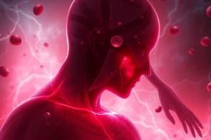
Anemia
- Commonly results from disabilities
- Main result is reduced oxygen carrying ability
- Symptoms: palpitations, dizziness, and fatigue
- Compensation involves the cardiovascular, respiratory, and hematologic systems
Acute Blood Loss
- Primarily involves the loss of intravascular volume
- Effects depend on the rate of hemorrhage
- Blood loss can lead to cardiovascular collapse, shock, and death
- Plasma is replaced within 24 hours if not severe
- Neutrophil and platelet counts rise if blood lost is not severe
- Normal erythrocyte count returns in 4-6 weeks if not severe
- Hemoglobin restoration may take 6-8 weeks if not severe
Types of Anemia
- Megaloblastic anemias are caused by nutritional deficiencies of vitamin B12
- Microcytic hypochromic anemia is characterized by small erythrocytes that cannot contain normal amounts of hemoglobin
- Iron deficiency anemia is the most common type of nutritional disorder worldwide
- Anemia of chronic disease results from decreased erythropoiesis and impaired iron utilization
- Aplastic anemia is a hematopoietic failure characterized by a reduction in the effective production of mature cells by the bone marrow
- Hemolytic anemia involves premature, accelerated destruction of erythrocytes
Polycythemia Vera (PV)
- One of several disorders from abnormal regulation of multipotent hematopoietic stem cells
- PV and similar disorders have overproduction of one or more of the formed elements of the blood
- PV is rare, with a peak incidence between 60 and 80 years
Platelet Disorders
- Platelet disorders are broken into quantitative and qualitative abnormalities
- Quantitative disorders involve an increase or decrease in the number of circulating platelets
- Qualitative disorders affect the structure or function of individual platelets
- Thrombocytopenia is defined as a platelet count less than 150,000/mm3
- Platelet counts less than 30,000/mm3 can cause bleeding from minor traumas
- Spontaneous bleeding can occur with counts less than 10,000/mm3
- Heparin-induced thrombocytopenia is an immune system IgG-mediated adverse drug reaction
- Thrombocythemia is defined as a platelet count greater than 450,000/mm3
- Thrombocythemia is usually caused by excessive platelet production
- Qualitative alterations in platelet function occur with an increased bleeding time in the presence of a normal platelet count
Hemostasis and Coagulation
- Hepatic cells produce most of the factors involved in hemostasis
- Liver disease can affect clotting
- Disseminated intravascular coagulation (DIC) is characterized by widespread activation of coagulation
- Results in formation of fibrin clots throughout the body
- This process may result in consumption of platelets and clotting factors, leading to severe bleeding
- Hypercoagulability, or thrombophilia, is primarily caused by defects in proteins involved in hemostasis
- Secondary caused by a variety of clinical disorders
- Acquired thrombophilia are often multifactorial
Aplastic Anemia (AA)
- Hematopoietic failure resulting in pancytopenia
- Causes include: acquired stem cell defects, immune-mediated destruction, chemical and physical agents, viral infections, inherited conditions such as Fanconi anemia
- Chemical agents: alkylating agents, benzene, chloramphenicol, inorganic and organic arsenicals
- Can be idiopathic (primary acquired) or secondary to known causes
- Characterized by hypocellular bone marrow replaced with fat
- Treated with bone marrow transplant or immunosuppression
Anemias Secondary to Drug Effects
- Various drugs can cause hemolytic, megaloblastic, sideroblastic, and aplastic anemias
- Chloramphenicol is associated with causing aplastic anemia
- Penicillin, sulfonamides and methyldopa are associated with hemolytic anemia
Anemia of Chronic Disease (ACD)
- Associated with chronic inflammatory conditions
- Characterized by impaired iron utilization and erythropoiesis
- Erythropoietin use has limited success in treating ACD
- Can be effective for severe anemia secondary to chronic kidney disease
Hemolytic Anemias
- Characterized by the premature destruction of erythrocytes, either extravascularly or intravascularly
- Can be congenital (intrinsic defects) or acquired (immunologic or mechanical)
- Congenital forms include hereditary spherocytosis, enzyme deficiencies, and hemoglobin synthesis defects
- Acquired forms include: autoimmune hemolytic anemias, drug-induced hemolytic anemias, those caused by mechanical injury or infectious agents
- Autoimmune hemolytic anemias (AIHA) are caused by autoantibodies or complement on red cells
- AIHAs are classified by the temperature at which the antibody binds, as warm reactive, cold agglutinin or cold hemolysin
- Drug-induced hemolytic anemia can occur through different mechanisms including the hapten model, immune complex formation, or the autoimmune model
- Clinical manifestations: jaundice, splenomegaly, and skeletal abnormalities in children
- Treatment can involve corticosteroids, splenectomy, monoclonal antibody therapies like rituximab
Myeloproliferative Red Blood Cell Disorders
- Characterized by an overproduction of cells in the bone marrow
- Includes polycythemia vera (PV), which is marked by the overproduction of red blood cells
- PV is a chronic neoplastic condition associated with a mutation in the JAK2 gene
- Clinical manifestations: enlarged spleen, increased blood viscosity, and aquagenic pruritus
- Treatment involves phlebotomy, low-dose aspirin, hydroxyurea, and potentially interferon or JAK2 inhibitors
Iron Overload
- Can be primary (hereditary hemochromatosis) or secondary (parenteral, dietary, or blood transfusions)
- Hereditary hemochromatosis (HH) is an autosomal recessive disorder of iron metabolism caused by mutations in the HFE gene
- Results in excessive iron absorption and deposition in organs, leading to tissue damage
- Treatment consists of phlebotomy and avoidance of iron supplements
Thrombocytopenia
- Defined as a platelet count below 150,000/mm³
- Can be caused by decreased platelet production, increased destruction, or sequestration
- Heparin-induced thrombocytopenia (HIT) is an immune-mediated drug reaction
- Characterized by a decrease in platelet counts beginning 5 to 10 days after administration of heparin
- HIT can cause both venous and arterial thrombosis
- Immune Thrombocytopenic Purpura (ITP) is an autoimmune condition where autoantibodies lead to platelet destruction
- ITP can be acute or chronic, and may manifest with bleeding problems
- Treatment can include glucocorticoids, intravenous immunoglobulin (IVIG), anti-D, thrombopoiesis-stimulating agents, or splenectomy
- Thrombotic Thrombocytopenic Purpura (TTP) is characterized by thrombocytopenia and thrombotic microangiopathy
- TTP is caused by a decrease or dysfunction of the ADAMTS13 enzyme
- Clinical features include thrombocytopenia, hemolytic anemia, and organ ischemia
- Treatment involves plasma exchange and potentially steroids or rituximab
Thrombocythemia
- Defined as a platelet count above 450,000/mm³
- Can be primary (essential) or secondary (reactive)
- Essential thrombocythemia (ET) is a myeloproliferative disorder caused by a defect in megakaryocyte progenitor cells
- Clinical manifestations include microvascular thrombosis, erythromelalgia, and arterial or venous thrombosis
- Treatment focuses on preventing thrombosis and involves drugs such as hydroxyurea, interferon, anagrelide, and aspirin
Alterations of Platelet Function
- Qualitative disorders of platelets can be congenital (thrombocytopathies) or acquired
- Congenital disorders affect platelet adhesion, interaction, secretion, and membrane phospholipid regulation
- Acquired disorders can result from drug effects, systemic inflammation, or hematologic conditions
- Aspirin is a drug that can irreversibly inhibit platelet function
Impaired Hemostasis
- Vitamin K Deficiency
- Necessary for the synthesis of coagulation factors
- Deficiency commonly caused by parenteral nutrition and broad-spectrum antibiotics
- Treatment involves vitamin K administration
- Liver Disease
- Can result in defects in clotting, fibrinolytic systems, and platelet function
- Hepatic parenchymal damage leads to decreased production of clotting factors
- Treatment involves fresh frozen plasma, exchange transfusions, and platelet concentrates
Disseminated Intravascular Coagulation (DIC)
- An acquired syndrome characterized by widespread activation of coagulation
- Results in the formation of fibrin clots in the microvasculature, leading to organ failure and consumption of clotting factors.
- Caused by conditions that activate the clotting cascade such as sepsis, trauma, or malignancy
- The common pathway appears to be excessive exposure of tissue factor
- Clinical manifestations include hemorrhage, thrombosis, and multisystem organ failure
- Diagnosis is based on clinical symptoms and confirmed by laboratory tests
- Treatment is directed at eliminating the underlying pathology, controlling thrombosis, and maintaining organ function
- Heparin, replacement therapy, and fluid replacement are treatments for DIC
Thromboembolic Disease
- Associated with the spontaneous development of clots in blood vessels
- A thrombus is a stationary clot attached to the vessel wall, while an embolus is a clot that circulates in the blood
- The risk factors for spontaneous thrombi are referred to as the Virchow triad:
- Injury to the blood vessel endothelium
- Abnormalities of blood flow
- Hypercoagulability of the blood
- Hypercoagulability can be primary (hereditary) or secondary (acquired)
- Hereditary thrombophilias are often caused by mutations in coagulation protein
- Factor V Leiden and prothrombin mutations are common hereditary thrombophilias
- Acquired thrombophilias include conditions like antiphospholipid syndrome (APS)
- APS is an autoimmune syndrome characterized by autoantibodies against plasma membrane phospholipids and phospholipid-binding proteins
Aplastic Anemia (AA)
- Hematopoietic failure characterized by a reduction in the production of mature cells in the bone marrow, leading to pancytopenia
- Causes: acquired (immune-mediated, chemical/physical agents, viral infections) or inherited (Fanconi anemia, telomerase defects)
- Idiopathic AA is the most common type
- Symptoms: Insidious onset, related to the rate of bone marrow destruction - includes hypoxemia, pallor, weakness, fever, dyspnea, hemorrhaging
- Diagnosis: Blood tests show diminished erythrocytes, leukocytes, and platelets. Bone marrow biopsy confirms the diagnosis.
- Treatment: Bone marrow transplant, immunosuppression (antithymocyte globulin and cyclosporine), and supportive care
Hemolytic Anemia
- Characterized by premature destruction of erythrocytes, either episodically or continuously
- Can be congenital (red cell membrane defects, enzyme deficiencies, hemoglobin synthesis defects) or acquired (immune hemolytic anemias, mechanical injury)
- Extravascular hemolysis occurs within phagocytes in lymphoid tissue
- Intravascular hemolysis occurs within blood vessels due to mechanical injury, complement fixation, or toxic factors
- Warm autoimmune hemolytic anemia is caused by IgG antibodies binding to erythrocytes at body temperature
- Cold agglutinin autoimmune hemolytic anemia is mediated by IgM antibodies that bind to erythrocytes at colder temperatures
- Cold hemolysin autoimmune hemolytic anemia (paroxysmal cold hemoglobinuria): Exposure to cold initiates acute intravascular hemolysis
- Drug-induced hemolytic anemia results from allergic reactions against foreign antigens or autoantibodies
- Symptoms vary with the degree of anemia and hemolysis, and jaundice may be present
- Diagnosis: Clinical manifestations, bone marrow studies, and blood tests
- Treatment: Removing the cause, corticosteroids, splenectomy, rituximab, eculizumab, fluid and electrolyte replacement, transfusions, and folate
Myeloproliferative Red Blood Cell Disorders
- Characterized by overproduction of cells in one or more hematopoietic lines
- Polycythemia: Excessive red blood cell production
- Relative polycythemia results from hemoconcentration due to dehydration
- Absolute polycythemia:
- Primary (Polycythemia Vera): Results from abnormal regulation of hematopoietic stem cells
- Secondary: Results from increased erythropoietin secretion in response to chronic hypoxia
Polycythemia Vera (PV)
- Chronic neoplastic condition with overproduction of red blood cells
- Often with increased white blood cells and platelets, and splenomegaly
- Most individuals have a JAK2 mutation
- Symptoms: Enlarged spleen, abdominal pain, increased blood viscosity, plethora, headache, visual disturbances, and aquagenic pruritus
- Diagnosis: Increased red blood cells and total blood volume, high hemoglobin and hematocrit, bone marrow examination and JAK2 mutation analysis
- Treatment: Phlebotomy, low-dose aspirin, hydroxyurea, radioactive phosphorus, interferon-alpha, and JAK2 inhibitors
Iron Overload
- Can be primary (hereditary hemochromatosis) or secondary (anemias with inefficient erythropoiesis, dietary iron overload, repeated transfusions)
Hereditary Hemochromatosis (HH)
- Autosomal recessive disorder with increased gastrointestinal iron absorption and tissue iron deposition
- Caused by mutations in the HFE gene (C282Y and H63D)
- Symptoms: Fatigue, malaise, abdominal pain, arthralgias, hepatomegaly, bronzed skin, altered glucose homeostasis, and cardiac dysfunction
- Diagnosis: Elevated serum iron, transferrin saturation, and ferritin levels along with genetic testing
- Treatment: Phlebotomy, avoidance of iron and vitamin C supplements, and moderation of alcohol consumption
Thrombocytopenia
- Defined as a platelet count less than 150,000 platelets/mm3
- Hemorrhage risk increases significantly below 50,000/mm3
- Pseudothrombocytopenia: In vitro artifact due to platelet agglutination
- Causes: Decreased platelet production, increased consumption, congenital conditions, or acquired conditions
Heparin-Induced Thrombocytopenia (HIT)
- Immune-mediated drug reaction caused by IgG antibodies against heparin-platelet factor 4 complex, leading to platelet activation
- Symptoms: Thrombocytopenia and risk for venous or arterial thrombosis
- Diagnosis: Dropping platelet counts after heparin treatment and tests for heparin-platelet factor 4 antibodies
- Treatment: Withdrawal of heparin and use of alternative anticoagulants (thrombin inhibitors)
Immune Thrombocytopenic Purpura (ITP)
- Thrombocytopenia secondary to increased platelet destruction caused by autoantibodies against platelet-specific antigens
- Acute ITP: Often seen in children, usually resolves
- Chronic ITP: More common in adults, tends to worsen progressively
- Symptoms: Petechiae, purpura, and hemorrhage from mucosal sites
- Diagnosis: History of bleeding, physical examination, CBC, and peripheral blood smear
- Treatment: Glucocorticoids, intravenous immunoglobulin (IVIG), anti-Rho(D) immune globulin, romiplostim, eltrombopag, and splenectomy
Thrombotic Thrombocytopenic Purpura (TTP)
- Characterized by thrombocytopenia and thrombotic microangiopathy, leading to platelet aggregation and occlusion of arterioles and capillaries
- Familial TTP: Rare, chronic, relapsing
- Acquired TTP: More common, acute, and severe
- Most cases are related to decreased or dysfunctional ADAMTS13, an enzyme responsible for digesting large vWF molecules
- Symptoms: Thrombocytopenia, intravascular hemolytic anemia, ischemic signs (CNS involvement), kidney failure, and fever
- Diagnosis: Blood smear reveals fragmented red cells (schistocytes), high LDH levels
- Treatment: Plasma exchange, steroids, rituximab, and caplacizumab
Thrombocythemia
- Platelet count greater than 450,000/mm3
- Secondary (reactive) thrombocythemia: Occurs after splenectomy, infections, or inflammatory conditions
- Essential (primary) thrombocythemia (ET): Chronic myeloproliferative disorder with excessive platelet production due to a defect in bone marrow megakaryocyte progenitor cells
Essential Thrombocythemia (ET)
- Thrombocythemia is secondary to increased plasma thrombopoietin levels resulting from defects in the thrombopoietin receptor
- Symptoms: Microvasculature thrombosis (erythromelalgia, headache, paresthesias), arterial or venous thrombosis
- Diagnosis: Sustained platelet count of at least 450 × 109/L, bone marrow biopsy, and exclusion of other myeloproliferative disorders
- Treatment: Hydroxyurea, interferon, anagrelide, and aspirin
Qualitative Platelet Function Alterations
- Increased bleeding time with normal platelet count
- Congenital alterations: Rare, affect platelet-vessel wall adhesion, platelet-platelet interactions, platelet granules and secretion, arachidonic acid pathways, and membrane phospholipid regulation
- Acquired disorders: More common, caused by drug effects, systemic inflammatory conditions, and hematologic conditions
Coagulation Disorders
- Usually caused by defects or deficiencies of clotting factors
- Impaired Hemostasis:
- Vitamin K Deficiency
- Liver Disease
- Consumptive Thrombohemorrhagic Disorders:
- Disseminated Intravascular Coagulation
- Impaired Hemostasis:
Disseminated Intravascular Coagulation (DIC)
- Acquired syndrome with widespread activation of coagulation, leading to fibrin clot formation in small vessels and consumption of platelets and clotting factors
- Causes: Sepsis, trauma, malignancy, obstetric complications, and blood transfusion
- Pathophysiology: Excessive exposure of tissue factor (TF)
- Symptoms: Hemorrhage (oozing from puncture sites, ecchymoses), thrombosis, and shock
- Diagnosis: Clinical symptoms and laboratory tests (thrombocytopenia, prolonged clotting times, presence of fibrin degradation products)
- Treatment: Eliminating the underlying pathology, controlling ongoing thrombosis (heparin), and maintaining organ function (fluid replacement)
Thromboembolic Disease
- Spontaneous clot formation within blood vessels
- Thrombus: Stationary clot attached to vessel wall
- Embolus: Thrombus that detaches and circulates
- Virchow triad: Injury to vessel endothelium, abnormalities of blood flow, and hypercoagulability of blood
Hypercoagulability (Thrombophilia)
Increased risk for thrombosis
- Primary (hereditary) causes: Defects in proteins involved in hemostasis
- Factor V Leiden
- Prothrombin mutation
- Secondary (acquired) causes: Clinical disorders or conditions
- Antiphospholipid syndrome
Anemia Classification
- Results in a reduction in the total circulating red cell mass, or a decrease in the quality or quantity of hemoglobin
- Results in a reduced oxygen-carrying capacity of the blood and tissue hypoxia
- Terms ending in "-cytic" refer to cell size, while "-chromic" refers to hemoglobin content
- Additional descriptors include anisocytosis (various sizes) and poikilocytosis (various shapes)
Classification of Anemia by Underlying Mechanism
- Blood Loss:
- Acute blood loss (trauma) leads to posthemorrhagic anemia
- Chronic blood loss (gastrointestinal lesions) can cause iron deficiency
- Increased Red Cell Destruction (Hemolysis): Can be inherited or acquired
- Inherited causes include red cell membrane disorders, enzyme deficiencies, and hemoglobin abnormalities
- Acquired causes include antibody-mediated destruction, mechanical trauma, infections, toxic or chemical injury, and membrane lipid abnormalities
- Decreased Red Cell Production: Can be inherited or acquired
- Inherited causes include defects leading to stem cell depletion or affecting erythroblast maturation
- Acquired causes include nutritional deficiencies, erythropoietin deficiency, immune-mediated injury, inflammation, primary hematopoietic neoplasms, space-occupying marrow lesions, and infections
Anemia General Symptoms
- Symptoms vary depending on the body’s ability to compensate.
- Compensation involves the cardiovascular, respiratory, and hematologic systems.
- Common symptoms include shortness of breath (dyspnea), rapid heartbeat (palpitations), dizziness, and fatigue
- Physical signs include pallor of the skin and mucous membranes, and in hemolytic anemia, the skin may become yellowish due to the accumulation of hemolysis products
- Severe or acute anemia can cause peripheral blood vessel constriction, salt and water retention, and decreased blood volume
- Chronic conditions trigger the movement of interstitial fluid into the intravascular space and cause a hyperdynamic circulatory state
Anemias of Blood Loss
- Acute Blood Loss (Posthemorrhagic Anemia):
- Is a normocytic-normochromic anemia caused by acute blood loss
- Volume loss reduces mean systemic filling pressure, decreasing venous return
- Initial treatment involves restoring blood volume with intravenous fluids; large losses may require transfusion
- Within 24 hours, plasma is replaced by mobilizing water and electrolytes.
- Chronic Blood Loss:
- Occurs when blood loss exceeds bone marrow replacement capacity, leading to iron deficiency anemia
Anemias of Diminished Erythropoiesis
Result from ineffective erythrocyte DNA synthesis, often due to nutritional deficiencies of vitamin B12 or folate
Megaloblastic Anemias
- Characterized by abnormally large erythroid precursors (megaloblasts) in the marrow that mature into large erythrocytes (macrocytes)
- Caused by vitamin B12 (cobalamin) or folate (folic acid) deficiencies
- Pernicious Anemia (PA)
- Caused by vitamin B12 deficiency, often associated with end-stage type A chronic atrophic (autoimmune) gastritis
- Autoimmune gastritis impairs the production of intrinsic factor (IF), required for vitamin B12 uptake Symptoms include weakness, fatigue, paresthesias, difficulty walking, loss of appetite, abdominal pains, weight loss, and a sore tongue
- Neurologic manifestations may result from nerve demyelination Treatment involves lifelong vitamin B12 replacement
- Folate Deficiency Anemia:
- Results in megaloblastic anemia due to impaired DNA synthesis
- Symptoms include cheilosis, stomatitis, painful ulcerations, and gastrointestinal issues
- Treatment: daily oral administration of folate
Microcytic-Hypochromic Anemias
- Characterized by abnormally small erythrocytes with reduced hemoglobin
Iron Deficiency Anemia (IDA)
- The most common nutritional disorder worldwide
- Caused by dietary deficiency, impaired absorption, increased requirement, and chronic blood loss
- Symptoms include fatigue, weakness, shortness of breath, and pale earlobes, palms, and conjunctivae
- Advanced symptoms include brittle nails (koilonychia), glossitis, and angular stomatitis
- Treatment: Identify/eliminate blood loss sources and iron replacement therapy
Anemia of Chronic Disease (ACD)
- A mild to moderate anemia resulting from decreased erythropoiesis and impaired iron utilization in individuals with chronic systemic disease or inflammation
- Commonly noted in hospitalized individuals and those with congestive heart failure
- Pathophysiology:
- Decreased erythrocyte lifespan
- Suppressed erythropoietin production
- Ineffective bone marrow erythroid progenitor response to erythropoietin
- Altered iron metabolism and iron sequestration in macrophages
- Characterized by abnormal iron metabolism with low circulating iron levels and high total body iron storage
- Treatment involves alleviating the underlying disorder
Aplastic Anemia (AA)
- Hematopoietic failure characterized by a reduction in the effective production of mature cells by the bone marrow, causing pancytopenia
- Mechanisms include immune-mediated destruction of hematopoietic stem cells
- Causes include chemical agents, physical agents, inherited factors, or unpredictable exposures
- Treatment may include bone marrow transplantation
Leukocyte Disorders
- Typically categorized as either quantitative or qualitative
- Quantitative disorders, such as infections and leukemias, usually result from decreased production in the bone marrow
- Qualitative disorders involve a disruption of function, such as phagocytic cells losing their capability to function or lymphocytes losing their capability to respond to antigens
- Leukocytosis is a leukocyte count that is higher than normal
- Occurs as a normal protective response to stressors such as infection, exercise, emotional changes, temperature changes, anesthesia, pregnancy, drugs, hormones, and toxins
- Neutrophilia describes granulocytosis because neutrophils are the most numerous of the granulocytes [See Previous Response]
- Cranial Psychosis is an increase in the number of granulocytes that are released from the bone marrow
- A shift to the left, or leftward shift, is when the demand for circulating mature neutrophils exceeds the supply
- The marrow begins to release immature neutrophils, increasing the number of immature leukocytes in the blood
- Eosinophilia is an absolute increase in the number of circulating eosinophils
- Eosinophilia can be associated with allergic disorders such as asthma, hay fever, and drug reactions, as well as parasitic infections
- Monocytosis is an increase in the number of circulating monocytes that is most commonly associated with a bacterial infection
- Lymphocytosis occurs most commonly in acute viral infections
Infectious Mononucleosis (IM)
- Is a benign, acute, self-limiting lymphoproliferative clinical syndrome characterized by acute viral infection of B lymphocytes and is caused by a type of herpes virus called EBV
- Transmission of EBV is usually by saliva through personal contact, which is the reason for naming it the kissing disease
Leukemia
- Is a disorder of leukocytes in the bone marrow, and usually, but not always, the blood
- There is an uncontrolled proliferation of malignant leukocytes with a decreased production and function of normal hematopoietic cells
Two Acute Leukemias:
- Acute lymphocytic leukemia (ALL)
- An aggressive, fast-growing leukemia with too many lymphoblasts
- Acute myelogenous leukemia (AML)
- An aggressive, fast-growing leukemia with an excessive number of myeloblasts found in the bone marrow and blood
Two Chronic Leukemias:
- Chronic myelogenous leukemia (CML)
- Chronic lymphocytic leukemia (CLL)
- CLL is a slow-growing cancer in which too many immature lymphocytes are found mostly in the bone and bone marrow
- There is a statistically significant tendency for leukemia to reappear in families
- There is an increased risk in adults with exposure to cigarette smoke, benzene, and ionizing radiation
Lymphoma
- Lymphadenopathy is caused by an increase in size and number of germinal centers of the lymph node
- Large neck nodes are characterized by being palpable and often tender
- Localized lymphadenopathy usually indicates drainage of an area associated with inflammation, a process, or infection
- Generalized lymphadenopathy is usually as a result of malignant or non-malignant disease
- Lymphomas develop from the proliferation of malignant lymphocytes in the lymphoid system
- Both Hodgkin lymphoma and non-Hodgkin's lymphoma occur in children and adults
- Hodgkin lymphoma is derived from a B cell in the germinal center that has not undergone successful immune immunoglobulin gene rearrangement and would normally undergo apoptosis
- Clinical manifestations of Hodgkin lymphoma include fever, weight loss, night sweats, pruritus, fatigue, and large, painless neck
- Clinical manifestations of non-Hodgkin's lymphoma are described as a progressive clonal expansion of B-cells, T cells, and natural killer cells
- These lymphomas most likely originate from mutations in cellular genes, which may have been environmentally induced
- Burkitt lymphoma: Is a highly aggressive B-cell non-Hodgkin lymphoma that accounts for 30% of childhood lymphomas worldwide
- EBV is associated with almost all cases, and suppression of the immune system increases the individual's susceptibility
- Lymphoblastic lymphoma: Is a relatively rare variant of non-Hodgkin lymphoma with a 2 to 4% incidence, but it accounts for 20% of cases in children and adolescents
Multiple Myeloma
- Multiple myeloma is a biologically complex disease with a wide range of genetic alterations in individual differences in responses
- Multiple myeloma has a poor prognosis
- Median survival is three years
- Although untreated, individuals with multiple bone lesions rarely survive more than 6 to 12 months
Splenic Function
- Splenomegaly, without specific ideology, is seen in 7 to 15% of individuals
- An overactive spleen results in hematologic alterations that affect all blood components, namely, the reduction in the number of circulating blood cells
- The spleen can sequester up to 50% of the red blood cell population, potentially creating anemia
Leukocyte Quantitative Alterations
- Can result from bone marrow dysfunction or premature cell destruction in circulation
- Leukocytosis is a higher-than-normal leukocyte count
- Often a response to physiologic stressors, malignancies, hematologic disorders, and microorganism invasion
- Leukopenia is a lower-than-normal leukocyte count
- Frequently associated with neutropenia and infection
- Can be caused by physiological stressors and pathological conditions such as malignancies and hematologic disorders
- Granulocytosis, particularly an increase in neutrophils, occurs in response to infection
- The bone marrow releases immature cells, leading to a shift-to-the-left
- Eosinophilia commonly results from parasitic invasion or inhalation/ingestion of toxic foreign particles
- Basophilia is observed in hypersensitivity reactions
- Monocytosis occurs during the late or recuperative phase of infection as macrophages phagocytose microorganisms and debris; it is often seen in chronic infections
- Granulocytopenia, a significant decrease in neutrophils, can be life-threatening if sepsis occurs
- Often caused by chemotherapeutic agents, severe infection, and radiation
- Lymphocytopenia occurs in most acute infections and some immunodeficiency syndromes
- Lymphocytosis occurs in viral infections, leukemia, lymphomas, and some chronic infections
Infectious Mononucleosis
- Is an acute infection of B lymphocytes commonly associated with EBV (Epstein-Barr virus), a type of herpesvirus
- Transmission of EBV occurs through personal contact, often via saliva
- Early manifestations include sore throat and fever caused by inflammation from viral entry
- Infectious mononucleosis is self-limiting, and treatment involves rest and symptom relief
- EBV is associated with tumors, including lymphomas
Lymphoid Neoplasms: Leukemias
- Leukemia is a clonal disorder of leukocytes in the bone marrow and blood
- Characterized by uncontrolled proliferation of leukocytes and overcrowding of the bone marrow, which leads to decreased production and function of other blood cell lines
- Leukemias are classified by cell type (B cells, T cells, NK cells) and onset (acute or chronic).
- *ALL, CLL, AML and CML the four major types of leukemia
- Leukemia may have a genetic predisposition and has been linked to exposure to cigarette smoke, benzene, and ionizing radiation
- Genetic aberrations in AML alter genes encoding transcription factors and affect the epigenome
- Major clinical manifestations include fatigue (anemia), bleeding (thrombocytopenia), fever (infection), anorexia, and weight loss
- Chemotherapy is the primary treatment for leukemia
- New drugs may possibly target leukemia stem cells
- Leukemias include precursor B-cell neoplasms (immature B cells), peripheral B-cell neoplasms (mature B cells), precursor T-cell neoplasms (immature T cells), peripheral T-cell and NK-cell neoplasms (mature T cells and NK cells)
Alterations of Lymphoid Function
- Lymphomas are tumors of primary lymphoid tissue (thymus, bone marrow) or secondary lymphoid tissue (lymph nodes, spleen, tonsils, intestinal lymphoid tissue)
- Hodgkin Lymphoma and Non-Hodgkin Lymphoma are the two major types of malignant lymphomas
- Hodgkin Lymphoma
- Reed-Sternberg (RS) cells in lymph nodes are the diagnostic marker for HL
- The RS cell is derived from a malignant B cell
- A virus might be involved in the pathogenesis of HL, and familial clustering suggests a genetic mechanism
- An initial sign is an enlarged, painless mass or swelling, commonly in the neck otherwise local symptoms arise from lymphadenopathy
- Treatment includes radiation therapy and chemotherapy
Non-Hodgkin Lymphoma
- The cause of lymph node enlargement and cancerous transformation in NHL is unknown
- Factors associated with industrialization and economic improvement may be linked to lymphoma incidence
- Lymphomas generally result from genetic mutations or viral infection
- Immunosuppressed persons have a higher incidence of NHL
- Swelling of lymph nodes is generally painless
- Includes Burkitt lymphoma
- Burkitt lymphoma involves the jaw and facial bones and occurs in children from east-central Africa and New Guinea
- In the United States, it is rare, usually involves the abdomen, and is characterized by extensive bone marrow invasion and replacement
- Lymphoblastic Lymphoma arises from immature T cells that become malignant in the thymus
Plasma Cell Malignancies
- Multiple Myeloma (MM)
- A neoplasm of B cells (immature plasma cells) and mature plasma cells characterized by multiple mmalignant tumors of plasma cells scattered throughout the skeletal system and occasionally in soft tissue
- Myeloma cells usually secrete monoclonal protein or M protein, an abnormal antibody molecule, and may also secrete free antibody light chain that is excreted in the urine
- Bence Jones protein
- The exact cause of MM is unknown, but risk factors include exposure to radiation or certain chemicals and a history of monoclonal gammopathy of undetermined significance
- Major clinical manifestations include recurrent infections (due to suppression of the humoral immune response) and renal disease (as a result of Bence Jones proteinuria)
- Chemotherapy is the primary treatment, along with stem cell transplantation, biologic therapy, radiation therapy, and sometimes surgery
- Waldenström Macroglobulinemia - Is a rare, slow-growing type
Alterations of Splenic Function
- Results in Splenomegaly (enlargement of the spleen)
- Can result from acute inflammatory or infectious processes, congestive disorders, infiltrative processes, and tumors or cysts
- Splenomegaly can cause hypersplenism (overactivity of the spleen), resulting in blood cell sequestration, destruction of red blood cells, and anemia
Pediatric Hematology
- During development, there are changes in hematopoietic sites
- Erythropoietin can increase site production, with the red marrow replacing the yellow marrow, and red blood cell production occurring in the liver and spleen
- Extramedullary hematopoiesis is more likely in children than adults, leading to enlargement of the spleen and liver
- Fetal hemoglobin has a greater affinity for oxygen due to less interaction with the enzyme 2,3-DPG
- Most blood cell counts are higher than adult levels at birth and decline throughout childhood
- Lymphocyte count is high at birth and rises during the first year of life
- Neutrophil count peaks 6-12 hours after birth and declines over the next few days
- Eosinophil count is elevated in the first year of life
- Platelet counts are comparable to adult values throughout infancy and childhood
Anemia in Children
- Is the most common blood disorder in children
Caused by:
- Ineffective erythropoiesis
- Premature destruction of red blood cells
- Iron deficiency the most common cause
Resulting from:
- Inefficient intake
- Chronic blood loss
- Iron deficiency anemia is the most common nutritional disorder worldwide, with the highest incidence between 6 months and 2 years
- Clinical manifestations are related to inadequate hemoglobin synthesis
- Chronic iron deficiency anemia can result from chronic intestinal blood loss due to milk allergy, producing hypochromic microcytic anemia
Clinical manifestations include:
- Renal abnormalities
- Widened skull fractures
- Decreased physical growth and development delays
Hemolytic Disease of the Fetus and Newborn
- Caused by antigens on fetal red blood cells differing from antigens on maternal red blood cells
- Most cases are caused by ABO incompatibility
- Rh incompatibility can cause issues in subsequent pregnancies due to the mother's immune system creating antigens
Other Anemias
- Infections acquired by mother and transmitted to the fetus may result in clinical manifestations similar to hemolytic diseases
Possibly related to direct injury on the erythrocyte membrane.
- Glucose-6-phosphate dehydrogenase (G6PD) deficiency is an inherited enzyme deficiency leading to damaged red blood cells that rupture prematurely
- Is a recessive disorder that occurs more often in tropical and subtropical regions of the Eastern Hemisphere
- Hereditary spherocytosis is an inherited disorder caused by defects in the membrane skeleton of red blood cells, leading to splenic sequestration and destruction
- Clinical manifestations are anemia, jaundice, and splenomegaly
Sickle Cell Disease
- Involves a mutation in the hemoglobin gene for beta-globin that can distort red blood cells into a sickle shape
- Deoxygenation is the most important variable in determining the occurrence of sickling
- Sickled red blood cells can cause damage to the vascular wall, leading to inflammation and clotting
- Treatment consists of supportive care aimed at preventing sickling episodes and the consequences of anemia
Thalassemia
- Involves mutations in the hemoglobin gene affecting the quantity of beta-globin
Severity
- Is affected by the type of mutation in the gene
- Beta thalassemia minor causes mild to moderate microcytic hypochromic hemolytic anemia
- Beta thalassemia major causes impaired physical growth and development with severe anemia, resulting in early death from cardiac failure
- Alpha thalassemia trait is symptom-free, alpha thalassemia minor causes traits similar to beta thalassemia minor
- Alpha thalassemia major causes intrauterine congestive heart failure and enlarged liver otherwise most affected babies are stillborn or die shortly after birth
- Treatment depends on severity but may involve regular transfusions and chelation therapy
Hemophilia
- Severe types are mutations that eliminate factor VIII or coagulation factor IX
- More than 2000 unique mutations have been identified for hemophilia A (factor VIII), and 1000 mutations for hemophilia B (factor IX)
- Often diagnosed when children become mobile due to injuries
- Those with hemophilia A or B will have prolonged PTT times
- Prophylaxis consists of regular infusions of factor VIII or IX
- Von Willebrand disease results from a deficiency or dysfunction of von Willebrand factor, leading to a disorder of clot formation
- Primary Immune Thrombocytopenia Is a disorder of platelet consumption where antiplatelet antibodies bind to platelets Causes sequestration and destruction in the spleen and other lymphoid tissues
Leukemia
- Is the most common malignancy of children and adolescents
- Chronic leukemias are rare in children, accounting for fewer than 5% of cases
- The cause of most childhood cancers is unknown, with about 5% caused by inherited mutations
- Some genetic diseases, such as Down syndrome, predispose a child to AML
- Ionizing radiation can lead to a higher risk
- Speculation exists that prenatal exposure to pesticides or benzene could be a factor
- Chemotherapy treatment has achieved remarkable success
Studying That Suits You
Use AI to generate personalized quizzes and flashcards to suit your learning preferences.