Podcast
Questions and Answers
Which of the following structures is part of the axial skeleton?
Which of the following structures is part of the axial skeleton?
- Scapula
- Femur
- Sternum (correct)
- Phalanges
Which section of the vertebral column articulates with the ribs?
Which section of the vertebral column articulates with the ribs?
- Thoracic (correct)
- Cervical
- Lumbar
- Sacral
What type of bone is the patella?
What type of bone is the patella?
- Flat bone
- Sesamoid bone (correct)
- Long bone
- Short bone
The squamous suture joins which two bones?
The squamous suture joins which two bones?
Which of the following is located on the radius?
Which of the following is located on the radius?
The acromion is a feature of which bone?
The acromion is a feature of which bone?
Which of the following is a feature of the humerus?
Which of the following is a feature of the humerus?
Which of the following is a carpal bone?
Which of the following is a carpal bone?
The iliac crest is found on which bone?
The iliac crest is found on which bone?
What bony landmark is located on the distal end of the femur?
What bony landmark is located on the distal end of the femur?
Which of the following is a part of the tibia?
Which of the following is a part of the tibia?
Which tarsal bone articulates with the tibia and fibula?
Which tarsal bone articulates with the tibia and fibula?
Where is the yellow bone marrow located?
Where is the yellow bone marrow located?
In a fetal skull, what is the area of fibrous connective tissue between the bones called?
In a fetal skull, what is the area of fibrous connective tissue between the bones called?
Which structure is a part of the respiratory system?
Which structure is a part of the respiratory system?
Which organ is part of the digestive system?
Which organ is part of the digestive system?
Which organ is part of the endocrine system?
Which organ is part of the endocrine system?
Which of the following is a primary component of the central nervous system?
Which of the following is a primary component of the central nervous system?
The suprapatellar bursa is associated with which joint?
The suprapatellar bursa is associated with which joint?
Which ligament is found on the medial side of the knee joint?
Which ligament is found on the medial side of the knee joint?
Which of the following structures contributes to the stability of the shoulder joint?
Which of the following structures contributes to the stability of the shoulder joint?
The iliofemoral ligament is associated with which joint?
The iliofemoral ligament is associated with which joint?
The deltoid ligament is associated with which joint?
The deltoid ligament is associated with which joint?
What movement occurs in the sagittal plane?
What movement occurs in the sagittal plane?
What is the movement when the palm is turned upwards?
What is the movement when the palm is turned upwards?
Which movement describes the scapula moving toward the spine?
Which movement describes the scapula moving toward the spine?
Which of the following muscles is located on the anterior aspect of the body?
Which of the following muscles is located on the anterior aspect of the body?
Which of the following muscles is located primarily on the posterior side of the body?
Which of the following muscles is located primarily on the posterior side of the body?
The gastrocnemius muscle is located in which region?
The gastrocnemius muscle is located in which region?
Which muscle is involved in flexing the wrist?
Which muscle is involved in flexing the wrist?
The iliopsoas muscle group is primarily involved in what action?
The iliopsoas muscle group is primarily involved in what action?
Which structure is responsible for the exchange of oxygen and carbon dioxide in the lungs?
Which structure is responsible for the exchange of oxygen and carbon dioxide in the lungs?
Which heart chamber receives deoxygenated blood from the superior and inferior vena cava?
Which heart chamber receives deoxygenated blood from the superior and inferior vena cava?
The left coronary artery primarily supplies blood to which part of the heart?
The left coronary artery primarily supplies blood to which part of the heart?
The brachial artery is a continuation of which major artery?
The brachial artery is a continuation of which major artery?
Which artery is located in the lower leg?
Which artery is located in the lower leg?
Which of the following veins drains directly into the superior vena cava?
Which of the following veins drains directly into the superior vena cava?
Which lobe of the cerebral cortex is primarily responsible for processing visual information?
Which lobe of the cerebral cortex is primarily responsible for processing visual information?
What is the role of the vertebral arch?
What is the role of the vertebral arch?
Which nerve innervates the anterior muscles of the arm, such as the biceps brachii?
Which nerve innervates the anterior muscles of the arm, such as the biceps brachii?
Which nerve is the largest in the human body and runs down the posterior thigh?
Which nerve is the largest in the human body and runs down the posterior thigh?
Which of the following bones articulates with the glenoid cavity?
Which of the following bones articulates with the glenoid cavity?
What feature is unique to the axis (C2) vertebra?
What feature is unique to the axis (C2) vertebra?
Which of the following bones is classified as irregular in shape?
Which of the following bones is classified as irregular in shape?
What is the primary function of the interosseous membrane between the radius and ulna?
What is the primary function of the interosseous membrane between the radius and ulna?
Which of the following bony landmarks is located on the posterior aspect of the scapula?
Which of the following bony landmarks is located on the posterior aspect of the scapula?
What is the functional significance of the greater and lesser tubercles on the humerus?
What is the functional significance of the greater and lesser tubercles on the humerus?
Which carpal bone articulates directly with the radius?
Which carpal bone articulates directly with the radius?
What is the acetabulum?
What is the acetabulum?
Which of the following is the main function of the tibial tuberosity?
Which of the following is the main function of the tibial tuberosity?
What anatomical structure is located on the talus?
What anatomical structure is located on the talus?
What is the primary function of red bone marrow?
What is the primary function of red bone marrow?
Which structure in the respiratory system directly precedes the alveoli?
Which structure in the respiratory system directly precedes the alveoli?
Which digestive organ is responsible for producing bile?
Which digestive organ is responsible for producing bile?
Which endocrine gland is located in the abdominal cavity and regulates blood sugar levels?
Which endocrine gland is located in the abdominal cavity and regulates blood sugar levels?
What is the main function of the cerebellum?
What is the main function of the cerebellum?
Which structure is found on the medial side of the knee and resists valgus forces?
Which structure is found on the medial side of the knee and resists valgus forces?
Which of the following structures primarily provides static stability to the shoulder joint?
Which of the following structures primarily provides static stability to the shoulder joint?
The ischiofemoral ligament primarily resists what type of movement at the hip joint?
The ischiofemoral ligament primarily resists what type of movement at the hip joint?
What is the movement that decreases the angle between the foot and the lower leg?
What is the movement that decreases the angle between the foot and the lower leg?
What movement is associated with the scapula moving away from the midline of the body?
What movement is associated with the scapula moving away from the midline of the body?
Flashcards
Manubrium
Manubrium
The main part of the sternum.
Xiphoid process
Xiphoid process
The inferior extension of the sternum.
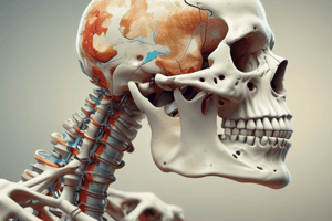
Skull
Skull
The bony enclosure for the brain.
Cranial portion
Cranial portion
Signup and view all the flashcards
Facial portion
Facial portion
Signup and view all the flashcards
Clavicle
Clavicle
Signup and view all the flashcards
Scapula
Scapula
Signup and view all the flashcards
Thoracic cage
Thoracic cage
Signup and view all the flashcards
Sternum
Sternum
Signup and view all the flashcards
Ribs
Ribs
Signup and view all the flashcards
Upper limb
Upper limb
Signup and view all the flashcards
Humerus
Humerus
Signup and view all the flashcards
Ulna
Ulna
Signup and view all the flashcards
Radius
Radius
Signup and view all the flashcards
Carpals
Carpals
Signup and view all the flashcards
Metacarpals
Metacarpals
Signup and view all the flashcards
Phalanges
Phalanges
Signup and view all the flashcards
Lower limb
Lower limb
Signup and view all the flashcards
Femur
Femur
Signup and view all the flashcards
Patella
Patella
Signup and view all the flashcards
Tibia
Tibia
Signup and view all the flashcards
Fibula
Fibula
Signup and view all the flashcards
Tarsals
Tarsals
Signup and view all the flashcards
Metatarsals
Metatarsals
Signup and view all the flashcards
Vertebral column
Vertebral column
Signup and view all the flashcards
Pelvic girdle
Pelvic girdle
Signup and view all the flashcards
Cervical vertebrae
Cervical vertebrae
Signup and view all the flashcards
Thoracic vertebrae
Thoracic vertebrae
Signup and view all the flashcards
Lumbar vertebrae
Lumbar vertebrae
Signup and view all the flashcards
Sacrum
Sacrum
Signup and view all the flashcards
Coccyx
Coccyx
Signup and view all the flashcards
Flat bone
Flat bone
Signup and view all the flashcards
Irregular bone
Irregular bone
Signup and view all the flashcards
Long bone
Long bone
Signup and view all the flashcards
Sesamoid bone
Sesamoid bone
Signup and view all the flashcards
Short bones
Short bones
Signup and view all the flashcards
Frontal bone
Frontal bone
Signup and view all the flashcards
Parietal bone
Parietal bone
Signup and view all the flashcards
Temporal bone
Temporal bone
Signup and view all the flashcards
Occipital bone
Occipital bone
Signup and view all the flashcards
Maxilla
Maxilla
Signup and view all the flashcards
Mandible
Mandible
Signup and view all the flashcards
Olecranon process
Olecranon process
Signup and view all the flashcards
Trochlear notch
Trochlear notch
Signup and view all the flashcards
Coronoid process
Coronoid process
Signup and view all the flashcards
radiale
radiale
Signup and view all the flashcards
Head of radius
Head of radius
Signup and view all the flashcards
Study Notes
Anatomy Requirements
- The slides illustrate the anatomy for which you are responsible.
- If the slide has red boxes, you are only responsible for the material within those boxes.
Unit One – Osteology and Arthrology
- Axial skeleton includes the skull, vertebral column, and thoracic cage.
- Appendicular skeleton includes the pectoral girdle, upper limb, pelvic girdle, and lower limb.
Vertebral Column
- 7 cervical vertebrae (C1-C7) form the cervical curve.
- 12 thoracic vertebrae (T1-T12) form the thoracic curve.
- 5 lumbar vertebrae (L1-L5) form the lumbar curve.
- Fused vertebrae of the sacrum and coccyx form the sacrococcygeal curve.
Bone Types
- Flat bones include the sternum.
- Irregular bones include vertebra.
- Long bones include the femur.
- Sesamoid bones include the patella.
- Short bones include lateral, intermediate, and medial cuneiforms.
Skull
- Parietal, frontal, temporal, zygomatic, maxilla, mandible, and occipital bones are key skull features.
Radius and Ulna
- Key features of the radius include the head, neck, radial tuberosity, and styloid process and the ulna features include the olecranon process, trochlear notch and styloid process.
Scapula
- Key features of the scapula: acromion, coracoid process, glenoid cavity, suprascapular notch, superior border, superior angle, supraspinous fossa, spine, infraspinous fossa, subscapular fossa, inferior angle, and lateral border.
Humerus
- Key features of the humerus include: greater tubercle, anatomical neck, head, surgical neck, lesser tubercle, intertubercular groove, deltoid tuberosity, body (shaft), lateral supracondylar ridge, radial fossa, coronoid fossa, capitulum, head of radius, olecranon fossa, coronoid process of ulna, medial epicondyle, and trochlea.
Hand Anatomy
- Carpal bones in the wrist
- Metacarpals in the palm
- Phalanges in the fingers
Pelvic girdle
- Ilium, pubis, and ischium are key features
- Iliac crest, anterior superior iliac spine, iliac fossa, acetabulum, obturator foramen, ischial tuberosity, pubic symphysis
Femur
- Head, neck, greater trochanter, intertrochanteric line and crest, lesser trochanter, body shaft, linea aspera, adductor tubercle, lateral epicondyle and condyle, medial epicondyle condyle, and patella articulate to form the knee.
Tibia and Fibula
- Key features of the tibia include lateral and medial condyles, tibial tuberosity, anterior border, medial malleolus, and articular surface
- Key features of the fibula include the head and lateral malleolus.
Foot and Ankle
- Key features of the foot: Tarsals, metatarsals, and phalanges
Bone Anatomy
- Articular cartilage is the hyaline cartilage covering the epiphysis.
- Proximal and distal epiphysis are the expanded ends of long bones.
- Metaphysis is the region between the diaphysis and epiphysis.
- Diaphysis is the shaft of a long bone
- The medullary cavity, or marrow cavity, is inside the diaphysis.
- Spongy and compact bone and endosteum, periosteum, nutrient artery, yellow bone marrow, red bone marrow are key component
- Compact bone is the dense outer layer of bone.
- Spongy bone is the inner, honeycomb-like bone tissue.
- Endosteum is the lining of the medullary cavity.
- Periosteum is the outer covering of bone.
- The nutrient artery supplies the diaphysis with blood
Adult vs Fetal Skull
- Key features of the adult and fetal skulls: Parietal bone, frontal bone, occipital bone, temporal bone (squamous portion), anterior and posterior fontanelles, and sphenoidal and mastoid fontanelles.
Unit Two – Anatomy of the Torso
Thoracic Cavity
- Trachea, esophagus, right and left lungs are key organs
- Diaphragm separates the thoracic and abdominal cavities
- The heart is located in the pericardial cavity.
Organ Systems
- Digestive System: processes food for use, removes waste from undigested food, and includes the esophagus, stomach, liver, gall bladder, large intestine, and small intestine.
- Urinary System: controls water balance, removes wastes from blood and excretes them include the kidneys, ureter, and urinary bladder.
- Endocrine System: secretes hormones and regulates processes, including the pancreas, adrenal glands, testes, and ovaries.
- Lymphatic System: returns fluids to the blood and defends against pathogens includes the thymus, lymph nodes, and spleen.
Central Nervous System
- Major components are the brain, spinal cord, and vertebral column
CardioRespiratory Anatomy
- The aorta is a major artery.
- The pulmonary trunk carries blood to the lungs.
Lab Seven – Arthrology
Knee Joint
-
Key features: Femur, tibia, patella, quadriceps femoris tendon, and patellar tendon
-
Menisci (medial and lateral) provide cushion and stability
-
Cruciate ligaments provide anterior-posterior, medial-lateral support
-
Collateral ligaments (tibial and fibular) provide medial and lateral support
Shoulder Joint
- Acromioclavicular ligament connects the acromion and clavicle.
- coracoacromialand glenohumeral ligament provide stability
Hip Joint
- Key features include the iliofemoral and ischiofemoral ligaments.
Ankle Joint
- Deltoid ligament is on the medial side.
- Talofibular and calcaneofibular ligaments are on the lateral side
Planes of Movement
- Anatomical directional terms: cranial, caudal, superior, inferior, anterior, posterior, medial, and lateral.
Movements
- Angular Movements: flexion, extension, abduction, adduction, circumduction
- Shoulder: shoulder joint inward rotation, outwrad rotation, hip joint flexion and extensions
- Special movements: pronation, supination, dorsiflexion, plantar flexion, eversion and inversion
- Shoulder girdle: elevation, depression, protraction, retraction, upward rotation, and downward rotation.
Lab Nine – Myology
Muscles on the Body
- Anterior: Deltoid, pectoralis major, rectus abdominis, abdominal external oblique, pectineus, adductor longus, sartorius, rectus femoris, vastus lateralis, fibularis longus, tibialis anterior, iliopsoas, gracilis, vastus medialis, soleus, and gastrocnemius
- Posterior: Levator scapulae, supraspinatus, teres minor, infraspinatus, teres major, triceps brachii, serratus posterior inferior, external oblique, gluteus medius, gluteus maximus, semimembranosus, peroneus longus, tibialis posterior, rhomboids, trapezius, deltoid, latissimus dorsi brachioradialis, extensors, flexor carpi ulnaris, gluteus minimus, gemellus muscles, biceps femoris, semitendinosus, gracilis, gastrocnemius and soleus
- Muscles of the neck: levator scapulae, trapezius, splenius, sternocleidomastoid, clavicle, Rhomboids
Distinguish Movements
- Distinguish between anterior, middle, and posterior portions of the deltoid.
Arm Muscles
- Anterior muscles are the Biceps brachii and Brachialis.
- Posterior muscles are the Triceps brachii.
Forearm Muscles
- Anterior muscles: Flexors, flexor carpi ulnaris
- Posterior muscles: Extensors, brachioradialis, extensor carpi ulnaris
Superficial Pelvic and Thigh Muscles
- Psoas major, iliacus, tensor fascia latae, rectus femoris, vastus lateralis, pectinius, adductor longus, gracilis, vastus medialis
Muscle Regions
- Deep Pelvic and Thigh Muscles light (anterior view): obturator, adductor brevis, adductor longus, Gracilis, adductor magnus
- Pelvic and thigh muscles of right leg (posterior view): gluteus medius (cut), gluteus minimus, Piriformis, superior gemellus, inferior gemellus, obturator externus, quadratus femoris, biceps femoris, semitendinosus, semimembranosus
Lower Leg Muscles
- Anterior Portion: tibialis anterior Plantar Portion: gastrocnemius, soleus, calcaneal, plantaris
Lab Sixteen – Cardiorespiratory Anatomy
- Key features: nasal cavity, oral cavity, pharynx, larynx, trachea, left and right lung, right and left main bronchus, and diaphragm.
- Larynx, trachea, terminal and respritory bronchiole, smooth muscles are key components
- Aveolar sac is a cluster of individual alevoli
- The heart is divided by the superior and inferior vena cava
Cardiac Circulation
- Pulmonary circuit: heart to lungs
- Systemic circuit: heart to the rest of the body
- Coronary Circulation: Oxygen rich blood supply to the heart
- Key componets: aorta, left pulmonary arteries, right pulmonary arteries
Vascular System
- Arteries: Right and left common carotid arteries, brachiocephalic trunk, right and left subclavian arteries, axillary artery, brachial artery and diaphragm
- Veins: internal jugular vein, subclavian vein, axillary vein, brachial vein, great saphenous vein
Lab Twenty – Neural Anatomy
- Key features: frontal lobe, parietal lobe, pre and primary motor cortex, somatosensory cortex, temporal lobe, occipital lobe, midbrain, pons, medulla, brain stem, and cerebellum.
- The spinal cord connect nerve pathways from all senses
- Key vertbral features: spinal cord, spinal process, transverse process and lamina.
Upper Body
- Nerves: musculocutaneous, median, radial, and ulnar
Lower Body
- Key nerves of the leg include the femoral, sciatic, common peroneal, and tibial
Vertebral Anatomy
- Spinal cord, vertebral arch and sympathetic trunk are key components
Studying That Suits You
Use AI to generate personalized quizzes and flashcards to suit your learning preferences.