Podcast
Questions and Answers
What is the function of the upper motor neuron?
What is the function of the upper motor neuron?
- Transmit orders from sensory fibers to the spinal cord
- Transmit orders from the brain to lower motor neurons (correct)
- Stimulate skeletal muscles to contract
- Connect with sensory fibers to form mixed nerves
Spinal reflexes are voluntary muscle movements.
Spinal reflexes are voluntary muscle movements.
False (B)
What are the three elements of a spinal reflex?
What are the three elements of a spinal reflex?
- Sensory neurons, 2) Connector neurons in the spinal cord, and 3) Lower motor neurons
The upper motor neuron sends axons through the _______________________ and medulla to reach the spinal cord.
The upper motor neuron sends axons through the _______________________ and medulla to reach the spinal cord.
Match the following types of reflexes with their descriptions:
Match the following types of reflexes with their descriptions:
What is the function of the lower motor neuron?
What is the function of the lower motor neuron?
The brain is involved in the transmission of pain stimuli in spinal reflexes.
The brain is involved in the transmission of pain stimuli in spinal reflexes.
What is the purpose of the association between the upper motor neuron and the lower motor neuron?
What is the purpose of the association between the upper motor neuron and the lower motor neuron?
The knee jerk is an example of a _______________________ reflex.
The knee jerk is an example of a _______________________ reflex.
What is the function of the connector neurons in the spinal cord?
What is the function of the connector neurons in the spinal cord?
Flashcards are hidden until you start studying
Study Notes
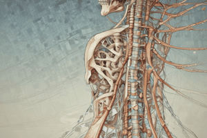
The Spinal Cord
- The spinal cord is an elongated, cylindrical structure suspended in the vertebral canal, surrounded by meninges and cerebrospinal fluid.
- It extends from the upper border of the atlas (1st cervical vertebra) to the lower border of the 1st lumbar vertebra, or specifically, to L1 vertebrae.
- The spinal cord links the brain and the body, and some activities are independent of the brain.
Structure of the Spinal Cord
- The spinal cord is composed of grey matter (in the center) and white matter (in the periphery), supported by neuroglia.
- Grey matter has the shape of an "H" and contains cell bodies of sensory neurons, lower motor neurons, and connector neurons.
- The grey matter columns include:
- Posterior column (dorsal): contains cell bodies of sensory neurons, stimulated by sensory impulses from the periphery and transmitting to the brain.
- Anterior column (ventral): contains cell bodies of lower motor neurons, stimulated by upper motor neurons or connector neurons.
White Matter
- White matter has three columns: anterior, posterior, and lateral.
- These tracts are formed by:
- Sensory nerve fibers ascending to the brain
- Motor nerve fibers descending from the brain
- Fibers of connector neurons
Sensory Nerve Tracts (Afferent)
- There are two sources of sensation transmitted to the brain via the spinal cord:
- From skin (cutaneous receptors)
- From muscles and tendons (proprioceptors)
- Nerve impulses from the skin are conducted by three neurons to the sensory area in the opposite hemisphere of the cerebrum, where the sensation and its location are perceived.
Motor Nerve Tracts (Efferent or Descending)
- Transmit impulses from the brain to the spinal cord, having a role in:
- Contraction of skeletal (striated) muscle (voluntary)
- Contraction of smooth muscle (involuntary), cardiac muscle, and secretion by glands
Voluntary Muscle Movement
- Contraction of muscles that move joints is under conscious (voluntary) control, originating at the level of consciousness in the cerebrum.
- Efferent nerve impulses are transmitted from the brain to the body via bundles of nerve fibers or tracts in the spinal cord.
- The motor pathways from the brain to the muscles are made up of two neurons:
- Pyramidal (corticospinal) tracts
- Extrapyramidal tracts
- The upper motor neuron (pyramidal) transmits the order from the brain to the lower motor neuron.
Spinal Reflexes
- A spinal reflex or reflex action is an involuntary and immediate motor response to a sensory stimulus.
- Spinal reflexes can occur without brain involvement, consisting of three elements:
- Sensory neurons
- Connector neurons in the spinal cord
- Lower motor neurons
- Examples of spinal reflexes include pain stimulus response and stretch reflexes (e.g., knee jerk).
Autonomic Reflexes
- Include the slight pupillary reflex in response to bright light, causing the pupil to constrict to prevent retinal damage.
Studying That Suits You
Use AI to generate personalized quizzes and flashcards to suit your learning preferences.