Podcast
Questions and Answers
Which part of the occipital bone is located anterior to the foramen magnum?
Which part of the occipital bone is located anterior to the foramen magnum?
- Condyle part
- Squamous part
- Lateral part
- Basilar part (correct)
What major structure passes through the hypoglossal canal of the occipital bone?
What major structure passes through the hypoglossal canal of the occipital bone?
- Vagus nerve
- Optic nerve
- Hypoglossal nerve (correct)
- Oculomotor nerve
Which feature of the occipital bone serves as a prominent external landmark and is easily felt?
Which feature of the occipital bone serves as a prominent external landmark and is easily felt?
- External occipital protuberance (correct)
- Pharyngeal tubercle
- Clivus
- Superior nuchal line
Which suture is located between the occipital bone and the parietal bones?
Which suture is located between the occipital bone and the parietal bones?
What anatomical feature is found on the superior surface of the basilar part of the occipital bone?
What anatomical feature is found on the superior surface of the basilar part of the occipital bone?
Which of the following bones are part of the neurocranium?
Which of the following bones are part of the neurocranium?
Which structure does the occipital condyle articulate with?
Which structure does the occipital condyle articulate with?
What is the primary function of the frontal bone?
What is the primary function of the frontal bone?
Which part of the occipital bone is positioned posterior to the foramen magnum?
Which part of the occipital bone is positioned posterior to the foramen magnum?
Which suture articulates the frontal bone with the parietal bones?
Which suture articulates the frontal bone with the parietal bones?
Which of the following bones contributes to the lateral surface of the cranium along with the occipital bone?
Which of the following bones contributes to the lateral surface of the cranium along with the occipital bone?
What is the primary function of the superciliary arches in the frontal bone?
What is the primary function of the superciliary arches in the frontal bone?
At what age do the two halves of the frontal bone typically fuse?
At what age do the two halves of the frontal bone typically fuse?
Which part of the frontal bone forms the superior border of the orbits?
Which part of the frontal bone forms the superior border of the orbits?
At what stage of development are the frontal sinuses typically fully developed?
At what stage of development are the frontal sinuses typically fully developed?
Which marking on the frontal bone is more pronounced in men?
Which marking on the frontal bone is more pronounced in men?
A persistent frontal suture that is visible in the midline of the glabella is known as what?
A persistent frontal suture that is visible in the midline of the glabella is known as what?
Which of the following structures can be found in the frontal bone?
Which of the following structures can be found in the frontal bone?
Which of the following functions does the lacrimal fossa serve?
Which of the following functions does the lacrimal fossa serve?
The landmark known as the glabella is located between which of the following features?
The landmark known as the glabella is located between which of the following features?
Which cranial bones does the frontal bone articulate with?
Which cranial bones does the frontal bone articulate with?
Which structure is found in the frontal bone that is an opening in the supraorbital margin?
Which structure is found in the frontal bone that is an opening in the supraorbital margin?
What separates the two frontal sinuses?
What separates the two frontal sinuses?
What is the nasal border in relation to the frontal bone?
What is the nasal border in relation to the frontal bone?
Flashcards
Frontal Bone
Frontal Bone
The bone forming the anterior portion of the cranium (forehead), articulating with parietal bones via the coronal suture.
Parietal Bone
Parietal Bone
Forms the sides and roof of the cranium.
Temporal Bone
Temporal Bone
Forms the sides and floor of the cranium.
Sphenoid Bone
Sphenoid Bone
Signup and view all the flashcards
Ethmoid Bone
Ethmoid Bone
Signup and view all the flashcards
Occipital Bone
Occipital Bone
Signup and view all the flashcards
Coronal Suture
Coronal Suture
Signup and view all the flashcards
Supraorbital Margin
Supraorbital Margin
Signup and view all the flashcards
Frontal Sinuses
Frontal Sinuses
Signup and view all the flashcards
Nasal Border
Nasal Border
Signup and view all the flashcards
Supraorbital Foramen/Notch
Supraorbital Foramen/Notch
Signup and view all the flashcards
Superciliary Arches
Superciliary Arches
Signup and view all the flashcards
Frontal Sinus
Frontal Sinus
Signup and view all the flashcards
Lacrimal Fossa
Lacrimal Fossa
Signup and view all the flashcards
Frontal Eminence
Frontal Eminence
Signup and view all the flashcards
Glabella
Glabella
Signup and view all the flashcards
Zygomatic Process
Zygomatic Process
Signup and view all the flashcards
Supraorbital Margin
Supraorbital Margin
Signup and view all the flashcards
Temporal line
Temporal line
Signup and view all the flashcards
Orbital Part
Orbital Part
Signup and view all the flashcards
Occipital Bone
Occipital Bone
Signup and view all the flashcards
Foramen Supraorbitale
Foramen Supraorbitale
Signup and view all the flashcards
Occipital Bone
Occipital Bone
Signup and view all the flashcards
Foramen Magnum
Foramen Magnum
Signup and view all the flashcards
Occipital Condyles
Occipital Condyles
Signup and view all the flashcards
Hypoglossal Canal
Hypoglossal Canal
Signup and view all the flashcards
External Occipital Protuberance
External Occipital Protuberance
Signup and view all the flashcards
Superior Nuchal Line
Superior Nuchal Line
Signup and view all the flashcards
Basilar Part of Occipital Bone
Basilar Part of Occipital Bone
Signup and view all the flashcards
Lateral Part of Occipital Bone
Lateral Part of Occipital Bone
Signup and view all the flashcards
Squamous Part of Occipital Bone
Squamous Part of Occipital Bone
Signup and view all the flashcards
Clivus
Clivus
Signup and view all the flashcards
Pharyngeal Tubercle
Pharyngeal Tubercle
Signup and view all the flashcards
Study Notes
Introduction to the Skeleton System
- The skeleton is a complex bony structure.
- The skeleton consists of two major divisions: axial and appendicular.
Divisions of the Skeleton
- Axial Skeleton
- Skull
- 22 bones and 6 auditory ossicles
- Protects brain and special senses
- Provides attachment for structures that take air, food, and water.
- The skull is made up of several cavities.
- Cranial cavity
- Nasal cavity
- Orbits
- Paranasal sinuses
- Ears
- Hyoid bone
- Vertebral column
- Thoracic (rib) cage
- Skull
- Appendicular Skeleton
- Limbs
- Girdles
Skull (Axial Skeleton)
- 22 Bones plus 6 auditory ossicles that function in hearing.
- Two portions:
- 1- Neurocranium (brainbox)
- Surrounds and protects brain
- Provides muscle attachment
- Contains sinuses to reduce weight of the skull
- 2- Viscerocranium (facial bones)
- Shapes the face
- Protects major sensory organs like eyes, nose, and tongue
- Provides attachment for muscles of mastication, facial expression, and eye movement
- 1- Neurocranium (brainbox)
- Bone List
- Neurocranium Bones (Cranium):
- Frontal (1)
- Parietal (2)
- Occipital (1)
- Temporal (2)
- Ethmoid (1)
- Sphenoid (1)
- Viscerocranium Bones:
- Inferior nasal concha (2)
- Lacrimal (2)
- Vomer (1)
- Nasal (2)
- Zygomatic (2)
- Palatin (2)
- Maxilla (2)
- Mandible (1)
- Neurocranium Bones (Cranium):
- Cranial Bones:
- Thin but remarkably strong for their weight
The Frontal Bone
- Forms the anterior portion of the cranium (forehead)
- Articulates posteriorly with parietal bones via the coronal suture
- Major markings include:
- Supraorbital margins
- Anterior cranial fossa
- Frontal sinuses (internal and lateral to glabella).
- Development: Forms in two halves, usually fused by ages 5 or 6
- Some adults have a metopic suture
- Parts of the frontal bone:
- Squamous part
- Orbital part
- Nasal part
- Supraorbital margin: Edge of the frontal part forming the superior border of the orbits
- Supraorbital foramen/notch: Opening or notch in the middle of the supraorbital margin
- Superciliary arches: Thickened ridges over the supraorbital margins supporting eyebrows
- Other features:
- Frontal eminence
- Glabella
- Frontal sinus
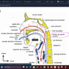
The Occipital Bone
- Contributes to the posterior, lateral and inferior surfaces of the cranium
- Encircled by four parts: basilar, lateral (2), squamous
- Foramen magnum: large circular opening for cranial and spinal cavities connection
- Clivus(superior surface);slightly concave and sloped upwards in continuity with the sphenoid
- Pharyngeal tubercle (inferior surface), site for muscle and raphe attachment
- Other features
- External occipital protuberance (inion)
- Superior nuchal line
- Occipital condyle
- Hypoglossal canal
The Parietal Bones
- Contribute to the superior and lateral surface of the cranium.
- Connected by immovable joints called sutures.
- Major Sutures:
- Lambdoid suture: Between occipital and parietal bones.
- Sagittal suture: Between the parietal bones.
- Coronal suture: Between the frontal and parietal bones.
- Squamous suture: Between the parietal and temporal bones.
The Temporal Bones
- Form the inferolateral aspects of the skull
- The temporal bone is divided into four parts:
- Squamous (flattened plate)
- Tympanic (surrounds external acoustic meatus or ear canal)
- Mastoid (posterior to the external acoustic meatus)
- Petrous (dense portion housing middle and inner ear structures)
- Major Features of the temporal bone:
- Zygomatic process:forms the zygomatic arch (cheek bone)
- Mandibular fossa: Forms a joint with the condyle of the mandible
- External acoustic meatus: Ear canal
- Styloid process: Projects inferiorly from the tympanic part.
- Mastoid process: Bony knob behind the earlobe.
- Stylomastoid foramen: Located between mastoid and styloid processes, passage for facial nerve.
- Mastoid foramen: Posterior to the mastoid process
- Other features in Petrous Part:
- Internal acoustic meatus
- Carotid canal
- Jugular foramen
The Sphenoid Bone
- Butterfly-shaped bone that spans the width of the middle cranial fossa
- Forms the central wedge that articulates with all other cranial bones.
- Consists of:
- Body (median portion)
- Two greater wings (lateral portion)
- Two lesser wings (anterior portion)
- Pterygoid processes (directed inferiorly)
- Other features:
- Sella turcica: Bony enclosure for the pituitary gland.
- Greater wings: Large, lateral wing-like processes that strengthen the sides of the skull; have openings(foramen rotundum, foramen ovale, and foramen spinosum)
- Lesser wings: small wing-like processes anterior to the sella turcica
- Superior orbital fissure: Irregular slit-like opening between lesser and greater wings.
- Pterygoid processes: Plates that permit muscle attachment
- Pterygoid canals
- Major Markings:
- Sella turcica
- Hypophyseal fossa
- Pterygoid processes
The Ethmoid Bone
- Lies between the sphenoid and nasal bones.
- Forms most of the bony area between nasal cavity and orbits
- Composed of:
- Cribriform plate: Perforated plate containing olfactory foramina
- Crista galli: Raised ridge that projects upward
- Perpendicular plate: Forms part of the nasal septum
- Superior and middle nasal conchae: Bony projections that increase surface area for airflow.
- Ethmoid sinuses: Air spaces within the bone
The Facial Bones
- The facial bones form the skeleton of the face.
- List of Facial Bones:
- Nasal bones (2)
- Lacrimal bones (2)
- Zygomatic bones (2)
- Maxillae (2)
- Palatine bones (2)
- Inferior nasal conchae (2)
- Vomer (1)
- Mandible (1)
The Mandible
- Largest and strongest bone of the face. Described as horseshoe shaped
- All muscles of mastication attach to the mandible
- Has different sections :
- Body
- Ramus
- Head of the mandible
- Neck of the mandible
- Angle of the mandible
- Coronoid process
- Condylar process
- Mandibular notch
- Mental tubercle
- Mental protuberance
- Mental foramen
- Alveolar process (supports mandibular teeth)
- Parts of the Mandible:
- Mylohyoid line
- Mylohyoid groove
- Sublingual fovea
- Submandibular fovea
- Mental spine
- The alveolar process supports the teeth
- Alveolar bone is resorbed when a tooth is lost
The Hyoid Bone
- Located at the C3 vertebral level, in front of the neck
- Unique as it does not articulate with any bone
- Attachment for hyoid muscles
- The upper and lower parts of the neck's hyoid muscles attach to this bone
Studying That Suits You
Use AI to generate personalized quizzes and flashcards to suit your learning preferences.
Related Documents
Description
This quiz covers key aspects of the occipital and frontal bones, including their anatomical features, functions, and relationships with other cranial structures. Test your knowledge on the important landmarks, sutures, and articulations of these cranial bones.