Podcast
Questions and Answers
Was ist die Hauptfunktion des somatischen Nervensystems?
Was ist die Hauptfunktion des somatischen Nervensystems?
- Übertragung motorischer Signale zu den inneren Organen.
- Regulation des inneren Milieus des Körpers.
- Interaktion mit der äußeren Umwelt. (correct)
- Leitung sensorischer Signale von inneren Organen zum ZNS.
Welche Aussage trifft auf efferente Nerven zu?
Welche Aussage trifft auf efferente Nerven zu?
- Sie leiten sensorische Signale zum ZNS.
- Sie lösen eher Affekte aus, indem sie Signale zum ZNS leiten.
- Sie erzeugen eher Effekte, da sie motorische Aktivitäten bedingen. (correct)
- Sie werden durch Afferenzen aktiviert.
Wo werden sympathische Nerven üblicherweise umgeschaltet, bevor sie ihr Erfolgsorgan erreichen?
Wo werden sympathische Nerven üblicherweise umgeschaltet, bevor sie ihr Erfolgsorgan erreichen?
- In einiger Entfernung von ihren Erfolgsorganen. (correct)
- In unmittelbarer Nähe ihres Erfolgsorgan.
- Im Rückenmark.
- Direkt im Gehirn.
Welche der folgenden Funktionen wird nicht dem Parasympathikus zugeschrieben?
Welche der folgenden Funktionen wird nicht dem Parasympathikus zugeschrieben?
Welcher der folgenden Hirnnerven ist der längste und projiziert zu den Eingeweiden?
Welcher der folgenden Hirnnerven ist der längste und projiziert zu den Eingeweiden?
Welche Reihenfolge der Hirnhäute ist korrekt, von außen nach innen?
Welche Reihenfolge der Hirnhäute ist korrekt, von außen nach innen?
Wo wird die Cerebrospinalflüssigkeit hauptsächlich produziert?
Wo wird die Cerebrospinalflüssigkeit hauptsächlich produziert?
Welche Struktur verbindet den dritten und vierten Ventrikel?
Welche Struktur verbindet den dritten und vierten Ventrikel?
Welche Eigenschaft der Blut-Hirn-Schranke ist für ihre Funktion wesentlich?
Welche Eigenschaft der Blut-Hirn-Schranke ist für ihre Funktion wesentlich?
Unter welchen Umständen kann es zu einem Hydrocephalus kommen?
Unter welchen Umständen kann es zu einem Hydrocephalus kommen?
Flashcards
Divisions of Nervous System
Divisions of Nervous System
CNS includes the brain and spinal cord; PNS is outside the skull and spine.
Afferent Nerves
Afferent Nerves
Sensory signals to the CNS.
Efferent Nerves
Efferent Nerves
Motor signals from the CNS to muscles.
Sympathetic Nerves
Sympathetic Nerves
Signup and view all the flashcards
Parasympathetic Nerves
Parasympathetic Nerves
Signup and view all the flashcards
Longest Cranial Nerve
Longest Cranial Nerve
Signup and view all the flashcards
The 3 Meninges?
The 3 Meninges?
Signup and view all the flashcards
Cerebrospinal Fluid (CSF)
Cerebrospinal Fluid (CSF)
Signup and view all the flashcards
Blood-Brain Barrier
Blood-Brain Barrier
Signup and view all the flashcards
Neurons and Glia
Neurons and Glia
Signup and view all the flashcards
Study Notes
- Chapter three is about the Anatomy of the Nervous System
- Chapter 3.1 focuses on the general structure of the nervous system
- Chapter 3.2 focuses on the cells of the nervous system
- Chapter 3.3 focuses on neuroanatomical methods and directional terms
- Chapter 3.4 focuses on the anatomy of the central nervous system
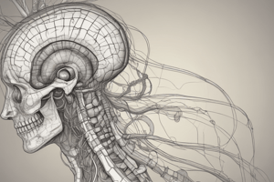
General Structure of the Nervous System
- Introduction to the general structure of the nervous system
- Begins with the two main divisions of the nervous system
- Touches on the importance of the meninges
- Discusses ventricles and cerebrospinal fluid
- Concludes with the blood-brain barrier
Divisions of the Nervous System
- The vertebrate nervous system consists of two parts: the central (CNS) and peripheral (PNS) nervous systems
- The central nervous system (CNS) resides in the skull and spinal cord
- The peripheral nervous system (PNS) exists outside the skull and spine
- The CNS consists of two parts: the brain and the spinal cord
- The brain (Encephalon) is the part of the CNS in the skull
- The spinal cord (Medulla spinalis) is the part in the spine
- The peripheral nervous system also has two parts: the somatic and autonomic nervous systems
- The somatic nervous system (SNS) interacts with external environment
- Afferent nerves carry sensory signals from the skin, skeletal muscles, joints, eyes, and ears to the CNS
- Efferent nerves carry motor signals from the CNS to the skeletal muscles
- The autonomic nervous system (ANS) regulates the body's internal environment
- Afferent nerves carry sensory signals from internal organs to the CNS
- Efferent nerves carry motor signals from the CNS to internal organs
- Efferent nerves achieve effects by initiating motor activities
- Afferent nerves trigger effects by sending signals to the CNS
- The autonomic nervous system has two types of efferent nerves: sympathetic and parasympathetic nerves
- Sympathetic nerves originate from the lumbar and thoracic regions of the spine in the body
- Parasympathetic nerves originate from the brain and sacral spine areas
- All sympathetic and parasympathetic nerves are switched once on their way to the target organ
- Sympathetic and parasympathetic neurons from the CNS travel only the first part to the target organ
- They form synaptic connections (synapses) with other neurons, which forward the signal the rest of the way
- In sympathetic nerves, projections from sympathetic neurons switch to the second neuron at a distance
- In parasympathetic nerves, the projections switch near their target organs to the second neuron
- The sympathetic nerves stimulate, organize, and mobilize energy reserves in threatening situations
- Parasympathetic nerves help conserve energy
- Each autonomic target organ receives opposite sympathetic and parasympathetic input
- Activity is controlled by the relative strength of sympathetic and parasympathetic activity
- Changes caused by the sympathetic nervous system indicate psychological activation
- Changes caused by the parasympathetic nervous system indicate psychological relaxation
- Most nerves in the peripheral nervous system originate in the spinal cord, but twelve "paired" exceptions exist.
- Those exceptions are the twelve pairs of cranial nerves
- Cranial nerves go directly from the brain, and they are numbered from front to back
- Cranial nerves include sensory and motor fibers
- The longest cranial nerve is the vagus nerve (X cranial nerve)
- The vagus nerve's motor and sensory fibers project from and to the viscera
- The twelve paired cranial nerves and their target organs are shown in Appendix III
- Appendix IV lists the cranial nerve functions
- The autonomic motor fibers of the cranial nerves belong to the parasympathetic nervous system
- An exam helps neurologists determine a diagnosis by testing cranial nerve function
- The loss of certain cranial nerve functions give reliable clues to the location and extent of a brain pathology
Meninges
- The brain and spinal cord (CNS) are the body's best-protected organs because they are surrounded by bone
- They are wrapped in membranes called meninges, which provide protection
- The outer meninx is a tough membrane called the dura mater (hard mother)
- Inside the dura mater sits the arachnoid mater (spider-like membrane) and subarachnoid space
- The subarachnoid space contains large blood vessels and cerebrospinal fluid
- Further down from the arachnoid mater is the innermost meninges called the pia mater (pious mother) on the CNS surface
Ventricles and Cerebrospinal Fluid
- Central nervous system is protected by cerebrospinal fluid that fills the subarachnoidal space
- Cerebrospinal fluid is in the brain ventricles and the spinal cord’s central canal
- The brain ventricles are the four large internal chambers of the brain: two lateral ventricles, the third ventricle, and the fourth ventricle
- The subarachnoid space, the central canal, and the brain ventricles are all interconnected and form a reservoir
- Cerebrospinal fluid cushions the brain
- Patients, who have had cerebrospinal fluid drained, have headaches because of pressure
- Over-production of cerebrospinal fluid leads to sinus durae matris
- Drainage can be prevented by tumors leading to hydrocephalus
- Hydrocephalus (Water Head) can be treated by draining extra fluid to relieve blockade
Blood-Brain Barrier
- The brain is a finely tuned electrochemical organ that is negatively affected by foreign chemicals
- A mechanism prevents toxic substances reaching the brain by way of the blood-brain barrier
- The special structure of cerebral blood vessels causes this barrier
- Cells that make up blood vessel walls are loosely connected, so molecules pass surrounding tissue easily
- Brain blood vessel tissue are tightly arranged and form molecules barrier
- The extent psychoactive substances has depends on ease
- Certain big molecules critical for normal brain function transports brain blood vessels
- The walls in brain regions allow unhindered passage of certain big molecules
Cells types of the nervous system
- The prominent cells types are neurons and glial cells
- They are discussed in this section
Studying That Suits You
Use AI to generate personalized quizzes and flashcards to suit your learning preferences.
Related Documents
Description
Explore the vertebrate nervous system's central (CNS) and peripheral (PNS) divisions. The CNS, located in the skull and spinal cord, includes the brain and spinal cord. The PNS exists outside these structures. Discover the significance of meninges, ventricles, and the blood-brain barrier.