Podcast
Questions and Answers
Where does the base of the renal pyramid originate?
Where does the base of the renal pyramid originate?
- At the border between the cortex and medulla (correct)
- At the outer border of the minor calyxes
- In the renal pelvis
- At the apex of the medulla
What structure does the renal pyramid's papilla project into?
What structure does the renal pyramid's papilla project into?
- Renal cortex
- Major calyxes
- Renal pelvis (correct)
- Minor calyxes
Which of the following correctly describes the division of major calyxes?
Which of the following correctly describes the division of major calyxes?
- They divide into renal pyramids
- They extend into the medulla
- They divide into lobes
- They divide into minor calyxes (correct)
Which part of the kidney is at the terminal point of the renal pyramids?
Which part of the kidney is at the terminal point of the renal pyramids?
What anatomical feature lies at the outer border of the renal pelvis?
What anatomical feature lies at the outer border of the renal pelvis?
What is the primary function of the glomerulus in the nephron?
What is the primary function of the glomerulus in the nephron?
How many nephrons are found in each human kidney?
How many nephrons are found in each human kidney?
What structural element in the walls of the calyxes and ureter helps move urine?
What structural element in the walls of the calyxes and ureter helps move urine?
Which of the following best describes the term 'nephron'?
Which of the following best describes the term 'nephron'?
Which component of the nephron extends out from the glomerulus?
Which component of the nephron extends out from the glomerulus?
What percentage of nephrons originate in the glomeruli located in the innermost cortex or juxtamedullary cortex?
What percentage of nephrons originate in the glomeruli located in the innermost cortex or juxtamedullary cortex?
What characterizes inner cortical nephrons?
What characterizes inner cortical nephrons?
Where are the glomeruli of the inner cortical nephrons primarily located?
Where are the glomeruli of the inner cortical nephrons primarily located?
What is the significance of the long loops of inner cortical nephrons?
What is the significance of the long loops of inner cortical nephrons?
Which region of the kidney is adjacent to the juxtamedullary cortex, where inner cortical nephrons are found?
Which region of the kidney is adjacent to the juxtamedullary cortex, where inner cortical nephrons are found?
What is the primary reason plasma proteins are not filtered during glomerular filtration?
What is the primary reason plasma proteins are not filtered during glomerular filtration?
Which statement best describes ultrafiltration in glomerular filtration?
Which statement best describes ultrafiltration in glomerular filtration?
Which of the following particles would most likely be filtered during glomerular filtration?
Which of the following particles would most likely be filtered during glomerular filtration?
What is a characteristic feature of ultrafiltration in the glomeruli?
What is a characteristic feature of ultrafiltration in the glomeruli?
How does the size of particles affect their filtration in the glomeruli?
How does the size of particles affect their filtration in the glomeruli?
What effect does increasing the thickness of the glomerular membrane have on Kf?
What effect does increasing the thickness of the glomerular membrane have on Kf?
What is the consequence of decreasing the number of functional glomerular capillaries due to disease?
What is the consequence of decreasing the number of functional glomerular capillaries due to disease?
Which of the following statements about Kf and GFR is true?
Which of the following statements about Kf and GFR is true?
In which situation would an increase in Kf occur?
In which situation would an increase in Kf occur?
Which factor is not considered a primary regulator of GFR?
Which factor is not considered a primary regulator of GFR?
Into what do the peritubular capillaries empty?
Into what do the peritubular capillaries empty?
Which of the following veins is NOT formed from the peritubular capillaries?
Which of the following veins is NOT formed from the peritubular capillaries?
What structure leaves the kidney alongside the renal artery and ureter?
What structure leaves the kidney alongside the renal artery and ureter?
In which order are the veins formed by the peritubular capillaries arranged?
In which order are the veins formed by the peritubular capillaries arranged?
Which vessels run parallel to the arteriolar vessels?
Which vessels run parallel to the arteriolar vessels?
Flashcards
Renal Pyramid Base
Renal Pyramid Base
The area where the renal pyramids start, marking the boundary between the outer cortex and the inner medulla.
Renal Papilla
Renal Papilla
The pointed tip of a renal pyramid that extends into the renal pelvis.
Renal Pelvis
Renal Pelvis
The funnel-shaped structure that collects urine from the renal papillae.
Major Calyxes
Major Calyxes
Signup and view all the flashcards
Minor Calyxes
Minor Calyxes
Signup and view all the flashcards
Calyxes, pelvis, and ureter
Calyxes, pelvis, and ureter
Signup and view all the flashcards
Nephron
Nephron
Signup and view all the flashcards
Glomerular filtration
Glomerular filtration
Signup and view all the flashcards
Glomerulus
Glomerulus
Signup and view all the flashcards
Renal tubule
Renal tubule
Signup and view all the flashcards
Peritubular Capillaries
Peritubular Capillaries
Signup and view all the flashcards
Interlobular Vein
Interlobular Vein
Signup and view all the flashcards
Arcuate Vein
Arcuate Vein
Signup and view all the flashcards
Interlobar Vein
Interlobar Vein
Signup and view all the flashcards
Renal Vein
Renal Vein
Signup and view all the flashcards
Juxtamedullary Nephrons
Juxtamedullary Nephrons
Signup and view all the flashcards
Inner Cortical Nephrons
Inner Cortical Nephrons
Signup and view all the flashcards
Urine Concentration
Urine Concentration
Signup and view all the flashcards
Concentration Segments
Concentration Segments
Signup and view all the flashcards
Loop of Henle's Role in Concentration
Loop of Henle's Role in Concentration
Signup and view all the flashcards
Why is glomerular filtration called ultrafiltration?
Why is glomerular filtration called ultrafiltration?
Signup and view all the flashcards
What prevents plasma proteins from being filtered?
What prevents plasma proteins from being filtered?
Signup and view all the flashcards
What is the filtration membrane?
What is the filtration membrane?
Signup and view all the flashcards
What is the importance of glomerular filtration?
What is the importance of glomerular filtration?
Signup and view all the flashcards
Glomerular Filtration Rate (GFR)
Glomerular Filtration Rate (GFR)
Signup and view all the flashcards
Kf (Filtration Coefficient)
Kf (Filtration Coefficient)
Signup and view all the flashcards
Kf and GFR Relationship
Kf and GFR Relationship
Signup and view all the flashcards
How Disease Affects Kf
How Disease Affects Kf
Signup and view all the flashcards
GFR Regulation
GFR Regulation
Signup and view all the flashcards
Study Notes
Physiology of the Urinary System
- The kidneys are vital organs for homeostasis, regulating water and electrolyte balance, excreting metabolic waste products, foreign chemicals, and regulating blood pressure
- The kidneys perform several essential functions, including regulating water and electrolyte balance, excreting metabolic waste products such as urea, creatinine, uric acid, bilirubin, and metabolites of hormones, excreting foreign chemicals like drugs, pesticides, and food additives, and regulating arterial blood pressure
- The kidneys are also involved in the regulation of erythrocyte production (EPO), Vitamin D activity (producing 1, 25-dihydroxyvitamin D3), gluconeogenesis (synthesizing glucose from amino acids) and acid-base balance.
Physiologic Anatomy of Kidneys
-
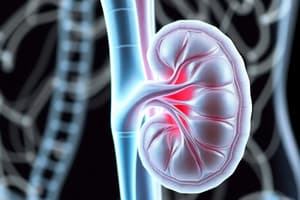
General Organization: The two adult human kidneys are bean-shaped, approximately 150 grams each and located outside the peritoneal cavity on the posterior abdominal wall. They consist of an outer cortex and inner medulla. Renal pyramids in the medulla project into the renal pelvis, which divides into major and minor calyces. The walls of the calyces, pelvis, and ureters contain contractile elements that move urine towards the bladder.
-
Nephron: The functional unit of the kidney is the nephron. The nephron is composed of a glomerulus (filtering component) and a tubule that extends from the glomerulus. The glomerulus is a tuft of capillaries which filters a protein-free filtrate from plasma into Bowman's capsule. The tubular portion extends into the medulla (for the loop of Henle) and then back into the cortex before draining further into collecting ducts that eventually form the ureter through the pelvis to the bladder.
-
Blood Supply: The renal artery branches into interlobar, arcuate, and interlobular arteries. These eventually lead to afferent arterioles, which supply the glomerular capillaries. The glomerular capillaries drain into efferent arterioles that form the peritubular capillaries. This arrangement allows for exchange of substances and water between the tubular lumen and capillaries. The renal circulation features two capillary beds (glomerular and peritubular) arranged in series, which are crucial to the nephron's function and fluid movement between the tubules and blood circulation.
-
Regional Differences (Nephron Structure): Approximately 80% of nephrons are outer cortical nephrons, with relatively short loops of Henle. The remaining 20% are juxtamedullary nephrons that have long loops of Henle extending deep into the medulla; this specialization is critical for concentrating urine.
The Juxtaglomerular Apparatus (JGA)
-
Interstitial Cells: Interstitial cells located between adjacent tubules and capillaries produce and release prostaglandins in response to appropriate stimuli.
-
Structure: The JGA includes the macula densa and juxtaglomerular cells. The macula densa, a short segment of the distal convoluted tubule, is adjacent to the afferent and efferent arterioles at the vascular pole of the glomerulus. Juxtaglomerular cells are in the walls of these arterioles.
-
Function: Macula densa cells monitor sodium chloride concentration in the filtrate, influencing renin release by juxtaglomerular cells. This release of renin starts the renin-angiotensin-aldosterone system (RAAS), which is a crucial mechanism for regulating blood pressure.
Urine Formation
-
Glomerular Filtration: The first step in urine formation, glomerular filtration, is the process of filtering plasma into Bowman's capsule. This fluid is essentially protein-free and lacks cellular elements. The filtration membrane consists of capillary endothelium, basement membrane, and epithelial cells (podocytes). The high filtration rate and selectivity of the glomerular membrane is critical in controlling which substances pass into the filtrate. The filtration process depends on size and charge of molecules.
- Ultrafiltration: The process is called ultrafiltration because even small particulates are filtered; however, the plasma protein's size prevents their filtration.
- Forces influencing Filtration: Forces involved in filtration include Glomerular capillary hydrostatic pressure (PG), Bowman's capsule hydrostatic pressure (PB), and Glomerular capillary colloid osmotic pressure (πG).
- Net Filtration Pressure (NFP): The final pressure determines the amount of fluid filtration.
-
Urinary Excretion Rate: The overall excretion rate is determined by glomerular filtration rate minus the rate of tubular reabsorption and plus the rate of tubular secretions.
Regulation of GFR
-
Factors Regulating GFR: GFR depends on glomerular capillary filtration coefficient (Kf), Net filtration pressure (NFP), total surface area available for filtration. Factors including changes in afferent or efferent arteriolar diameter, vasoconstriction, and resistance play a significant role in regulating GFR.
- Changes in Afferent/Efferent Arterioles: Vasodilation in the afferent arteriole increases RBF, PG, and GFR; vasoconstriction in the afferent arteriole reduces RBF, PG, and GFR. Vasodilation in the efferent arteriole decreases RBF, increases πG, and decreases GFR; vasoconstriction in the efferent arteriole increases RBF, decreases πG, and increases GFR.
-
Other Controls: Kidney stones can influence GFR. Changes in plasma protein concentration also affect GFR.
Renal Handling of Substances
- Waste products (e.g., creatinine)
- Electrolytes
- Nutritional substances (e.g., glucose, amino acids)
- Organic acids/bases and foreign compounds/drugs are handled by different mechanisms in their movement through each part of the nephron and in the tubular system in the kidney.
Studying That Suits You
Use AI to generate personalized quizzes and flashcards to suit your learning preferences.


