Podcast
Questions and Answers
Which ligament provides lateral stability in the ankle joint?
Which ligament provides lateral stability in the ankle joint?
- Calcaneofibular ligament (correct)
- Anterior talofibular ligament
- Posterior talofibular ligament
- Tibiocalcaneus ligament
Which muscle is responsible for dorsiflexion of the foot?
Which muscle is responsible for dorsiflexion of the foot?
- Tibialis anterior (correct)
- Peroneus longus
- Tibialis posterior
- Gastrocnemius
What is the name of the joint that connects the talus and calcaneus?
What is the name of the joint that connects the talus and calcaneus?
- Talocalcaneal joint (correct)
- Tibiocalcaneus joint
- Tibiotalar joint
- Talofibular joint
Which bone is part of the midfoot?
Which bone is part of the midfoot?
What is the function of the subtalar joint?
What is the function of the subtalar joint?
What type of joint is the subtalar joint?
What type of joint is the subtalar joint?
What is the name of the muscle that helps to plantarflex the foot?
What is the name of the muscle that helps to plantarflex the foot?
What is the name of the joint that connects the talus and navicular bones?
What is the name of the joint that connects the talus and navicular bones?
Which of the following is NOT a function of the foot?
Which of the following is NOT a function of the foot?
What is the name of the muscle that helps to dorsiflex the big toe?
What is the name of the muscle that helps to dorsiflex the big toe?
What is the function of the interosseous talocalcaneal ligament?
What is the function of the interosseous talocalcaneal ligament?
What is the result of pronation in the subtalar joint?
What is the result of pronation in the subtalar joint?
What is the concave rule of the anterior facet of the calcaneus?
What is the concave rule of the anterior facet of the calcaneus?
Which bone forms part of the internal arch?
Which bone forms part of the internal arch?
What is the function of the plantar calcaneonavicular ligament?
What is the function of the plantar calcaneonavicular ligament?
Which muscle supports the internal arch?
Which muscle supports the internal arch?
What is the function of the windlass mechanism?
What is the function of the windlass mechanism?
What is the function of the metatarsophalangeal and interphalangeal joints?
What is the function of the metatarsophalangeal and interphalangeal joints?
Which ligament supports the external arch?
Which ligament supports the external arch?
Which muscle supports the external arch?
Which muscle supports the external arch?
What is the primary function of the ankle and foot?
What is the primary function of the ankle and foot?
What type of joint is the proximal tibiofibular joint?
What type of joint is the proximal tibiofibular joint?
What is the movement of the talus during dorsiflexion in the open kinematic chain?
What is the movement of the talus during dorsiflexion in the open kinematic chain?
What is the resting position of the talocrural joint?
What is the resting position of the talocrural joint?
What is the type of joint that connects the concave face of the tibia to the convex face of the fibula?
What is the type of joint that connects the concave face of the tibia to the convex face of the fibula?
During plantar flexion in the open kinematic chain, what is the movement of the talus?
During plantar flexion in the open kinematic chain, what is the movement of the talus?
What is the primary static stabilizer of the talocrural joint?
What is the primary static stabilizer of the talocrural joint?
What is the movement of the proximal tibiofibular joint during dorsiflexion?
What is the movement of the proximal tibiofibular joint during dorsiflexion?
What is the role of the interosseous membrane in the proximal tibiofibular joint?
What is the role of the interosseous membrane in the proximal tibiofibular joint?
What is the rule that applies to the talocrural joint during dorsiflexion in the open kinematic chain?
What is the rule that applies to the talocrural joint during dorsiflexion in the open kinematic chain?
Flashcards are hidden until you start studying
Study Notes
Ankle and Foot Function
- Provide a stable base while conforming to uneven surfaces
- Flexible to absorb stress and adapt to the ground
- Rigid to withstand propulsive forces and push off
- Weight-bearing during walking, running, and standing
- Allows positioning of the foot
Tibiofibular Joints
- Proximal tibiofibular joint (PTFJ)
- Formed by the head of the fibula and the posterolateral aspect of the tibia
- Plane joint with anterior and posterior proximal tibiofibular ligaments
- Interosseous membrane connects the tibia and fibula
- Distal tibiofibular joint (DTFJ)
- Formed by the concave face of the tibia and the convex face of the fibula
- Syndesmosis with anterior and posterior distal tibiofibular ligaments
- Fibrous fat tissue connects the tibia and fibula
Arthrokinematics of the Tibiofibular Joints
- Slight gliding motion in the PTFJ during osteokinematics of the ankle and knees
- Dorsiflexion: cranial glide
- Plantar flexion: caudal glide
- Knee flexion: forward glide
- Knee extension: posterior glide
- Trochlea of the talus is wider anteriorly than posteriorly
- In the DTFJ:
- Dorsiflexion: separation
- Plantar flexion: approximation
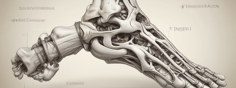
Talocrural Joint
- Mortise formed by the medial and lateral malleoli
- Hinge joint with 1 degree of freedom
- Rest position: 10° plantar flexion
- Close pack: maximal dorsiflexion
- Concave/convex rule:
- Open kinematic chain: convex rule
- Close kinematic chain: concave rule
Osteokinematics and Arthrokinematics of the Talocrural Joint
- During dorsiflexion in the open kinematic chain, the dome of the talus is convex, and the distal tibia/fibula is concave
- Concave rule applies during dorsiflexion in the closed kinematic chain
- Talus (convex): anterior roll, posterior glide
- Mortise (concave): anterior roll, anterior glide
Talocrural Joint Stability
- Static stabilizers (passive structures):
- Mortise and talar shape
- Gravity
- Capsule and ligaments
- Medial stability:
- Deltoid ligament
- Tibionavicular, tibiotalar, and tibiocalcaneal ligaments
- Lateral stability:
- Anterior talofibular ligament
- Posterior talofibular ligament
- Calcaneofibular ligament
- Dynamic stabilizers: musculature
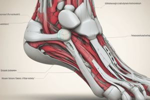
Kinetics of the Ankle and Foot
- Dorsiflexion:
- Tibialis anterior
- Extensor digitorum longus
- Extensor hallucis longus
- Plantar flexion:
- Gastrocnemius
- Soleus
- Tibialis posterior
- Peroneus longus and brevis
- Plantaris
- Flexor hallucis longus
Foot Anatomy
- Rearfoot: calcaneus, talus
- Midfoot: navicular, cuboid, 3 cuneiforms
- Forefoot: metatarsals and phalanges
Foot Joints
- Subtalar/talocalcaneal joint
- Mediotarsal (Chopart's) joint:
- Talonavicular
- Calcaneocuboid
- 5 tarsometatarsal joints
- Cubonavicular and cuneonavicular joints
Subtalar/Talocalcaneal Joint
- Synovial, gliding joint
- Three separate plane articulations between the talus and calcaneus
- Allows supination and pronation of the ankle
- Pronation: loose pack position, allows foot to absorb shock during loading phase of gait
- Supination: closed pack, creates rigid lever
Subtalar Joint Stability
- Interosseous talocalcaneal ligament
- Lateral talocalcaneal ligament
- Posterior talocalcaneal ligament
- Medial talocalcaneal ligament
Osteokinematics and Arthrokinematics of the Subtalar Joint
- Osteokinematics: oblique axis, hence pronation and supination
- Pronation: dorsiflexion, eversion, and abduction
- Supination: plantar flexion, inversion, and adduction
- Arthrokinematics:
- Posterior facet of calcaneus: convex rule (OKC)
- Anterior facet of calcaneus: concave rule (OKC)
Midtarsal/Chopart's Joint
- Talonavicular and calcaneocuboid joints
- Osteokinematics: combined with subtalar joint, contributes to pronation, supination, and simplified arthrokinematics
Arch Support
- Internal arch:
- Formed by calcaneus, talus, navicular, medial cuneiform, and 1st metatarsal
- Support ligaments: plantar calcaneonavicular ligament, interosseous talocalcaneal ligament
- Support muscles: tibialis posterior, peroneus longus, flexor hallucis longus, abductor hallucis
- External arch:
- Formed by calcaneus, cuboid, and 5th metatarsal
- Support ligament: plantar calcaneocuboid ligament
- Support muscles: peroneus longus, peroneus brevis, abductor digiti minimi
- Anterior arch:
- Formed by metatarsal heads
- Support ligaments: intermetatarsal ligament
- Support muscles: adductor hallucis
Metatarsophalangeal and Interphalangeal Joints
- Function in the closed kinematic chain
- Windlass plantar mechanism:
- The plantar aponeurosis acts similarly to a windlass mechanism
- Hyperextended MTPs tighten the plantar aponeurosis
- Joints the metatarsal and tarsal bones, making them a rigid structure and eventually raising the longitudinal arches
Studying That Suits You
Use AI to generate personalized quizzes and flashcards to suit your learning preferences.