Podcast
Questions and Answers
Which vein receives blood from the posterior chest wall?
Which vein receives blood from the posterior chest wall?
- Azygos vein (correct)
- Internal thoracic vein
- Accessory hemiazygos vein
- Hemiazygos vein
The upper left side of the thoracic cage drains to which vein?
The upper left side of the thoracic cage drains to which vein?
- Azygos vein
- Hemiazygos vein
- Accessory hemiazygos vein (correct)
- Left brachiocephalic vein
The azygos vein drains into which structure?
The azygos vein drains into which structure?
- Right brachiocephalic vein
- Superior vena cava (correct)
- Left brachiocephalic vein
- Inferior vena cava
Blockage of the azygos vein will result in?
Blockage of the azygos vein will result in?
The aorta supplies blood to the posterior chest wall via which arteries?
The aorta supplies blood to the posterior chest wall via which arteries?
The thoracic duct begins at which location?
The thoracic duct begins at which location?
The azygos system is characterized by?
The azygos system is characterized by?
Infections can easily spread along the azygos vein to which structure?
Infections can easily spread along the azygos vein to which structure?
What is the anatomical boundary in front of the mediastinum?
What is the anatomical boundary in front of the mediastinum?
What is the name of the imaginary plane that divides the mediastinum into superior and inferior parts?
What is the name of the imaginary plane that divides the mediastinum into superior and inferior parts?
Which of the following structures is NOT located in the middle mediastinum?
Which of the following structures is NOT located in the middle mediastinum?
What is the name of the artery that is located in the superior mediastinum?
What is the name of the artery that is located in the superior mediastinum?
What is the most posterior structure in the superior mediastinum?
What is the most posterior structure in the superior mediastinum?
What is the purpose of examining the superior mediastinum through axial scans such as CT or MRI?
What is the purpose of examining the superior mediastinum through axial scans such as CT or MRI?
What is the convention when viewing cross-sections of the superior mediastinum?
What is the convention when viewing cross-sections of the superior mediastinum?
Which of the following structures is located in the anterior part of the superior mediastinum?
Which of the following structures is located in the anterior part of the superior mediastinum?
In the cross-sectional view of the inferior mediastinum, which chamber of the heart is positioned behind the others?
In the cross-sectional view of the inferior mediastinum, which chamber of the heart is positioned behind the others?
What is the relationship between the thickness of the muscular walls of the left and right ventricles?
What is the relationship between the thickness of the muscular walls of the left and right ventricles?
In the superior mediastinum, what is the position of the oesophagus relative to the trachea?
In the superior mediastinum, what is the position of the oesophagus relative to the trachea?
What is the relationship between the oesophagus and the aorta in the posterior mediastinum?
What is the relationship between the oesophagus and the aorta in the posterior mediastinum?
What is the final destination of the oesophagus after passing through the left muscular hemi-diaphragm?
What is the final destination of the oesophagus after passing through the left muscular hemi-diaphragm?
In the cross-sectional view of the body of the T4 vertebra, which vein is visible as it projects forwards to join the superior vena cava?
In the cross-sectional view of the body of the T4 vertebra, which vein is visible as it projects forwards to join the superior vena cava?
In the schematic diagram, what is the relationship between the trachea and oesophagus?
In the schematic diagram, what is the relationship between the trachea and oesophagus?
What is the deviation of the oesophagus seen in the CT scan?
What is the deviation of the oesophagus seen in the CT scan?
Which nerve is responsible for reducing heart rate?
Which nerve is responsible for reducing heart rate?
What is the main function of the phrenic nerves in the thorax?
What is the main function of the phrenic nerves in the thorax?
What is the effect of sympathetics on the smooth muscle in the blood vessels of the lungs?
What is the effect of sympathetics on the smooth muscle in the blood vessels of the lungs?
Where do the vagus nerves arise from?
Where do the vagus nerves arise from?
Which nerve is responsible for the parasympathetic supply to the oesophagus?
Which nerve is responsible for the parasympathetic supply to the oesophagus?
What is the location of the phrenic nerves in the thoracic inlet?
What is the location of the phrenic nerves in the thoracic inlet?
What is the main function of the sympathetic chains in the thorax?
What is the main function of the sympathetic chains in the thorax?
What is the effect of the sympathetic nervous system on the oesophageal glands?
What is the effect of the sympathetic nervous system on the oesophageal glands?
Which plexus supplies the lungs?
Which plexus supplies the lungs?
What is the location of the vagus nerves in the thoracic inlet?
What is the location of the vagus nerves in the thoracic inlet?
What is the main component of the vagus nerves?
What is the main component of the vagus nerves?
What is the origin of the parasympathetic supply to the lungs?
What is the origin of the parasympathetic supply to the lungs?
What is the location of the phrenic nerves in the chest?
What is the location of the phrenic nerves in the chest?
What is the origin of the phrenic nerves?
What is the origin of the phrenic nerves?
Flashcards are hidden until you start studying
Study Notes
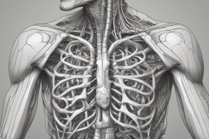
Definition of the Mediastinum
- The mediastinum is the space between the lungs, within the thoracic cavity, bounded by the sternum and vertebral column.
Divisions of the Mediastinum
- The mediastinum can be divided into superior and inferior mediastinum by an imaginary plane, the plane of the sternal angle.
- The inferior mediastinum can be further divided into three parts: anterior, middle, and posterior mediastinum.
Superior Mediastinum
- The superior mediastinum lies behind the manubrium of the sternum, making it difficult to visualize from the front.
- The superior mediastinum contains several structures, including the thymus gland, great veins, branches of the arch of the aorta, trachea, and oesophagus, and the thoracic duct.
Cross-section of Superior Mediastinum
- The section passes through the T3 vertebra, and anteriorly passes through the sternum and 1st costal cartilages.
- The image is presented as though the viewer is standing at the patient's feet, looking upwards.
Cross-section of Inferior Mediastinum
- The cross-section shows the positions of the chambers of the heart, the aorta, oesophagus, and azygos vein.
Relations of the Oesophagus
- The oesophagus is a major organ of the posterior mediastinum, lying behind the trachea in the superior mediastinum.
- The oesophagus moves to the left as it approaches the plane of the sternal angle, passing behind the left main bronchus, and then moves anterior to the aorta.
- The oesophagus ultimately passes through the left muscular hemi-diaphragm to reach the abdomen, where it enters the stomach.
Relationship of the Trachea and Oesophagus
- The trachea and oesophagus are in the midline, but the oesophagus deviates to the left.
Veins of the Posterior Mediastinum
- The azygos vein receives blood from the posterior chest wall, and drains to the superior vena cava.
- The hemiazygos vein drains the lower left side of the thoracic cage, and the upper left side drains to the accessory hemiazygos vein.
Arteries of Posterior Mediastinum
- The aorta supplies blood to the posterior chest wall via the posterior intercostal arteries, and gives visceral branches to the lungs and oesophagus.
Lymphatics of Posterior Mediastinum
- The thoracic duct is the most posterior structure in the mediastinum, starting at the upper end of the cisterna chyli at the upper border of T12, and terminating in the root of the neck.
Nerves of the Chest
- The phrenic nerves are the motor control for the diaphragm, arising from C3-5 and passing down in front of the subclavian artery at the thoracic inlet.
- The vagus nerves (Xth cranial nerves) are complex nerves with both somatic and autonomic elements, providing a wide variety of functions in both the head and neck, thorax, and abdomen.
Autonomic Supply to the Heart
- The parasympathetic supply to the heart is via the vagus nerves, which reduce heart rate.
Autonomic Supply to Lungs
- The pulmonary plexus supplies the lungs, with sympathetics from T1-T5, and parasympathetics from the vagus.
Autonomic Supply to the Oesophagus
- The oesophageal plexus supplies the oesophagus, with sympathetics from T2-T6, and parasympathetics from the vagus.
Studying That Suits You
Use AI to generate personalized quizzes and flashcards to suit your learning preferences.