Podcast
Questions and Answers
Which ligament becomes tense during extension?
Which ligament becomes tense during extension?
- ISCHIOFEMORAL
- PUBOFEMORAL
- Iliofemoral (correct)
- Tranverse acetubular ligament
Which artery is responsible for blood supply to the hip joint?
Which artery is responsible for blood supply to the hip joint?
- Superior gluteal artery
- Inferior gluteal artery
- Obturator artery
- All of the above (correct)
What is the function of the ligamentum teres?
What is the function of the ligamentum teres?
- To reinforce the posterior aspect of the fibrous membrane
- To connect the transverse acetubular ligament to the fovea of the head of femur (correct)
- To attach medially to the ischium just below the acetabulum
- To prevent hyperextension of the joint
What type of joint is the knee joint?
What type of joint is the knee joint?
Which nerve supplies the quadratus femoris?
Which nerve supplies the quadratus femoris?
What is the weakest ligament of the hip joint?
What is the weakest ligament of the hip joint?
Where is the ISCHIOFEMORAL ligament attached medially?
Where is the ISCHIOFEMORAL ligament attached medially?
Which ligament reinforces the posterior aspect of the fibrous membrane?
Which ligament reinforces the posterior aspect of the fibrous membrane?
What structure is the posterior cruciate ligament attached to on the posterior surface of the tibia?
What structure is the posterior cruciate ligament attached to on the posterior surface of the tibia?
What is the function of the anterior cruciate ligament?
What is the function of the anterior cruciate ligament?
In which plane do the cruciate ligaments cross each other?
In which plane do the cruciate ligaments cross each other?
Where does the posterior cruciate ligament attach to on the femur?
Where does the posterior cruciate ligament attach to on the femur?
What is the relationship between the cruciate ligaments and the fibrous membrane?
What is the relationship between the cruciate ligaments and the fibrous membrane?
What is the function of the posterior cruciate ligament?
What is the function of the posterior cruciate ligament?
Where does the anterior cruciate ligament attach to on the tibia?
Where does the anterior cruciate ligament attach to on the tibia?
In which region of the knee are the cruciate ligaments located?
In which region of the knee are the cruciate ligaments located?
What type of joint is the ankle joint?
What type of joint is the ankle joint?
What muscles are involved in flexion of the knee joint?
What muscles are involved in flexion of the knee joint?
What is the function of the locking mechanism in the knee joint?
What is the function of the locking mechanism in the knee joint?
Which nerve supplies the knee joint?
Which nerve supplies the knee joint?
What type of joint is the hip joint classified as?
What type of joint is the hip joint classified as?
What is the type of movement that occurs in the knee joint during locking?
What is the type of movement that occurs in the knee joint during locking?
Which of the following muscles is involved in the flexion of the hip joint?
Which of the following muscles is involved in the flexion of the hip joint?
What is the function of the ligamentum teres of the head of femur?
What is the function of the ligamentum teres of the head of femur?
What is the name of the ligament that provides medial stability to the ankle joint?
What is the name of the ligament that provides medial stability to the ankle joint?
Which of the following ligaments is the strongest in the body?
Which of the following ligaments is the strongest in the body?
What is the movement of the hip joint that involves the gluteus medius and minimus muscles?
What is the movement of the hip joint that involves the gluteus medius and minimus muscles?
Which of the following muscles is involved in the extension of the hip joint?
Which of the following muscles is involved in the extension of the hip joint?
What is the articular surface of the hip joint that forms the socket?
What is the articular surface of the hip joint that forms the socket?
What is the type of movement that involves a combination of flexion, abduction, extension, and adduction of the hip joint?
What is the type of movement that involves a combination of flexion, abduction, extension, and adduction of the hip joint?
Which of the following ligaments connects the talus to the navicular bone?
Which of the following ligaments connects the talus to the navicular bone?
What is the movement that turns the sole of the foot medially?
What is the movement that turns the sole of the foot medially?
Which muscle is responsible for the movement of eversion?
Which muscle is responsible for the movement of eversion?
What is the name of the joint that articulates between the talus and the calcaneus?
What is the name of the joint that articulates between the talus and the calcaneus?
Which ligament connects the talus to the fibula?
Which ligament connects the talus to the fibula?
What is the name of the ligament that connects the calcaneus to the navicular bone?
What is the name of the ligament that connects the calcaneus to the navicular bone?
Which muscle is responsible for the movement of inversion?
Which muscle is responsible for the movement of inversion?
Where do the movements of eversion and inversion occur?
Where do the movements of eversion and inversion occur?
Flashcards are hidden until you start studying
Study Notes
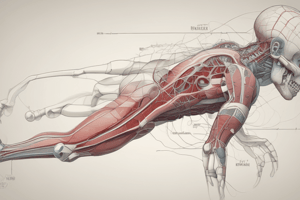
Hip Joint
- Type: Synovial, polyaxial, ball and socket joint
- Articular surfaces: spherical head of the femur and lunate surface of the acetabulum
- Movements:
- Flexion (iliopsoas, sartorius, rectus femoris, and pectineus)
- Extension (gluteus maximus, hamstrings)
- Abduction (gluteus medius and minimus)
- Adduction (adductor longus, brevis, and magnus)
- Medial and lateral rotation (gluteus medius and minimus, small lateral rotators)
- Circumduction
- Ligaments:
- Iliofemoral ligament (strongest ligament in the body, becomes tense during extension, prevents hyperextension)
- Pubofemoral ligament
- Ischiofemoral ligament (weakest ligament of the hip joint)
- Transverse acetabular ligament
- Ligamentum teres of the head of the femur
- Blood supply:
- Superior gluteal artery
- Inferior gluteal artery
- Obturator artery
- Lateral circumflex femoral artery
- Medial circumflex femoral artery
- Nerve supply:
- Superior gluteal nerve
- Nerve to quadratus femoris
Knee Joint
- Type: Synovial, biaxial, condyloid joint
- Articular surfaces: condyles of the femur and tibia
- Movements:
- Flexion (hamstrings, sartorius, and gracilis)
- Extension (quadriceps femoris)
- Rotation (limited, medial and lateral)
- Cruciate ligaments:
- Anterior cruciate ligament (prevents anterior displacement of the tibia on the femur)
- Posterior cruciate ligament (restricts posterior displacement)
- Locking mechanism:
- Medial rotation of the femur on the tibia during extension
- Tightens associated ligaments, locking the knee into position
- Blood supply:
- Femoral artery
- Popliteal artery
- Nerve supply:
- Anterior tibial nerve
- Common peroneal nerve
Ankle Joint
- Type: Synovial, uniaxial, hinge joint
- Articular surfaces: talus and tibia, fibula
- Movements:
- Dorsiflexion and plantarflexion
- Ligaments:
- Medial (deltoid) ligament
- Lateral ligaments (anterior and posterior talofibular ligaments, calcaneofibular ligament)
- Blood supply:
- Anterior and posterior tibial arteries
- Nerve supply:
- Anterior tibial nerve
- Posterior tibial nerve
Subtalar Joint
- Articulation between the talus and calcaneus
- Allows gliding and rotation movements
Talocalcaneonavicular Joint
- Complex joint between the talus, calcaneus, and navicular
- Allows gliding and rotation movements
Eversion and Inversion
- Movements occur in the subtalar and talocalcaneonavicular joints
- Inversion: movement that turns the sole of the foot medially (done by tibialis anterior and posterior muscles)
- Eversion: movement that turns the sole of the foot laterally (done by peroneus longus, brevis, and tertius muscles)
Studying That Suits You
Use AI to generate personalized quizzes and flashcards to suit your learning preferences.