Podcast
Questions and Answers
What is the primary function of a bursa?
What is the primary function of a bursa?
- To decrease friction between structures (correct)
- To produce red blood cells
- To support bone strength
- To store energy
Where are major bursae typically found in the body?
Where are major bursae typically found in the body?
- Brain and spinal cord
- Shoulder, elbow, hip, and knee (correct)
- Heart and lungs
- Stomach and intestines
What is bursitis?
What is bursitis?
- The growth of new bone tissue
- A disease affecting the heart valves
- A condition characterized by bone weakening
- Inflammation of a bursa due to excessive friction (correct)
What substance is contained within a bursa to help decrease friction?
What substance is contained within a bursa to help decrease friction?
Why are bursae typically difficult to palpate?
Why are bursae typically difficult to palpate?
What is the primary function of the protein keratin in the epidermis?
What is the primary function of the protein keratin in the epidermis?
Which layer of the skin contains hair follicles and glands?
Which layer of the skin contains hair follicles and glands?
What type of tissue makes up the hypodermis?
What type of tissue makes up the hypodermis?
Where is melanin produced?
Where is melanin produced?
What anatomical feature gives us tactile sensitivity on the fingertips?
What anatomical feature gives us tactile sensitivity on the fingertips?
What is another name for the hypodermis?
What is another name for the hypodermis?
Which of the following organs is included in the lymphoid organs?
Which of the following organs is included in the lymphoid organs?
What helps propel lymph along in the lymphatic system?
What helps propel lymph along in the lymphatic system?
What can cause swelling and fullness in lymph nodes?
What can cause swelling and fullness in lymph nodes?
Which of the following regions is NOT mentioned as having dense clusters of lymph nodes?
Which of the following regions is NOT mentioned as having dense clusters of lymph nodes?
Which organ is NOT a lymphoid organ?
Which organ is NOT a lymphoid organ?
What condition results from inefficient circulation of lymph?
What condition results from inefficient circulation of lymph?
What type of fibers does fibrous cartilage have?
What type of fibers does fibrous cartilage have?
Where is fibrous cartilage found in the body?
Where is fibrous cartilage found in the body?
The collagen network in fibrous cartilage helps it resist which types of forces?
The collagen network in fibrous cartilage helps it resist which types of forces?
Why is fibrous cartilage considered an ideal cushion?
Why is fibrous cartilage considered an ideal cushion?
What function do the structures cushioned by fibrous cartilage have?
What function do the structures cushioned by fibrous cartilage have?
Which type of cartilage has the highest proportion of elastic fibers?
Which type of cartilage has the highest proportion of elastic fibers?
Where is hyaline cartilage commonly found?
Where is hyaline cartilage commonly found?
What is a key function of hyaline cartilage?
What is a key function of hyaline cartilage?
What can result from damage to hyaline cartilage?
What can result from damage to hyaline cartilage?
What unique property of cartilage limits its ability to heal following injury?
What unique property of cartilage limits its ability to heal following injury?
How does hyaline cartilage respond to increased activity?
How does hyaline cartilage respond to increased activity?
Which structures are mechanically responsible for human movement?
Which structures are mechanically responsible for human movement?
Which structures help protect, nourish, regulate, and support the function of movement-related structures?
Which structures help protect, nourish, regulate, and support the function of movement-related structures?
What function does the skin NOT perform?
What function does the skin NOT perform?
How does the skin help us interact with the outside environment?
How does the skin help us interact with the outside environment?
What is one of the roles of blood vessels mentioned in the content?
What is one of the roles of blood vessels mentioned in the content?
What is the primary component of tendons that contributes to their strength and elasticity?
What is the primary component of tendons that contributes to their strength and elasticity?
Which statement accurately describes the function of tendons?
Which statement accurately describes the function of tendons?
What features distinguish tendons from bones and ligaments?
What features distinguish tendons from bones and ligaments?
What type of tendons would you likely find in the arm and wrist?
What type of tendons would you likely find in the arm and wrist?
In what way do tendons change similar to muscles?
In what way do tendons change similar to muscles?
What is the primary function of blood vessels?
What is the primary function of blood vessels?
Which type of blood vessels are the smallest in size?
Which type of blood vessels are the smallest in size?
What should you be cautious of when palpating near blood vessels?
What should you be cautious of when palpating near blood vessels?
What does palpating a pulse under your fingers indicate?
What does palpating a pulse under your fingers indicate?
What substances are exchanged in capillaries?
What substances are exchanged in capillaries?
What type of muscle tissue is responsible for movements at joints?
What type of muscle tissue is responsible for movements at joints?
Which muscle type is primarily found in the walls of hollow organs and vessels?
Which muscle type is primarily found in the walls of hollow organs and vessels?
Which muscle tissue is classified as involuntary and located in the heart?
Which muscle tissue is classified as involuntary and located in the heart?
What type of muscle tissue creates the pulsing action to circulate blood?
What type of muscle tissue creates the pulsing action to circulate blood?
Which muscle type cannot be consciously controlled?
Which muscle type cannot be consciously controlled?
To which main type of body tissue does muscle belong?
To which main type of body tissue does muscle belong?
What are the primary movement structures that are considered connective tissues?
What are the primary movement structures that are considered connective tissues?
What type of connective tissue has high levels of ground substance and fewer fibers?
What type of connective tissue has high levels of ground substance and fewer fibers?
Which connective tissue is characterized by its high collagen fiber content and supportive function around bones?
Which connective tissue is characterized by its high collagen fiber content and supportive function around bones?
What percentage of plasma in the extracellular matrix makes fluid connective tissue watery?
What percentage of plasma in the extracellular matrix makes fluid connective tissue watery?
What are examples of fluid connective tissue?
What are examples of fluid connective tissue?
Which type of connective tissue is strong due to the additional calcium salts deposited in its ground substance?
Which type of connective tissue is strong due to the additional calcium salts deposited in its ground substance?
In what direction do nerve impulses travel?
In what direction do nerve impulses travel?
What does each nerve bundle into according to microscopic observation?
What does each nerve bundle into according to microscopic observation?
Flashcards are hidden until you start studying
Study Notes
Bursae
- Bursae are small, flattened sacs that contain synovial fluid, a lubricant that helps decrease friction and create gliding movement between structures.
- They are located in areas of friction in the body, such as where muscles or tendons have to glide over bony prominences.
- Major bursae are found around the shoulder, elbow, hip, and knee.
- Bursae are fibrous, soft, and pillow-like when palpated, but normally difficult to palpate because they reside between bones and large tendons.
Cartilage
- Cartilage is a type of supporting connective tissue that varies in consistency and function by the proportion of proteins distributed through its matrix.
- Because cartilage does not contain blood vessels or nerves, it has a limited ability to heal following injury.
- There are three types of cartilage in the body: elastic cartilage, hyaline cartilage, and fibrous cartilage.
- Elastic cartilage has the highest proportion of elastic fibers and is found in the nose and ears, where it creates a structure that is self-supporting but flexible.
- Hyaline cartilage is found in the voice box, between the ribs and the sternum, and on the surfaces of bones where they form joints.
- Hyaline cartilage is smooth and rubbery and helps reduce friction during movement.
- Damage to hyaline cartilage can result in chronic inflammation of the joint, commonly termed osteoarthritis.
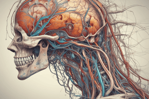
Blood Vessels
- Blood vessels are part of the circulatory system, the pathway by which blood flows throughout the body.
- The circulation of blood is necessary to deliver oxygen and nutrients to the body's tissues and remove wastes.
- Blood vessels vary in size from large arteries and veins to smaller arterioles and venules to the smallest capillaries.
- The network of blood vessels is woven throughout the body, existing side by side with lymphatic structures, nerves, and the structures of movement.
- Use caution when palpating near these structures to avoid damaging them.
Lymphoid Organs
- Lymphoid organs include the lymph nodes, as well as larger organs such as the spleen, thymus gland, tonsils, and Peyer's patches of the intestine.
- All of these organs are critical to the body's immune system.
- The lymphatic system is not pressurized in the same way as the circulatory system, as it has no pump comparable to the heart.
- The circulation of lymph relies heavily on skeletal muscle contraction and body movement.
- Breathing and the pulsation of nearby arteries also help propel lymph along.
- When lymph does not circulate efficiently, the tissue develops edema, an abnormal accumulation of fluid.
Lymph Nodes
- Lymph nodes cluster in certain areas of the body, such as the cervical, axillary, and inguinal regions.
- Lymph nodes are usually small, shaped like a kidney bean, and pliable when healthy.
- Diseases such as viral or bacterial infections can prompt enlargement of the associated lymph nodes, making them feel swollen and full.
Structure of the Skin
- The skin is composed of three tissue layers: the epidermis, dermis, and hypodermis.
- The epidermis is epithelial tissue, containing several thin layers of cells, which produce a protective protein called keratin and a pigment protein called melanin.
- The epidermis also contains defensive cells that protect against foreign substances.
- Beneath the epidermis is an underlying dermis, which is mostly dense connective tissue.
- The dermis contains hair follicles, glands, nerves, blood vessels, and tiny muscles.
- The hypodermis lies beneath the dermis, containing adipose cells that cushion and protect underlying structures.
SPECIAL STRUCTURES
- Skin, blood vessels, lymphatic vessels and lymph nodes, nerves, cartilage, and bursae are all special structures that contribute to healthy and efficient movement.
- Each of these structures plays a critical role in supporting the mechanical function of bones, muscles, tendons, ligaments, and fascia.
Tendons
- The dense connective tissue that surrounds muscles converges to form a tendon, which connects the muscle to a bone.
- Tendons contain abundant collagen fibers, giving them strength and elasticity as they transmit the forces produced by muscles into joint movement.
- Tendons come in a variety of shapes and sizes depending on their function and location.
- Like muscles, tendons change shape as they stretch and contract, helping us differentiate them from bones and ligaments.
- They are also denser and smoother than muscles.
Connective Tissue
- Connective tissue is the most abundant type of tissue in the body, found in nearly all structures involved in human movement.
- Primary movement structures, such as bone, tendons, ligaments, and fascia, are considered connective tissues.
- Support tissues, such as cartilage, adipose tissue, and blood, are also classified as connective tissue.
- There are four types of connective tissue: loose, dense, fluid, and supporting connective tissue.
- Loose connective tissue has high levels of ground substance and fewer fibers.
- Dense connective tissue is thicker and stronger with more collagen fibers and less ground substance.
- Fluid connective tissue is watery because of the presence of plasma.
- Supporting connective tissue is strong and solid because of the additional calcium salts deposited in its ground substance.
Nerves
- Nerves are parts of the nervous system, including the central nervous system and the peripheral nervous system.
- Nerves carry impulses, such as sensory impulses and motor nerve impulses.
- The function of nerves is to conduct impulses from one part of the body to another part.
- The impulse will always travel in a single direction, from dendrite to axon.
Muscle
- Muscle is one of the four main types of body tissue.
- There are three types of muscle in the human body: smooth muscle, cardiac muscle, and skeletal muscle.
- Smooth muscle is present in the walls of hollow organs, vessels, and respiratory passageways, and functions in digestion, urinary excretion, reproduction, circulation, and breathing.
- Cardiac muscle makes up the wall of the heart and creates the pulsing action necessary to circulate blood through the body.
- Skeletal muscles are connected to bones and create movements at joints, and are under our conscious control.
Studying That Suits You
Use AI to generate personalized quizzes and flashcards to suit your learning preferences.