Podcast
Questions and Answers
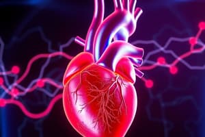
What is the structure of the heart?
What is the structure of the heart?
Located in mediastinum, with four chambers (left atrium and ventricle, right atrium and ventricle), two atrioventricular valves, two semilunar valves, and three layers (epicardium, myocardium, endocardium).
Where is the heart located in relation to surrounding structures?
Where is the heart located in relation to surrounding structures?
The heart is posterior to the sternum, medial to the lungs, anterior to the vertebral column, with the base beneath the 2nd rib and the apex at the 5th intercostal space.
What is the epicardium?
What is the epicardium?
The outer layer of the heart, also called the visceral pericardium.
What is the myocardium?
What is the myocardium?
What is the endocardium?
What is the endocardium?
What is the function of the right atrium?
What is the function of the right atrium?
What is the function of the right ventricle?
What is the function of the right ventricle?
What is the role of the left atrium?
What is the role of the left atrium?
What is the function of the left ventricle?
What is the function of the left ventricle?
Where is the tricuspid valve located and what is its function?
Where is the tricuspid valve located and what is its function?
What is the function of the pulmonary valve?
What is the function of the pulmonary valve?
What is the location and function of the mitral valve?
What is the location and function of the mitral valve?
What is the aortic valve's function?
What is the aortic valve's function?
What constitutes the skeleton of the heart?
What constitutes the skeleton of the heart?
The path of blood through the heart starts with the ______
The path of blood through the heart starts with the ______
What supplies blood to the heart?
What supplies blood to the heart?
What is atrial systole?
What is atrial systole?
What is ventricular diastole?
What is ventricular diastole?
What occurs during ventricular systole?
What occurs during ventricular systole?
What happens during atrial diastole?
What happens during atrial diastole?
What happens during atrial systole and ventricular diastole?
What happens during atrial systole and ventricular diastole?
What occurs during ventricular systole and atrial diastole?
What occurs during ventricular systole and atrial diastole?
Flashcards are hidden until you start studying
Study Notes
Structure of the Heart
- Located in the mediastinum with four chambers: left atrium, left ventricle, right atrium, and right ventricle.
- Contains two atrioventricular (AV) valves: tricuspid valve between the right atrium and right ventricle, and bicuspid (mitral) valve between the left atrium and left ventricle.
- Equipped with two semilunar valves: pulmonic valve at the pulmonary trunk exit and aortic valve at the aorta exit.
- Composed of three layers: epicardium (outer), myocardium (middle), and endocardium (inner).
Heart Positioning
- Positioned posterior to the sternum and medial to the lungs.
- Anterior to the vertebral column, with the base beneath the 2nd rib and apex at the 5th intercostal space.
- Sits just above the diaphragm.
Heart Layers
- Epicardium: The outermost layer, also called visceral pericardium; consists of connective tissue with epithelium, blood capillaries, lymph capillaries, and nerve fibers.
- Myocardium: The middle layer made of cardiac muscle tissue; contains blood capillaries, lymph capillaries, and nerve fibers.
- Endocardium: The innermost layer, consisting of epithelium and underlying connective tissue, along with blood vessels and specialized muscle fibers.
Chambers of the Heart
- Right Atrium: Receives deoxygenated blood from the inferior and superior vena cava, as well as the coronary sinus.
- Right Ventricle: Pumps deoxygenated blood from the right atrium into the pulmonary artery.
- Left Atrium: Receives oxygenated blood from the pulmonary veins and pumps it into the left ventricle.
- Left Ventricle: Pumps oxygenated blood from the left atrium into the aorta for systemic distribution.
Valves of the Heart
- Tricuspid Valve: Located at the right atrioventricular orifice; prevents backflow into the right atrium during ventricular relaxation.
- Pulmonary Valve: Located at the entrance to the pulmonary trunk; prevents backflow into the right ventricle during relaxation.
- Mitral Valve: Separates left atrium from left ventricle; prevents backflow into the left atrium during contraction.
- Aortic Valve: Situated at the entrance to the aorta; prevents backflow into the left ventricle during relaxation.
Heart Skeleton
- Consists of fibrous rings and dense connective tissue in the inter-ventricular septum, providing structure and support.
Blood Flow Pathway
- Blood circulates from the body to the heart via the superior and inferior vena cava, entering the right atrium.
- It passes through the tricuspid valve into the right ventricle, then through the pulmonary valve into the pulmonary arteries.
- Oxygenated blood returns via pulmonary veins to the left atrium, passes through the mitral valve into the left ventricle, and exits through the aortic valve to the descending aorta.
Coronary Circulation
- Left and right coronary arteries supply oxygenated blood to the heart muscle tissues.
Cardiac Cycle Phases
- Atrial Systole: Contraction of the atria to push blood into the ventricles.
- Ventricular Diastole: Relaxation of the ventricles, allowing them to fill with blood.
- Ventricular Systole: Contraction of the ventricles to pump blood out of the heart.
- Atrial Diastole: Relaxation of the atria, receiving blood from the venae cavae and pulmonary veins.
Blood Flow Dynamics
- During atrial systole and ventricular diastole, blood flows passively into the ventricles; the AV valves are open, while semilunar valves are closed.
- The remaining 30% blood in the atria is pushed into the ventricles, causing a rise in ventricular pressure.
- During ventricular systole and atrial diastole, AV valves close to prevent backflow while chordae tendineae maintain valve integrity. Atria relax, enabling blood inflow.
Studying That Suits You
Use AI to generate personalized quizzes and flashcards to suit your learning preferences.