Podcast
Questions and Answers
What is the approximate size of the heart?
What is the approximate size of the heart?
Approximately the size of a fist.
Describe the shape of the heart.
Describe the shape of the heart.
Conical, with a pointed apex.
Where is the heart located?
Where is the heart located?
Located in the thoracic cavity.
The heart is tilted to the right side of the body.
The heart is tilted to the right side of the body.
What are the heart coverings?
What are the heart coverings?
Name the layers of the heart wall.
Name the layers of the heart wall.
Identify the outer layer of the heart wall.
Identify the outer layer of the heart wall.
Which layer of the heart wall is responsible for contraction?
Which layer of the heart wall is responsible for contraction?
What is the inner layer lining heart chambers?
What is the inner layer lining heart chambers?
How many chambers does the heart have, and what are they called?
How many chambers does the heart have, and what are they called?
What does the right atrium do?
What does the right atrium do?
What is the function of the right ventricle?
What is the function of the right ventricle?
What is the role of the left atrium?
What is the role of the left atrium?
What does the left ventricle do?
What does the left ventricle do?
Name some of the major blood vessels connected to the heart.
Name some of the major blood vessels connected to the heart.
What is the function of heart valves?
What is the function of heart valves?
Name the atrioventricular valves.
Name the atrioventricular valves.
What are the semilunar valves?
What are the semilunar valves?
Describe the blood flow pathway from the right atrium.
Describe the blood flow pathway from the right atrium.
What type of muscle is cardiac muscle?
What type of muscle is cardiac muscle?
Describe cardiac muscle cells.
Describe cardiac muscle cells.
What are intercalated discs?
What are intercalated discs?
What function do desmosomes perform in cardiac cells?
What function do desmosomes perform in cardiac cells?
What is the purpose of gap junctions in cardiac cells?
What is the purpose of gap junctions in cardiac cells?
What is a functional syncytium in the heart?
What is a functional syncytium in the heart?
What percentage of cardiac muscle cell volume is comprised of mitochondria?
What percentage of cardiac muscle cell volume is comprised of mitochondria?
What is the primary energy source for cardiac muscle?
What is the primary energy source for cardiac muscle?
What are autorhythmic cells?
What are autorhythmic cells?
What does the intrinsic conduction system do?
What does the intrinsic conduction system do?
Where is the sinoatrial node located, and what is its function?
Where is the sinoatrial node located, and what is its function?
What is the role of the atrioventricular node?
What is the role of the atrioventricular node?
What does the atrioventricular bundle do?
What does the atrioventricular bundle do?
What is the function of bundle branches?
What is the function of bundle branches?
What is the function of Purkinje fibers?
What is the function of Purkinje fibers?
What are arrhythmias?
What are arrhythmias?
What is ventricular fibrillation?
What is ventricular fibrillation?
What is defibrillation?
What is defibrillation?
What does the cardioacceleratory center do?
What does the cardioacceleratory center do?
How does the cardioinhibitory center affect heart rate?
How does the cardioinhibitory center affect heart rate?
What are action potentials?
What are action potentials?
What is the role of voltage-gated Na+ channels in cardiac action potentials?
What is the role of voltage-gated Na+ channels in cardiac action potentials?
When do calcium channels open during the cardiac action potential?
When do calcium channels open during the cardiac action potential?
What is the refractory period in cardiac muscle?
What is the refractory period in cardiac muscle?
What does an electrocardiogram (ECG) record?
What does an electrocardiogram (ECG) record?
What does the P wave indicate on an ECG?
What does the P wave indicate on an ECG?
What does the QRS complex represent on an ECG?
What does the QRS complex represent on an ECG?
What is excitation-contraction coupling?
What is excitation-contraction coupling?
What do cardiac pacemaker cells do?
What do cardiac pacemaker cells do?
Which part of the brain regulates autonomic control of heart rate?
Which part of the brain regulates autonomic control of heart rate?
Flashcards
Heart Size
Heart Size
Roughly the size of your closed fist.
Heart Shape
Heart Shape
Cone-shaped with a pointed bottom.
Heart Location
Heart Location
Found in the chest cavity.
Heart Orientation
Heart Orientation
Signup and view all the flashcards
Heart Coverings
Heart Coverings
Signup and view all the flashcards
Heart Wall Layers
Heart Wall Layers
Signup and view all the flashcards
Epicardium
Epicardium
Signup and view all the flashcards
Myocardium
Myocardium
Signup and view all the flashcards
Endocardium
Endocardium
Signup and view all the flashcards
Heart Chambers
Heart Chambers
Signup and view all the flashcards
Right Atrium
Right Atrium
Signup and view all the flashcards
Right Ventricle
Right Ventricle
Signup and view all the flashcards
Left Atrium
Left Atrium
Signup and view all the flashcards
Left Ventricle
Left Ventricle
Signup and view all the flashcards
Major Blood Vessels
Major Blood Vessels
Signup and view all the flashcards
Heart Valves
Heart Valves
Signup and view all the flashcards
Atrioventricular Valves
Atrioventricular Valves
Signup and view all the flashcards
Semilunar Valves
Semilunar Valves
Signup and view all the flashcards
Blood Flow Pathway
Blood Flow Pathway
Signup and view all the flashcards
Cardiac Muscle
Cardiac Muscle
Signup and view all the flashcards
Cardiac Muscle Cells
Cardiac Muscle Cells
Signup and view all the flashcards
Intercalated Discs
Intercalated Discs
Signup and view all the flashcards
Desmosomes
Desmosomes
Signup and view all the flashcards
Gap Junctions
Gap Junctions
Signup and view all the flashcards
Functional Syncytium
Functional Syncytium
Signup and view all the flashcards
Mitochondria in Cardiac Muscle
Mitochondria in Cardiac Muscle
Signup and view all the flashcards
Aerobic Respiration
Aerobic Respiration
Signup and view all the flashcards
Autorhythmic Cells
Autorhythmic Cells
Signup and view all the flashcards
Intrinsic Conduction System
Intrinsic Conduction System
Signup and view all the flashcards
Sinoatrial Node
Sinoatrial Node
Signup and view all the flashcards
Study Notes
- Heart size is about the size of a fist.
- The heart has a conical shape, featuring a pointed apex.
- The heart resides in is the thoracic cavity.
- The heart is oriented with a tilt towards the left side of the body.
- The heart is protected by the pericardium, which is a double-walled sac.
- The wall of the heart is composed of three layers: the epicardium, myocardium, and endocardium.
Heart Wall Layers
- The epicardium is the heart wall's outer layer.
- The myocardium is the muscular middle layer responsible for contraction.
- The endocardium is the inner layer lining the heart chambers.
Heart Chambers and Vessels
- The heart contains four chambers: two atria and two ventricles.
- The right atrium receives deoxygenated blood from the body.
- The right ventricle pumps deoxygenated blood to the lungs.
- The left atrium receives oxygenated blood from the lungs.
- The left ventricle pumps oxygenated blood to the body.
- Major blood vessels connected to the heart include the vena cavae, pulmonary arteries, and the aorta.
Heart Valves
- Heart valves prevent the backflow of blood.
- Atrioventricular valves include the tricuspid and mitral valves.
- Semilunar valves include the pulmonary and aortic valves.
- Blood flows in the sequence: right atrium → right ventricle → lungs.
Cardiac Muscle
- Cardiac muscle is striated and unique to the heart.
- Cardiac muscle cells have a branched structure and typically contain one or two nuclei.
- Intercalated discs connect cardiac muscle cells and synchronize contraction.
- Desmosomes provide structural strength between cardiac cells.
- Gap junctions facilitate ion flow between cardiac cells.
- Heart cells function as a single unit, known as a functional syncytium.
- Mitochondria make up 25-35% of cardiac muscle cell volume, providing energy.
- Aerobic respiration serves as the primary energy source for cardiac muscle.
- Autorhythmic cells are self-excitable and initiate heartbeats.
- The intrinsic conduction system controls heart rhythm independently of the nervous system.
Heart Nodes
- The sinoatrial node is the primary pacemaker, located in the right atrium.
- The atrioventricular node delays impulses to allow for atrial contraction.
- The atrioventricular bundle conducts impulses from the atria to the ventricles.
- Bundle branches conduct impulses down the interventricular septum.
- Purkinje fibers distribute impulses throughout the ventricular walls.
- Arrhythmias are irregular heart rhythms resulting from conduction defects.
- Ventricular fibrillation involves rapid, ineffective contractions that prevent blood pumping.
- Defibrillation restores a normal heart rhythm after fibrillation.
- The cardioacceleratory center increases heart rate via sympathetic neurons.
- The cardioinhibitory center decreases heart rate via parasympathetic impulses.
- Action potentials are electrical signals that trigger cardiac muscle contraction.
Channels and Waves
Voltage-gated Na+ channels open during the rapid depolarization phase.
- Calcium channels open during the plateau phase of the action potential.
- The refractory period prevents tetanus in cardiac muscle.
- An electrocardiogram (ECG) records the heart's electrical activity.
- The P wave indicates atrial depolarization on an ECG.
- The QRS complex indicates ventricular depolarization on an ECG.
- The T wave indicates ventricular repolarization on an ECG.
- Excitation-contraction coupling is the process that links electrical stimulation to muscle contraction.
- Cardiac pacemaker cells generate rhythmic heartbeats.
- The medulla oblongata regulates autonomic control of heart rate.
Studying That Suits You
Use AI to generate personalized quizzes and flashcards to suit your learning preferences.
Description
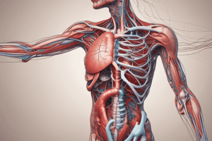
The heart, about the size of a fist, lies in the thoracic cavity and is protected by the pericardium. Its wall comprises the epicardium, myocardium, and endocardium. The heart includes four chambers and is connected to major blood vessels.