Podcast
Questions and Answers
The pinna contributes to hearing by performing which function?
The pinna contributes to hearing by performing which function?
- Converting sound waves into physical vibrations.
- Transmitting sound waves into the skull.
- Collecting and amplifying sound. (correct)
- Equalizing pressure differences between the middle ear and external atmosphere.
What transformation occurs at the tympanic membrane?
What transformation occurs at the tympanic membrane?
- Sound waves are converted to physical vibrations. (correct)
- Fluid pressure is converted to mechanical movement.
- Sound waves are converted to nerve signals.
- Physical vibrations are converted to sound waves.
Which of the following is true regarding the ossicles?
Which of the following is true regarding the ossicles?
- They decrease the pressure of vibrations entering the inner ear.
- They include the malleus, incus, and stapes. (correct)
- They directly convert sound waves into nerve impulses.
- They are located within the outer ear.
What is the role of the Eustachian tube in the middle ear?
What is the role of the Eustachian tube in the middle ear?
Where are the physical vibrations of sound waves converted into nerve signals?
Where are the physical vibrations of sound waves converted into nerve signals?
Which structures are responsible for the sense of balance and spatial orientation?
Which structures are responsible for the sense of balance and spatial orientation?
The semi-circular canals detect movement in how many planes?
The semi-circular canals detect movement in how many planes?
What is the gelatinous structure that encases hair cells within the crista ampullaris?
What is the gelatinous structure that encases hair cells within the crista ampullaris?
Which inner ear structure is primarily responsible for detecting linear acceleration?
Which inner ear structure is primarily responsible for detecting linear acceleration?
What is the function of the scala media in the cochlea?
What is the function of the scala media in the cochlea?
Sound vibrations are transmitted to the oval window by the stapes. What is the next step in the process of hearing?
Sound vibrations are transmitted to the oval window by the stapes. What is the next step in the process of hearing?
What supports the basilar membrane?
What supports the basilar membrane?
The bases and sides of the hair cells are enmeshed by a network of cochlear nerve endings which lead to the:
The bases and sides of the hair cells are enmeshed by a network of cochlear nerve endings which lead to the:
What critical event occurs when stereocilia bend?
What critical event occurs when stereocilia bend?
How is sound frequency or pitch determined in the cochlea?
How is sound frequency or pitch determined in the cochlea?
What is the primary role of keratinocytes in the epidermis?
What is the primary role of keratinocytes in the epidermis?
Which epidermal layer contains a clear homogenous layer of cells without nuclei or organelles and is only found in thick skin?
Which epidermal layer contains a clear homogenous layer of cells without nuclei or organelles and is only found in thick skin?
Which of the following is a primary function of melanin in the skin?
Which of the following is a primary function of melanin in the skin?
What is the term for a patch of dark pigmentation on the skin that is commonly referred to as a mole?
What is the term for a patch of dark pigmentation on the skin that is commonly referred to as a mole?
Hair, nails, claws, and hooves are composed of which of the following?
Hair, nails, claws, and hooves are composed of which of the following?
In early fetal development, what layer of tissue does the epidermis grow down into to form the cellular shaft of a hair follicle?
In early fetal development, what layer of tissue does the epidermis grow down into to form the cellular shaft of a hair follicle?
What is the function of the arrector pili muscle?
What is the function of the arrector pili muscle?
What is the correct order of the phases of a hair cycle?
What is the correct order of the phases of a hair cycle?
What is the mechanism by which secretion released via hair follicles makes its way into the hair follicle shaft?
What is the mechanism by which secretion released via hair follicles makes its way into the hair follicle shaft?
Ceruminous glands are modified apocrine glands that secrete what substance?
Ceruminous glands are modified apocrine glands that secrete what substance?
From what structures are mammary glands derived?
From what structures are mammary glands derived?
What is the main type of cell found in the dermis?
What is the main type of cell found in the dermis?
Which layer of skin is composed predominately of loose connective tissue and contains large blood and lymphatic vessels and nerves?
Which layer of skin is composed predominately of loose connective tissue and contains large blood and lymphatic vessels and nerves?
What types of sensory receptors are muscle spindles and Golgi tendon organs considered to be?
What types of sensory receptors are muscle spindles and Golgi tendon organs considered to be?
What are sensory receptors responsible for?
What are sensory receptors responsible for?
The five functional categories of sensory receptors are mechanoreceptors, thermoreceptors, nociceptors, photoreceptors, and:
The five functional categories of sensory receptors are mechanoreceptors, thermoreceptors, nociceptors, photoreceptors, and:
Which receptors are responsible for the senses of taste and smell?
Which receptors are responsible for the senses of taste and smell?
Which receptor type includes free nerve endings, tactile hair cells and encapsulated end-organs?
Which receptor type includes free nerve endings, tactile hair cells and encapsulated end-organs?
Pacinian corpuscles transmit pressure on naked nerve endings. What does this pressure cause?
Pacinian corpuscles transmit pressure on naked nerve endings. What does this pressure cause?
Consisting of a central non-myelinated tip of a nerve fibre surrounded by up to 30 concentric layers of connective tissue describes what structure?
Consisting of a central non-myelinated tip of a nerve fibre surrounded by up to 30 concentric layers of connective tissue describes what structure?
Concentrated in the dermal papillae of fingertips, lips, palms of the hands, soles of the feet and other sensitive areas of skin describes?
Concentrated in the dermal papillae of fingertips, lips, palms of the hands, soles of the feet and other sensitive areas of skin describes?
Taste is sensed by small organs on the tongue:
Taste is sensed by small organs on the tongue:
Both the gustatory and sustentacular cells have long:
Both the gustatory and sustentacular cells have long:
About how many genes code for olfactory receptors?
About how many genes code for olfactory receptors?
Flashcards
Pinna
Pinna
The external visible part of the ear; collects and amplifies sound.
Auditory canal
Auditory canal
Canal linking the outer and middle ear, transmitting sound waves.
Tympanic membrane
Tympanic membrane
Thin connective tissue blocking the ear canal; converts soundwaves to physical vibrations.
Tympanic cavity
Tympanic cavity
Signup and view all the flashcards
Ossicles
Ossicles
Signup and view all the flashcards
Eustachian tube
Eustachian tube
Signup and view all the flashcards
Inner ear
Inner ear
Signup and view all the flashcards
Bony labyrinth
Bony labyrinth
Signup and view all the flashcards
Membranous labyrinth
Membranous labyrinth
Signup and view all the flashcards
Vestibular system
Vestibular system
Signup and view all the flashcards
Semi-circular canals
Semi-circular canals
Signup and view all the flashcards
Crista ampullaris
Crista ampullaris
Signup and view all the flashcards
Otolith organs
Otolith organs
Signup and view all the flashcards
Cochlea
Cochlea
Signup and view all the flashcards
Organ of Corti
Organ of Corti
Signup and view all the flashcards
Epidermis
Epidermis
Signup and view all the flashcards
Dermis
Dermis
Signup and view all the flashcards
Hypodermis
Hypodermis
Signup and view all the flashcards
Stratum corneum
Stratum corneum
Signup and view all the flashcards
Stratum lucidum
Stratum lucidum
Signup and view all the flashcards
Stratum granulosum
Stratum granulosum
Signup and view all the flashcards
Stratum spinosum
Stratum spinosum
Signup and view all the flashcards
Stratum basale
Stratum basale
Signup and view all the flashcards
Melanocytes
Melanocytes
Signup and view all the flashcards
Keratinised Structures
Keratinised Structures
Signup and view all the flashcards
Hair
Hair
Signup and view all the flashcards
Hair follicle
Hair follicle
Signup and view all the flashcards
Sebaceous glands
Sebaceous glands
Signup and view all the flashcards
Apocrine glands
Apocrine glands
Signup and view all the flashcards
Eccrine glands
Eccrine glands
Signup and view all the flashcards
Ceruminous glands
Ceruminous glands
Signup and view all the flashcards
Mammary glands
Mammary glands
Signup and view all the flashcards
Dermis
Dermis
Signup and view all the flashcards
Functional Types of Sensory Receptor
Functional Types of Sensory Receptor
Signup and view all the flashcards
Mechanoreceptors
Mechanoreceptors
Signup and view all the flashcards
Pacinian Corpuscles
Pacinian Corpuscles
Signup and view all the flashcards
Ruffini's End Organs
Ruffini's End Organs
Signup and view all the flashcards
Meissner's Corpuscles
Meissner's Corpuscles
Signup and view all the flashcards
Merkel's Discs
Merkel's Discs
Signup and view all the flashcards
Sense of position
Sense of position
Signup and view all the flashcards
Study Notes
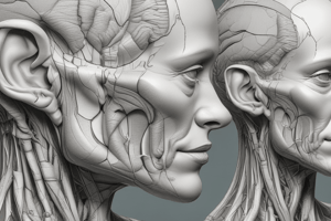
The Structure Of The Ear
- The outer, middle, and inner ear structures are described.
- The role of the vestibular apparatus is discussed in detecting movement and angular rotation.
- The structure and function of the cochlea and the organ of Corti is explained in relation to hearing.
The Ear
- Mammals have two sense organs in the ear for hearing and balance.
- The ear is divided into the outer, middle, and inner ear.
Outer Ear
- The outer ear includes the pinna, which collects and amplifies sound, and the auditory canal.
- The tympanic membrane, or ear drum, blocks the ear canal.
- The ear drum converts soundwaves into physical vibrations with membrane movement.
- The ear drum can rupture from trauma, leading to hearing loss. Small perforations can heal spontaneously.
Middle Ear
- This consists of the tympanic cavity between the eardrum and the oval window of the inner ear.
- It contains three small bones called the ossicles.
- The Latin names for these bones are malleus, incus, and stapes, which translate to "the hammer, anvil, and stirrup".
- The stapes is the smallest bone in the human body.
- The middle ear bones amplify and transmit vibrations from the tympanic membrane to the inner ear.
- They translate the low-pressure movement of the large eardrum into high-pressure movement of the small oval window.
- The malleus connects to the eardrum, and the incus bone connects to the other side.
- Vibrational energy transfers from the eardrum to the incus.
- The incus transfers energy to the stapes, which connects to the oval window of the inner ear.
- The middle ear also contains the Eustachian tube, which links the back of the nose to the middle ear.
- The Eustachian tube equalizes the pressure between the middle ear and external atmosphere.
- Also helps drain mucous from the middle ear.
Inner Ear
- Innermost section of the ear where physical vibrations of sound waves convert to nerve signals.
- Inner Ear is contained within a hollow cavity in the temporal bone of the skull called the bony labyrinth.
- The membranous labyrinth is a system of fluid-filled chambers and channels found within the bony labyrinth.
- These two structures are separated by a layer of perilymph fluid.
- The inner ear includes the vestibular system and the cochlea.
- The vestibular system provides balance and spatial awareness.
- The vestibular system consists of semicircular canals which detect rotation, and otolith organs which detect linear acceleration.
Semi-Circular Canals
- There are three semi-circular canals that lie at approximate right angles to each other: lateral, anterior, and posterior.
- Each canal contains a fluid called endolymph and ends with a sac called the osseous ampulla.
- Head movements causes endolymph to flow within the canals.
- Different canals detect movement within three planes.
Ampullae and Crista Ampullaris
- Each ampule contains a crista ampullaris.
- Crista ampullaris translate endolymph flow into nerve signals.
- The crista ampullaris consists of a cluster of hair cells encased within a gelatinous, cupula structure.
- When the head turns, endolymph within the semi-circular canals pushes the cupula in the opposite direction of the head.
- This movement triggers membrane depolarization of hair cells.
- Vestibular systems in both ears work together, this allows the body to sense all movement directions.
- Excessive triggering of this system results in motion sickness.
Otolith Organs
- The inner ear includes two otolith organs: the utricle and the smaller saccule.
- The utricle and saccule are filled with endolymph and detect linear acceleration.
- The utricle responds to horizontal movement, and the saccule detects vertical acceleration.
- Both organs contain a sensitive hair cell patch called the macula, which is enclosed within an otolithic membrane.
- Calcium carbonate crystals, or otoliths, cover the top of this membrane.
- Head tilting allows gravity to pull the otoliths forward.
- The process triggers hair cell activation.
Cochlea - Hearing
- The organ of hearing is a coiled tube that resembles a snail shell with three canals separated by thin membranes.
- The upper canal, the scala vestibuli (vestibular canal), connects to the oval window and thus the stapes.
- The lower canal, the scala tympani (tympanic canal) connects to the round window which opens on the middle ear.
- At cochlear apex, the helicotrema connects the scala tympani to the scala vestibuli.
Scala Media
- The scala media separates from the scala vestibuli by a thin vestibular or Reissner's membrane.
- The scala media is separated from the scala tympani by thicker basilar membrane, which does impede sound.
- About 20,000 reed-like thin spines, called basilar fibers, protrude most of the basilar membrane distance.
- The organ of Corti is on scala media basilar membrane , this is the organ of hearing.
Organ of Corti
- The organ of Corti is on the basilar membrane in the scala media.
- The organ of Corti has one row of internal hair cells (3,500) and three to four rows of external hair cells (20,000).
- Cochlear nerve endings enmesh bases and sides of the hair cells.
- This network leads to of the spiral ganglion of Corti in the core of the cochlea.
- Axons from the spiral ganglion travel through the cochlear nerve to the brain, via the VIII auditory nerve.
- Minute hairs or stereocilia project from each hair cell.
- Stereocilia either touch or embed in the surface gel of the tectorial membrane overhanging flap of tissue.
- The tectorial membrane lies free in the scala media, attached on one side.
- Stereocilia arrange with the shortest hairs on the outer rows and the longest in the center.
- Sound vibrations from the oval window are transmitted by the stapes, creating pressure waves in the scala vestibuli and scala media.
- Causes the basilar membrane vibrates which move the hair cells against the tectorial membrane.
- Bending stereocilia causes ion channels to open, this allows potassium and calcium to enter the cell.
- This triggers release of glutamate neurotransmitter with subsequent activation of postsynaptic cochlear nerves
- Also triggers creation of a nerve impulse.
Soundwave Frequency or Pitch
- The basilar membrane composes of 20,000+ basilar fibres from the oval and round windows toward the helicotrema.
- The basilar membrane varies from 0.04mm at the windows to 0.5mm at the apex, giving a distance of 35mm.
- Fibres are fixed at their basal ends but free at the distal end, loosely embedded in the basilar membrane.
- Fibre diameter decreases from base to helicotrema.
- Short, stiff fibres at the windows vibrate at high frequency, and long, thin fibres at the helicotrema vibrate at low frequency.
- Sounds pitch/ frequency is based on the place principle in the activated part of the basilar membrane.
Integument: Epidermis, Dermis
- The role of the skin as a significant organ in the integumentary system is emphasized.
- Epidermis's histology is described, focusing on cell types, layers, and processes like keratinization.
- Various epidermal derivatives and keratinized structures including nails, hair, sweat glands, and mammary glands are discussed.
- The dermis and subcutaneous layers of the skin are described.
- Skin constitutes an integument for the entire body. Plays roles in protection, thermoregulation, sensation, and metabolism.
- Skin protects by its tough hide, pathogens defense, water regulation, and defending against dangerous UV light.
- Skin provides insulation via hair and sweating, playing a major role in temperature regulation.
- Skin origin is from the area of sensory and nervous structures in the ancestral metazoan.
- Skin originates continuity embryologically with the nervous system.
- It produces vitamin D which is used calcium and phosphate metabolism.
- Sensational sensory nerve endings interpret external environment, temperature, and pain.
- Skin equals 12.5% to 16.7% of the mass of a normal person.
- It varies in thickness - very thin eyelids, thick on soles of feet.
- Continuous integument with the digestive tract, mouth, anus, urinary, genitals, penis, vagina, and urethra.
- Composed of two layers - outer epidermis and an inner dermis with a hypodermis, the underlayer of loose connective tissue.
- Glabulous skin: thick, hairless skin. Ex: palms and soles of feet.
- Hairy skin: thinner skin - Most skin areas.
Epidermis Strata and Protection
- Protective barrier that guards against chemical, physical, and microbial factors.
- It is 0.06-0.1 mm thick.
- This arises from embryonic ectoderm and gives rise to sweat glands, sebaceous glands, and mammary glands.
- Keratinocytes arrange into strata, creating stratified, squamous epithelium.
- Keratinocytes start at the basal membrane and move progressively outwards.
- Cells mature to corneocytes and are removed by desquamation, which produces a stratum comeum.
- The entire epidermal cycle takes about 27 days.
- A thick, outer layer of flattened, fused cell remnants made of keratin.
- This is covered with an oily secretion from sebaceous glands (sebum).
- Found only in thick skin; a clear of cells homogenous, and lacks nuclei or organelles.
- Shows internal granules within the cell.
- Prickle layer, where cells grow, showcasing early keratin synthesis.
- This contains a proliferative layer of columnar or cuboidal cells.
Skin 3-Factor Pigmentation
- Carotene creates Yellowish that derives from subcutaneous fat.
- Hemoglobin affects capillary dermal concentration and oxygenation.
- Special branched cells also affect pigment that is scattered throughout the stratum basale.
- Melanin is the cells pigment produce melanocytes.
- Pigment able to dissipate over 99.9% of uv radiation to protect from uv radiation.
- Melanocytes number constant irrespective of skin color.
- Melanosomes are vesicles where melanocytes package melanin.
- Keratinocytes take these into the nucleus to protect the DNA from ionizing radiation.
- A patch of dark pigmentation, for example, a mole, is a melanocytic nevus that produces melanoma in percentage.
Keratinised Structures
- Mammals and foot pads primates, and humans, feature friction ridges on the palms and soles, fingerprint dermatoglyph.
- Hair, Nails, claws, hooves, make most of keratinised structures.
- Hair (shaft), which grow from a root (hair bulb) lying in the dermal layer that forms from epidermal cells.
- Hair bulb are located at a base sunk in a pit, the dermis, called the hair follicle that is made by epidermal cells, thus continuous.
- Epidermis cells grow down into forming a cellular shaft during life.
- Hairshaft or dead epidermal matrix with the growing matrix cell exists.
- The vas plug of dermis that the matrix forms is called the dermal matrix.
- Germinal matrix cells are connected via dermis cellular layer(s).
- The structure makes a pilosebaceous unit.
- Germinal matrix gives hair growth.
- Proliferation starts with cells pushed through the follicle.
- Cells supply become keratinized and die.
- Hair follicle is organized cells that are initial.
Hair Structure
- They perform to create the cortex single and cell single.
- Glands/cell/hairs enter and exit on basis of the cycles from:
- permanent.
- Bulge.
- Exogen.
- Bulb phases grow around hair bulb and the muscle arrector pili with some sebaceous activity.
Hair Follicle Structure
- Hair Follicle is in position the of the hair sheaths with:
- Medulla: seldom seen.
- Cortex: keratin.
- Cuticle single-layer over position on follicles, but needs outer sheath.
- Germination needs external and internal.
- Cells perform the cortex with outer/cuticles.
Hair Growth Cycle - Anagen, Catagen, and Telogen
- Hair grows in cycles, so phases are active, and rest stages are required depending on sebaceous for proper maintenance and regeneration of the hair follicles.
- Hair can be elevated via muscle.
- Glands produce and move.
- Cycle involves bulge to grow hair as well to the matrix or matrix and with cell/glands phases and muscle for elevation.
- Telogen is then entered when old or hair shaft loosens.
- In baldness, the matrix phases don't resume.
Sudoriferous & Sebaceous Glands
- Sweat and Oil Glands are found in Hair Follicles and the Skin
- Types;
- sudoriferous helps with oil with holocrine for oil
- for skin and hair production
- the palms of feet and hand
- Most areas of body and the limbs is regulated with skin, oil and glands/unit.
- Ducts open for thermoregulation, and sweat at most areas outside lip and eardrums with fascia connected to the skin.
- Apocrine Ducts also exist in the hair or epidermis through axillary/arm pits (breast).
Ceruminous and Mammary Integument Structure
- Ceram is related to audio structure where is drains external sudatory and also contains secretion with lubricants, bacteria, water protection through sand, wax.
- Mammae are what's called glands of with derivatives for their control such as:
- Composition, protein milk from animals.
- Pig dog cow is similar, but can have lines.
Dermis Properties Description
-
Cells in dermis, most fibers secrete and organize fibroblasts called: ECM
-
Major, fibrillar, 1, 2,3, v elastin
-
Papillary dermis that is Rich in vasculature and can Thermo/ Nerve Endings
-
Reticular – dermis contains elastin and provide with structural skin.
Properties of Dermis Skin
- Properties with fibers includes/involves connective tissue and vessels 2 mm thickness for the most parts in skin structure:
- collagen is elastic
- Vessels maintain blood
- Cells include fiber formation (collagin elastin).
- Appendix retains through hyaluronic in a mesoderm.
Subcutis and Tissue Structure
- Structure is mostly in adipocytes, tissue-based loose connective for cells and with fiber/fat areas, as with collagen/fat. It has large layers of tissues along with nervous supply.
Sense Organs: Mechanoreceptors and Chemoreceptors
- Functional sensory receptor is defined, including 5 functional types
- Free nerve mechano receptors explained with 3 encapsulated including:
- Pacinian corpuscles
- Meissner's corpuscles
- Ruffini's end organs
- Merkel's discs.
- muscle spindles
- Golgi tendon organs as proprioreceptors are defined.
- Structure of the taste bud chemoreceptors and their mechanism is recalled.
- The olfactory sense operation and membrane/function, is described.
- Sensory receptors include those for light touch, pressure, temperature, pain and the senses. These enable homeostasis
Major Senses Role
- Input into nervous system is receptors.
- Nerve endings/ cells that convert stimuli are signals in the external, internal. They're like, "afferent". Organs are auditory/optic for the information needed.
- the main senses provide part of sensory senses, but for general purposes, taste + body is important for senses.
Types of Functional & Sensory Receptors
- Categories classified into:
- Five functional categories
- "Mechano" mechanical detections
- "Thermo" temperatures change
- "Nociceptors" free/tear pain
- "Photo" retinal rods
- "Chemo" smell
- Not always perfect classification.
Mechanoreceptors and Functions & Role
- Function most and nerve endings,
- Detect pain temperature through free cells/tissues.
- Nerve ending in skin muscles is surrounded
Tactile Receptors in Sensory Endings
- Types:
- Pacinian deep
- Meissner: Touch + vibration
- Merkel is lighting, and ruffini has tissue.
Pacinian Corpuscles Detailes
- Transmit via open
- Depolarize through pressure for signal, tissue detection is great with vibration. onion, 1 + meter is size.
- Found:
- abdominal
- external, tendons, and joints.
Ruffini Receptor Functions
- Tissue detect via nerve, for deformation in joints for the role/action of:
- pacinian through pressure
- thermo
- adapting is slow
- Capsule/Fiber with the "deeper".
Receptor Sizes and Characteristics
- Meissner's
- Has stacked lamellae.
- 75m long.
- It has the ability to detect touch that is light and for recognization. This is most acute at the lips, palms and face.
- Merkel has discs with epidermis (stratum) base, (light).
- Vesicles that are synapsed transmit without the neural (touch + small).
- Muscle for organ joints are Golgi/spindles. Adaptation, like for receptors, is slow.
Types of Response and Muscle Role
- Spindles are adaptation is slow and have no adaptation for continuous monitoring of tonic tone while being for position. The fibers can even be enclosed to impulses/sensory.
Muscle Fibers and Characteristics
- Muscles have attachment and immediate dendrite fibers, capsule/tendon.
- Spindles have a length and all have a fiber and signal and for sensory for control.
Adaptation Types and Nerves
- Rapid adaptation occurs between nerve stimulus is constant with the thalmic that has movement related.
Chemoreceptors Functions
- Taste and receptors
- Taste via organ with (3,000) that are 3,000 buds and arranged for 50/modified. roof in ororal, bud/ laryx.
- Two main types.
Types of Chemoreceptors
- Parts:
- epoglottis
- tonsils: lingual, etc.
- Regions: bitter, salty, sweet.
- Area bud responds: and is taste the is tested for that receptor but taste olfactory when have covid, cold, etc.
Taste Receptor Shapes
Sensation of Taste Types
- Hair type cells connect 50 or less near cell that taste.
- Sugar like G protein, can activate.
Gustatory Cells
Taste has cell types of base, gutaory long microvilli in brain
Taste Molecules and Cheminals
- The G protein interacts.
- This activates a protein kinase and closes K which opens Na+.
- High-Level: Ion is neuron and high.
Olfactory Sense/Function
- Epthelum can detect tetrapods smell, main sources of information are developed and the sensitive receptor can adapt.
Sensory Function of Small and Tissue
- The olfactory bulbs have sensors in cavity/tissue(wall) nose and Olfactory bulbs(tissue wall) in sinus.
Olfaction Mechanism and Properties
- Has membrane (contains the olfactory cells).
- Simple and
- Three cell types Bipolar: Support mucosa + ethmoid sensory bone.
- Fibers get through ethmoid .
- Mucosa: Can connect soma in the tissue where the project/cell in axons project (axon). Microvili + Epithelium with odor receptors. Then open channels (G), cause to enter and act. 7 scent classes. Very sensitive one per 30B.
Studying That Suits You
Use AI to generate personalized quizzes and flashcards to suit your learning preferences.