Podcast
Questions and Answers
What is the medical term for the joint that connects the humerus to the scapula?
What is the medical term for the joint that connects the humerus to the scapula?
Shoulder joint
Which of the following joints make up the elbow joint?
Which of the following joints make up the elbow joint?
- Shoulder joint Elbow joint Wrist joint
- Superior radioulnar joint Middle radio-ulnar joint Inferior radio-ulnar joint
- Inferior radio-ulnar joint (correct)
- Superior radioulnar joint (correct)
- Humerus Radius Ulna
- Middle radio-ulnar joint (correct)
- Elbow joint (correct)
Match the following terms to the correct anatomical location on the clavicle:
Match the following terms to the correct anatomical location on the clavicle:
Lateral end = Acromion Medial end = Sternal Inferior surface = Trapezoid line Conoid tubercle = Subclavius groove Impression for costo-clavicular ligament = Conoid tubercle
How many borders does the scapula have?
How many borders does the scapula have?
What are the three angles of the scapula?
What are the three angles of the scapula?
How many processes does the scapula have?
How many processes does the scapula have?
What are the three fossae of the scapula?
What are the three fossae of the scapula?
Which of the following is NOT a process found on the scapula?
Which of the following is NOT a process found on the scapula?
What is the name of the depression on the scapula that articulates with the humerus?
What is the name of the depression on the scapula that articulates with the humerus?
Which of the following is NOT a tubercle found at the head of the humerus?
Which of the following is NOT a tubercle found at the head of the humerus?
The surgical neck of the humerus is the same as the anatomical neck of the humerus.
The surgical neck of the humerus is the same as the anatomical neck of the humerus.
The humerus has a spiral groove that houses the radial nerve.
The humerus has a spiral groove that houses the radial nerve.
What is the name of the most distal bony projection found on the ulna?
What is the name of the most distal bony projection found on the ulna?
The ulna has a supinator crest.
The ulna has a supinator crest.
What is the purpose of the styloid process on the radius?
What is the purpose of the styloid process on the radius?
What is the name of the depression on the radius that articulates with the ulna?
What is the name of the depression on the radius that articulates with the ulna?
The anterior oblique line is a feature of the humerus.
The anterior oblique line is a feature of the humerus.
What is the name of the joint that connects the femur to the hip bone?
What is the name of the joint that connects the femur to the hip bone?
What is the name of the cartilaginous joint that connects the two pubic bones?
What is the name of the cartilaginous joint that connects the two pubic bones?
What is the name of the joint between the femur, tibia, and patella?
What is the name of the joint between the femur, tibia, and patella?
What are the two joints that connect the tibia and fibula?
What are the two joints that connect the tibia and fibula?
What are the three parts that make up the hip bone?
What are the three parts that make up the hip bone?
What is the name of the socket on the hip bone that articulates with the femur?
What is the name of the socket on the hip bone that articulates with the femur?
Which of the following is NOT a feature found on the ilium?
Which of the following is NOT a feature found on the ilium?
What is the name of the prominent ridge along the superior border of the ilium?
What is the name of the prominent ridge along the superior border of the ilium?
What is the name of the large opening found on the hip bone, formed by the ilium, ischium, and pubis?
What is the name of the large opening found on the hip bone, formed by the ilium, ischium, and pubis?
What is the name of the depression on the ilium that houses the iliacus muscle?
What is the name of the depression on the ilium that houses the iliacus muscle?
What is the name of the roughened area on the ilium that serves as an attachment site for the gluteal muscles?
What is the name of the roughened area on the ilium that serves as an attachment site for the gluteal muscles?
What is the name of the bony prominence on the ischium that you sit on?
What is the name of the bony prominence on the ischium that you sit on?
What is the purpose of the intertrochanteric line and intertrochanteric crest of the femur?
What is the purpose of the intertrochanteric line and intertrochanteric crest of the femur?
What is the name of the prominent ridge on the posterior aspect of the femur, which runs down the shaft?
What is the name of the prominent ridge on the posterior aspect of the femur, which runs down the shaft?
What are the two condyles of the femur?
What are the two condyles of the femur?
What is the name of the depression on the distal femur that separates the two condyles?
What is the name of the depression on the distal femur that separates the two condyles?
What is the name of the bony prominence on the tibia, located just below the condyles, where the patellar ligament attaches?
What is the name of the bony prominence on the tibia, located just below the condyles, where the patellar ligament attaches?
The fibula is a weight-bearing bone.
The fibula is a weight-bearing bone.
What is the name of the suture that joins the two parietal bones?
What is the name of the suture that joins the two parietal bones?
What is the name of the intersection of the sagittal suture and the coronal suture?
What is the name of the intersection of the sagittal suture and the coronal suture?
What is the name of the opening in the skull through which the spinal cord passes?
What is the name of the opening in the skull through which the spinal cord passes?
What are the names of the bony projections on the occipital bone that articulate with the first cervical vertebra?
What are the names of the bony projections on the occipital bone that articulate with the first cervical vertebra?
What are the two main parts of a vertebra?
What are the two main parts of a vertebra?
What are the two structures that make up the vertebral arch?
What are the two structures that make up the vertebral arch?
What is the name of the bony projection that extends posteriorly from the vertebral arch?
What is the name of the bony projection that extends posteriorly from the vertebral arch?
What is the name of the bony projection that extends laterally from the vertebral arch?
What is the name of the bony projection that extends laterally from the vertebral arch?
What is different about the structure of a thoracic vertebra compared to a cervical vertebra?
What is different about the structure of a thoracic vertebra compared to a cervical vertebra?
What is distinct about the structure of a lumbar vertebra?
What is distinct about the structure of a lumbar vertebra?
What is the name of the bone formed by the fusion of five sacral vertebrae?
What is the name of the bone formed by the fusion of five sacral vertebrae?
The coccyx is a single bone.
The coccyx is a single bone.
Flashcards
Articulation
Articulation
The connection between two or more bones, allowing movement.
Humerus
Humerus
The bone on the upper arm.
Ulna
Ulna
The bone on the lower arm, on the pinky side.
Radius
Radius
Signup and view all the flashcards
Femur
Femur
Signup and view all the flashcards
Tibia
Tibia
Signup and view all the flashcards
Fibula
Fibula
Signup and view all the flashcards
Clavicle
Clavicle
Signup and view all the flashcards
Scapula
Scapula
Signup and view all the flashcards
Acromion
Acromion
Signup and view all the flashcards
Coracoid process
Coracoid process
Signup and view all the flashcards
Elbow joint
Elbow joint
Signup and view all the flashcards
Wrist joint
Wrist joint
Signup and view all the flashcards
Knee joint
Knee joint
Signup and view all the flashcards
Hip joint
Hip joint
Signup and view all the flashcards
Hip bone
Hip bone
Signup and view all the flashcards
Ilium
Ilium
Signup and view all the flashcards
Pubis
Pubis
Signup and view all the flashcards
Ischium
Ischium
Signup and view all the flashcards
Acetabulum
Acetabulum
Signup and view all the flashcards
Obturator foramen
Obturator foramen
Signup and view all the flashcards
Iliac crest
Iliac crest
Signup and view all the flashcards
Linea aspera
Linea aspera
Signup and view all the flashcards
Greater and lesser trochanter
Greater and lesser trochanter
Signup and view all the flashcards
Tibial tuberosity
Tibial tuberosity
Signup and view all the flashcards
Frontal bone
Frontal bone
Signup and view all the flashcards
Parietal bones
Parietal bones
Signup and view all the flashcards
Occipital bone
Occipital bone
Signup and view all the flashcards
Nasal bone
Nasal bone
Signup and view all the flashcards
Zygomatic bone
Zygomatic bone
Signup and view all the flashcards
Maxillary bone
Maxillary bone
Signup and view all the flashcards
Study Notes
Anatomical Structures
- Shoulder Joint: A complex ball-and-socket joint connecting the upper arm bone (humerus) to the shoulder blade (scapula).
- Elbow Joint: A hinge joint formed by the humerus, radius, and ulna.
- Superior Radio-ulnar Joint: A pivot joint located above the elbow.
- Middle Radio-ulnar Joint: A pivot joint located in the middle of the forearm.
- Inferior Radio-ulnar Joint: A pivot joint located below the elbow.
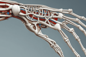
- Wrist Joint: A complex joint between the forearm bones (radius and ulna) and the carpal bones of the hand.
- Clavicle: Collarbone, a long, S-shaped bone connecting the sternum (breastbone) to the acromion of the scapula. It has a lateral (acromial) and medial (sternal) end.
- Clavicle Features: Inferior surface, trapezoid line, subclavius groove, conoid tubercle, impression for costo-clavicular ligament.
- Scapula: Shoulder blade, a flat, triangular bone in the posterior thorax. It has 3 borders (superior, medial, lateral) and 3 angles (superior, inferior, lateral).
- Scapula Features: Superior border, lateral border, medial border, superior angle, lateral angle, inferior angle, acromion, spine, coracoid process.
- Fossae of Scapula: Subscapular fossa, supraspinous fossa, infraspinous fossa.
- Humerus Features: Upper end (head, anatomical neck, greater tuberosity, lesser tuberosity, surgical neck), bicipital groove, shaft (deltoid tuberosity, spiral groove),lower end( lateral supracondylar ridge, medial supracondylar ridge, radial fossa, coronoid fossa, lateral epicondyle, medial epicondyle, capitulum, trochlea, olecranon fossa).
- Ulna: One of the two forearm bones, located medially. It has an upper end with a radial notch, olecranon, coronoid process and an ulnar tuberosity. The shaft has a interosseous border. The lower end displays an ulnar notch.
- Radius: The other forearm bone, located laterally. It has an upper end with a head, neck, radial tuberosity, and shaft with an interosseous border and a lower end with a styloid process.
- Hip Joint: Ball-and-socket joint between the head of the femur and the acetabulum of the hip bone (formed by the ilium, ischium, and pubis).
- Knee Joint: Hinge joint between the femur, tibia, and patella.
- Superior Tibiofibular Joint: Plane joint between the head of the fibula and the lateral condyle of the tibia.
- Inferior Tibiofibular Joint: Syndesmosis joint between the distal ends of the tibia and fibula, allowing limited movement.
- Hip Bone (Coxal Bone): Ilium, ischium, pubis.
- Hip Bone Parts: Iliac crest, anterior superior iliac spine, anterior inferior iliac spine, posterior superior iliac spine, posterior inferior iliac spine, greater sciatic foramen.
- Hip Bone Other Features: Acetabulum, pubis, obturator foramen, iliac fossa, iliac tuberosity, auricular surface.
- Femur: Thigh bone, long bone in the thigh. It has a head, neck, greater trochanter, lesser trochanter, intertrochanter line, intertrochanter crest, shaft (posterior border, linea aspra).
- Femur Features: Gluteal tuberosity, lateral lip, lateral supracondylar ridge, popliteal surface, medial lip, medial supracondylar ridge. Lower end( medial condyle, lateral condyle, intercondyler fossa, patellar surface),
- Tibia: Shinbone, long bone in the lower leg. It displays an upper end with a head, lateral condyle, fibular facet, medial condyle, and tibial tuberosity. The shaft (anterior border, soleal line); the lower end.
- Skull: A complex structure composed of several bones.
- Cranial Bones: Frontal, parietal, occipital, temporal, sphenoid, ethmoid.
- Cranial Sutures: Coronal, sagittal, lambdoid.
- Cranial Features: Bregma, lambda, parietal eminence, parietal emissary foramen, anterior fontanelle, posterior fontanelle, norma verticalis, norma frontalis, norma occipitalis, supraorbital notch / foramen, infra-orbital foramen, zygomatic arch, maxilla, nasal bone, temporal fossa, external auditory meatus, mastoid process, styloid process, infratemporal fossa, occipital condyle, foramen magnum
Vertebral Column Structures:
-
Vertebrae: Cervical, thoracic, lumbar, sacrum, coccyx.
-
Vertebral Body: Anterior portion of a vertebra.
-
Vertebral Arch: Posterior portion of a vertebra.
-
Pedicle: Part of the vertebral arch connecting the lamina to the vertebral body.
-
Lamina: Part of the vertebral arch, located posterior to the pedicle.
-
Vertebral Features: Superior articular process, transverse process, spine, inferior articular process, vertebra foramen, foramen transversarium, costal facet.
-
Types of vertebrae:
- Cervical vertebrae (7): Typically have a bifid spinous process, prominent foramen transversarium.
- Thoracic vertebrae (12): Have costal facets for articulation with ribs.
- Lumbar vertebrae (5): Large, robust, without costal facets or foramen transversarium; large size & robust
- Typical vs Atypical: Some vertebrae have features that deviate from the typical pattern, e.g., the 1st (Atlas) and 2nd (Axis) cervical vertebrae.
-
Sacrum and Coccyx: Form the inferior end of the vertebral column.
-
Sacrum: Large triangular shaped bone.
-
Coccyx: Small triangular bone.
Studying That Suits You
Use AI to generate personalized quizzes and flashcards to suit your learning preferences.