Podcast
Questions and Answers
What is one primary function of epithelial tissue?
What is one primary function of epithelial tissue?
- Forming a protective lining or membrane (correct)
- Regulating bodily movements
- Providing structural support to organs
- Transmitting electrical impulses
Which type of connective tissue is specifically characterized by its ability to transport nutrients and gases?
Which type of connective tissue is specifically characterized by its ability to transport nutrients and gases?
- Adipose tissue
- Cartilage
- Fascia
- Blood (correct)
What distinguishes cardiac muscle tissue from other types of muscle tissue?
What distinguishes cardiac muscle tissue from other types of muscle tissue?
- It contains intercalated discs. (correct)
- It is found exclusively in the limbs.
- It is responsible for movement of the digestive tract.
- It operates under voluntary control.
Which component of nervous tissue is primarily responsible for transmitting electrical impulses?
Which component of nervous tissue is primarily responsible for transmitting electrical impulses?
Why is studying histology important in veterinary practice?
Why is studying histology important in veterinary practice?
What is the primary purpose of fixation in histology?
What is the primary purpose of fixation in histology?
Why must tissues be dehydrated before processing with paraffin wax?
Why must tissues be dehydrated before processing with paraffin wax?
Which staining technique specifically helps differentiate between various cell types and their components?
Which staining technique specifically helps differentiate between various cell types and their components?
What is required to achieve contrast in the electron microscopy of cellular ultrastructure?
What is required to achieve contrast in the electron microscopy of cellular ultrastructure?
What are the three primary germ layers formed during embryonic development?
What are the three primary germ layers formed during embryonic development?
Flashcards
Histology
Histology
The study of cells and tissues.
Fixation (Histology)
Fixation (Histology)
Preserving cellular structure using chemicals to prevent decay.
Embedding (Histology)
Embedding (Histology)
Setting tissue in wax for slicing.
Sectioning (Histology)
Sectioning (Histology)
Signup and view all the flashcards
H&E Stain
H&E Stain
Signup and view all the flashcards
Epithelial Tissue Function
Epithelial Tissue Function
Signup and view all the flashcards
Connective Tissue Purpose
Connective Tissue Purpose
Signup and view all the flashcards
Connective Tissue Types
Connective Tissue Types
Signup and view all the flashcards
Muscle Tissue Types
Muscle Tissue Types
Signup and view all the flashcards
Nervous Tissue Role
Nervous Tissue Role
Signup and view all the flashcards
Study Notes
AGRC1041: Introduction to Cell Ultrastructure & Histology
- Vertebrate animals are a collection of organ systems.
- Organs are composed of tissues.
- Tissues consist of cells and their products.
- Cells are functional units varying in form, size, and function.
- Histology studies cells and tissues.
Histology
- Examining cells and tissues requires preparation for light microscopy.
- Preparation steps include fixation, processing, embedding, sectioning, and staining.
Fixation
- Fixatives preserve cellular structure by halting autolysis and decay and stabilizing proteins.
- Fixatives must be buffered to match the pH and osmolarity of the tissue.
- Tissue is cut into small pieces.
- Use a 10x volume of 10% neutral buffered formalin (NBF).
Processing
- Tissue is kept rigid for sectioning by infiltration with paraffin wax (including plasticising polymers).
- Wax is not miscible with water, so tissue is dehydrated in increasing alcohol concentrations.
- Tissue is cleared of alcohol using xylene (or similar), which is miscible with wax.
- Tissue is infiltrated with paraffin wax under a vacuum.
Embedding
- Paraffin-infiltrated tissue is embedded in a wax block.
Sectioning
- Using a microtome, 3-10 µm thick sections are cut from the block and mounted onto glass slides.
Staining
- Wax is removed by reversing the prior steps: xylene → decreasing alcohol series → water.
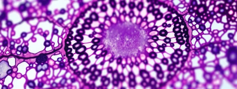
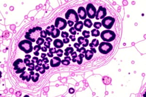
- Sections are stained to identify cell types and components (e.g., nucleus and cytoplasm).
- Examples of stains include haematoxylin & eosin (H&E), Masson's trichrome, and Wright Giemsa.
- Haematoxylin binds to acids, giving a blue/purple color.
- Eosin binds to alkali, producing a pink/orange color.
Ultrastructure
- Transmission electron microscopy (TEM) magnifies up to 1.5 million times, with a resolution of 0.14 nm (5x diameter of H atom).
- TEM uses a beam of electrons instead of light.
- Electron beam is produced by heating a tungsten filament.
- TEM optical system must be evacuated of air to prevent distortion.
- TEM uses multiple lenses to magnify the image produced by electrons interacting with the tissue section.
- Tissue is fixed with formaldehyde + glutaraldehyde + osmium tetroxide.
- Tissue is dehydrated.
- Tissue is embedded in resin and sectioned at 60-500 nm using an ultramicrotome with glass or diamond knife with water trough.
- Sections are mounted onto copper grids.
- Stained with electron-absorbing heavy metal salts (e.g., uranyl acetate & lead citrate) for contrast.
Four Main Tissue Types
- Epithelial Tissue: sheets of cells forming linings (externally – skin, internally – digestive, urogenital, and respiratory tracts, blood vessels); forms glands.
- Connective Tissue: provides support (physically – cartilage and bone, physiologically – blood); packing between tissues (adipose, fascia, ligaments, tendons). Types include: Connective Tissue Proper (ligaments, tendons, adipose tissue), Cartilage & Bone, and Blood.
- Muscle Tissue: three main types: skeletal (striated), smooth, and cardiac.
- Nervous Tissue: brain, spinal cord, nerves, and sensory receptors; enables information processing and responses from external and internal environments; consists of neurons (transmit impulses) and support cells.
Embryonic Germ Layers
- After fertilization and multiple cell divisions, gastrulation occurs (cell rearrangements).
- Germ layers: ectoderm (outer), mesoderm (middle), and endoderm (inner).
- Ectoderm gives rise to epidermis, nervous system, and other structures.
- Mesoderm gives rise to skeletal, muscular systems, and other structures.
- Endoderm gives rise to lining of digestive tract and other structures.
Why study histology?
- Veterinary Practice: determine tissue health (disease), fine-needle aspirates and biopsies (suspicious lumps), and pathology.
- Research: study development, gene function, and regenerative medicine.
Studying That Suits You
Use AI to generate personalized quizzes and flashcards to suit your learning preferences.


