Podcast
Questions and Answers
What are the key differences between hard drusen and soft drusen in the context of AMD progression?
What are the key differences between hard drusen and soft drusen in the context of AMD progression?
Hard drusen are small and well-defined with no significant visual symptoms, while soft drusen are larger and are associated with a greater risk of severe visual loss and progression to wet AMD.
How does the classification of drusen influence the progression of AMD according to NICE guidelines?
How does the classification of drusen influence the progression of AMD according to NICE guidelines?
The classification of drusen determines the risk of developing advanced stages of AMD, with many large soft drusen indicating a higher likelihood of atrophy and severe visual loss.
What are the two main classifications of late AMD as per NICE guidelines?
What are the two main classifications of late AMD as per NICE guidelines?
The two main classifications of late AMD are 'dry (geographic atrophy)' and 'wet (active or inactive)' AMD.
Describe the main clinical features of dry AMD.
Describe the main clinical features of dry AMD.
What percentage of individuals with dry AMD experience only mild or moderate vision loss?
What percentage of individuals with dry AMD experience only mild or moderate vision loss?
Identify two additional conditions that have been included under neovascular AMD in recent years.
Identify two additional conditions that have been included under neovascular AMD in recent years.
What role do pigmentary abnormalities play in the clinical features of dry AMD?
What role do pigmentary abnormalities play in the clinical features of dry AMD?
Why might individuals with hard drusen be classified as having 'Normal eyes' under NICE guidelines?
Why might individuals with hard drusen be classified as having 'Normal eyes' under NICE guidelines?
What are drusen and how do they relate to the classification of AMD?
What are drusen and how do they relate to the classification of AMD?
What do the NICE guidelines recommend regarding the referral of patients with AMD to ophthalmology?
What do the NICE guidelines recommend regarding the referral of patients with AMD to ophthalmology?
How is AMD classified, and what distinguishes dry AMD from wet AMD?
How is AMD classified, and what distinguishes dry AMD from wet AMD?
What are the symptoms of wet AMD compared to dry AMD?
What are the symptoms of wet AMD compared to dry AMD?
What role does visual acuity play in the management of AMD?
What role does visual acuity play in the management of AMD?
How does a patient's medical history influence the management of AMD?
How does a patient's medical history influence the management of AMD?
What are the implications of a family history of AMD for patient management?
What are the implications of a family history of AMD for patient management?
What investigations are recommended to assess a patient suspected of having AMD?
What investigations are recommended to assess a patient suspected of having AMD?
What are drusen, and how do they relate to age-related macular degeneration?
What are drusen, and how do they relate to age-related macular degeneration?
Describe the two main types of age-related macular degeneration.
Describe the two main types of age-related macular degeneration.
What does the NICE guideline state regarding risk factors for AMD?
What does the NICE guideline state regarding risk factors for AMD?
How does family history influence the risk of developing late AMD?
How does family history influence the risk of developing late AMD?
What is the significance of the retinal pigment epithelium (RPE) in the development of AMD?
What is the significance of the retinal pigment epithelium (RPE) in the development of AMD?
Define the term 'oxidative stress' in the context of AMD.
Define the term 'oxidative stress' in the context of AMD.
Which dietary components are associated with a lower risk of AMD according to current research?
Which dietary components are associated with a lower risk of AMD according to current research?
What role does light exposure play in the risk of AMD?
What role does light exposure play in the risk of AMD?
Explain the classification of age-related macular degeneration.
Explain the classification of age-related macular degeneration.
What are some lifestyle factors that have a protective effect against AMD?
What are some lifestyle factors that have a protective effect against AMD?
What imaging technique is commonly used to visualize AMD abnormalities, and how does it work?
What imaging technique is commonly used to visualize AMD abnormalities, and how does it work?
Identify the primary clinical findings that characterize age-related macular degeneration.
Identify the primary clinical findings that characterize age-related macular degeneration.
Discuss the connection between hypertension and AMD.
Discuss the connection between hypertension and AMD.
List two potential inflammatory response elements involved in the development of AMD.
List two potential inflammatory response elements involved in the development of AMD.
Flashcards are hidden until you start studying
Study Notes
Age-related Macular Degeneration (AMD)
- AMD is a progressive disease that affects the macula, the central area of the retina responsible for detailed and color vision.
- The macula is located in the central part of the eye, and it’s responsible for our detailed vision.
- Two main types of AMD are dry AMD (non-exudative, non-neovascular) and wet AMD (exudative, neovascular).
Dry AMD
- Most common form of AMD (90%).
- Progresses slower than wet AMD and usually affects both eyes.
- The advanced stage of dry AMD is called geographic atrophy (GA).
- Characterized by the build-up of debris under the retina causing drusen formation and degeneration of the Retinal Pigment Epithelium (RPE).
- Slow and progressive thinning and degeneration of the photoreceptors and the RPE gradually leads to failure of central vision.
- 90% of people with dry AMD will only have mild or moderate gradual visual loss.
- Despite being the less aggressive form, it still causes about 20% of AMD-related severe visual loss.
Wet AMD
- Less common than dry AMD.
- Progresses more rapidly, leading to advanced sight loss.
- Characterized by Choroidal Neovascularization (CNV) and Pigment Epithelium Detachment (PED) as primary manifestations.
- In recent years, Retinal Angiomatous Proliferation (RAP) and Polypoidal Choroidal Vasculopathy (PCV) were included under the umbrella of neovascular AMD.
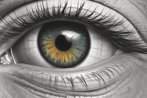
Drusen
- A clinical feature of dry AMD.
- Yellow spots around the macula.
- People with many large soft drusen are more likely to develop atrophy and severe visual loss.
- They are at greater risk of developing wet AMD.
- Most people over the age of 40 years have at least one druse.
Hard Drusen
- Small and well defined.
- In early stages, patients are likely to have no visual symptoms.
- Even in cases with obvious and widespread hard drusen, people may not experience any visual symptoms.
- Using the NICE guidance [NG82] classification, a person with hard drusen and no other anomalies would be classified as 'Normal eyes'.
Symptoms
- Important in guiding the investigations and management of a person with AMD.
- Wet AMD:
- Sudden onset of reduced vision or distortion.
- Some types of wet AMD can develop slowly over a few months.
- Dry AMD:
- Slower progression and more gradual, usually bilateral, reduced vision.
- Patients may report a significant change in vision if they develop wet AMD.
Why Need to Know About AMD?
- You will encounter patients with age-related macular degeneration.
- Age-related macular degeneration can have a significant impact on a patient's vision and quality of life.
- Important to identify the different types of age-related macular degeneration.
- It’s important to understand the clinical presentation, risk factors, and potential complications associated with AMD.
Risk Factors
-
Multi-factorial causes including:
- Genetically susceptible individuals.
- Increasing age.
- Increasing vulnerability due to oxidative stress.
-
Other risk factors:
- Older age.
- Presence of AMD in the other eye.
- Family history of AMD.
- Smoking.
- Hypertension.
- BMI of 30 kg/m2 or higher.
- Diet low in omega 3 and 6, vitamins, carotenoid and minerals.
- Diet high in fat.
- Lack of exercise.
Retinal Pigment Epithelium (RPE)
- Highly pigmented layer of the retina.
- Sits between the neurosensory retina and Bruch's membrane.
- Maintains the photoreceptors by:
- Absorbing stray light.
- Forming the outer blood retinal barrier.
- Regenerating the visual pigment.
Anatomy for AMD Evaluation
- Retina: Layered structure with ganglion (nerve cells) nearest the vitreous and photoreceptors rods and cones nearest the choroid.
- Choroid: Sandwiched between sclera and retina; provides 2/3 nutrition to the retina and RPE.
- Macula: Highest concentration of photoreceptors, responsible for high central resolution, color vision, and detailed vision. It has a diameter of around 5mm and is horizontally oval.
- Foveola: The central floor of the macula, with a diameter of 0.35mm, is the thinnest part of the retina. The entire thickness consists only of cone photoreceptors, providing the most acute vision.
- Bruch’s membrane: Located in between the choroid and the RPE, involved in the controlled passage of nutrients and waste products to and from the retina.
- Choroid: Sits below Bruch's membrane and is made up of choroidal vessels that supply the outer retina.
Optical Coherence Tomography (OCT)
- Non-invasive and non-contact imaging system.
- Provides high-resolution cross-sectional images.
- Measures light reflectance of structures.
- High reflectivity structures appear red, intermediate reflectivity appear green-yellow, and low reflectivity appear blue-black in a pseudo-color image.
Aetiology
- Part of the aging process, where undigested metabolic debris and lipofuscin accumulate beneath the retina.
- Accumulation occurs between Bruch's membrane and the RPE.
- In younger healthy eyes, the debris is removed, but in older eyes, it is not possible to be removed and forms drusen.
- There may also be an inflammatory response to this process.
Additional Important Points
- Family history: Patients with a family history of AMD are around 4 to 6 times more likely to develop late AMD than those without such family history.
- Smoking: Those who smoke are 2 to 3 times more likely to develop late AMD than those who do not smoke. Stopping smoking lowers the relative risk of AMD.
- Hypertension: An association between hypertension and late AMD exists.
- BMI and exercise: BMI of 30 kg/m2 or higher and lack of exercise are risk factors for AMD. Active lifestyle is shown to have a protective effect against developing wet AMD.
- Nutrition: Low intake of omega 3 and 6, vitamins, carotenoid and minerals, and a diet high in fat increase the risk of AMD.
- Light exposure: No significant evidence suggests that light exposure increases the risk of AMD.
- Race: Previous studies suggested that Caucasians are at greater risk of AMD than people of African American ethnicity. This is not included as a risk factor in current guidelines.
Studying That Suits You
Use AI to generate personalized quizzes and flashcards to suit your learning preferences.