Podcast
Questions and Answers
What is the primary cause of Acute Coronary Syndromes (ACS)?
What is the primary cause of Acute Coronary Syndromes (ACS)?
- Hypertension
- Hyperlipidemia
- Atherosclerosis (correct)
- Diabetes
What is the atheroma composed of in atherosclerosis?
What is the atheroma composed of in atherosclerosis?
- Fatty lipid-rich gruel (correct)
- Tunica intima
- Collagen fibers
- Platelet aggregations
What is the outcome if a blood clot occludes the entire coronary artery?
What is the outcome if a blood clot occludes the entire coronary artery?
- Myocardial infarction (heart attack) (correct)
- Hypertension
- Cardiac arrhythmia
- Angina pectoris
Which of the following is NOT a common symptom of ACS?
Which of the following is NOT a common symptom of ACS?
What is anginal equivalence in ACS?
What is anginal equivalence in ACS?
What is the primary purpose of a 12-lead ECG in diagnosing ACS?
What is the primary purpose of a 12-lead ECG in diagnosing ACS?
What increases the pre-test probability of ACS?
What increases the pre-test probability of ACS?
What is the clinical event responsible for triggering ACS?
What is the clinical event responsible for triggering ACS?
What is one of the primary goals of initial management in acute STEMI?
What is one of the primary goals of initial management in acute STEMI?
What is a key aspect of identifying acute inferior STEMI on a 12-lead ECG?
What is a key aspect of identifying acute inferior STEMI on a 12-lead ECG?
What is a benefit of aspirin administration in ACS patients?
What is a benefit of aspirin administration in ACS patients?
What is a characteristic of a patient with right ventricular infarction?
What is a characteristic of a patient with right ventricular infarction?
What is a sign of posterior infarction on a 12-lead ECG?
What is a sign of posterior infarction on a 12-lead ECG?
What can cause ST elevation mimicking STEMI?
What can cause ST elevation mimicking STEMI?
What is a limitation of the STEMI/Non-STEMI framework?
What is a limitation of the STEMI/Non-STEMI framework?
What can help identify changes and dynamic myocardial oxygen supply vs. demand?
What can help identify changes and dynamic myocardial oxygen supply vs. demand?
What is a reciprocal change seen in lead AVL when identifying acute inferior STEMI?
What is a reciprocal change seen in lead AVL when identifying acute inferior STEMI?
What can nitroglycerin administration do in patients with right ventricular infarction?
What can nitroglycerin administration do in patients with right ventricular infarction?
Study Notes
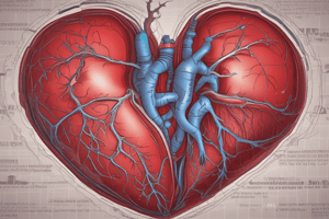
Acute Coronary Syndromes (ACS)
Underlying Pathology
- ACS is primarily caused by atherosclerosis, a disease that affects coronary arteries
- Atherosclerosis consists of two main components: atheroma (fatty lipid-rich gruel) and tunica intima (innermost lining of coronary arteries)
- Atheroma grows within the walls of the tunica intima and can cause a pathological thickening of the tunica intima, separating it from circulating blood
- The atheroma's contents are highly thrombogenic, meaning they can trigger a blood clot if exposed to circulating blood
Clinical Event Triggering ACS
- The clinical event responsible for triggering ACS is often plaque rupture, which exposes the atheroma to circulating blood
- Platelets are activated, leading to platelet aggregation and the formation of a blood clot
- If the clot occludes the entire coronary artery, it can cause a myocardial infarction (heart attack)
Signs and Symptoms of ACS
- Chest discomfort or pain (poorly localized, often in the center of the chest, lasting 15 minutes or more)
- Radiation of pain to the arm, neck, back, or jaw
- Shortness of breath, diaphoresis, nausea, vomiting, palpitations, and anxiety or sense of impending doom
- Denial or minimization of symptoms due to fear of hospital bills or previous misdiagnosis
Anginal Equivalence
- ACS can present with anginal equivalence, such as:
- Jaw, neck, ear, or arm pain
- New-onset exertional dyspnea
- Palpitations without chest discomfort
- Syncope (fainting)
- Unexplained fatigue, especially in diabetics
Pre-Test Probability and 12-Lead ECG
- The pre-test probability of ACS is high if the patient presents with typical chest discomfort and signs of acute illness
- A 12-lead ECG is essential in diagnosing ACS, with a focus on:
- ST segment elevation ( STEMI) or depression (NSTEMI)
- Q waves and cardiac biomarkers
- Serial ECGs to monitor changes and dynamic myocardial oxygen supply vs. demand
Goals of Initial Management
- Restore balance between myocardial oxygen supply and demand
- Optimize blood pressure and oxygen supply
- Provide pain control and anxiety relief
- Prepare for reperfusion therapy (PCI or fibrinolytics)
- Defibrillate if necessary
Identifying Acute STEMI on 12-Lead ECG
- Look for ST segment elevation in two or more anatomically contiguous leads
- Consider pre-test probability and rule of proportionality (repolarization proportional to depolarization)
- Normal ECG understanding is essential for recognizing abnormal patterns
- Serial ECGs can help identify changes and dynamic myocardial oxygen supply vs. demand
Secondary Prevention and Reperfusion Therapy
- Aspirin administration has a 25% mortality benefit in ACS patients
- Nitroglycerin is a potent coronary vasodilator that can help restore balance between myocardial oxygen supply and demand
- Reperfusion therapy (PCI or fibrinolytics) is crucial in preventing irreversible heart damage
- Post-PCI outcome data can help improve systems of care and reduce first medical contact to balloon time### Identifying Acute Inferior STEMI
- Criteria for sinus rhythm: p waves, QRS complexes in a one-to-one relationship, constant PR interval between 120-200 milliseconds
- In lead 2, PR interval measures 132 milliseconds, indicating sinus rhythm borderline sinus bradycardia at a rate of 60
- Presence of ST segment elevation in leads 2, 3, and AVF, indicating acute inferior STEMI
- Reciprocal change in lead AVL (down-sloping ST segment) supports the diagnosis of acute inferior STEMI
Reciprocal Changes
- Inferior leads (2, 3, and AVF) are reciprocal to high lateral leads (1 and AVL)
- Right precordial leads (V1, V2, and V3) are reciprocal to the posterior wall of the left ventricle
- Look for reciprocal changes in the right precordial leads when identifying acute inferior STEMI to potentially spot acute isolated posterior STEMI
Right Ventricular Infarction
- Can be a complication of acute inferior STEMI, especially with proximal occlusion of the right coronary artery
- Patients with right ventricular infarction are preload dependent, relying on central venous pressure to maintain cardiac output
- Nitroglycerin can drop central venous pressure, exacerbating the condition
- Suspect right ventricular infarction in patients with acute inferior STEMI, especially if the culprit artery is the right coronary artery
Case Study
-
79-year-old female with chest discomfort, history of previous heart attack
-
Initial 12-lead ECG shows sinus rhythm, ST elevation in leads 2, 3, and AVF, and reciprocal change in lead AVL, indicating acute inferior STEMI
-
No reciprocal changes in the right precordial leads; however, a modified lead V4R (placed on the right side of the chest) shows ST elevation, indicating right ventricular infarction
-
Initial blood pressure was 100/55, which dropped to 90/40 after a trial dose of nitroglycerin, highlighting the importance of caution when administering nitroglycerin in such cases### Recognizing Myocardial Infarction Patterns on the ECG
-
Worsening ST elevation in lead V1 indicates right ventricular infarction
-
Extensive anterior ST elevation in leads V1-V6 suggests left anterior descending (LAD) artery occlusion
-
Reciprocal ST changes in inferior leads with anterior ST elevation help confirm diagnosis
-
High lateral ST elevation may be subtle, look for reciprocal changes in lead III to identify
-
Inferior lead ST elevation with reciprocal changes in aVL indicates lateral wall infarction
Isolated Posterior Infarction
- Posterior infarction can present without obvious ST elevation on a 12-lead ECG
- Look for isolated ST depression in right precordial leads (V1-V3) as a sign of posterior infarction
- The "carousel pony" sign - tall R waves and down-upsloping ST depression in V2 - can indicate posterior involvement
- Flipping the ECG over to view a "mirror image" can reveal posterior ST elevation
Mimics of Acute MI
- Left bundle branch block, pacemaker rhythms, and left ventricular hypertrophy can all cause ST elevation mimicking STEMI
- These represent secondary ST-T wave changes from abnormal ventricular depolarization, not acute ischemia
- Differentiating these from true STEMI requires assessing concordance of ST changes with QRS, clinical presentation, and serial ECG changes
Limitations of the STEMI/Non-STEMI Framework
- The current STEMI/Non-STEMI paradigm misses some patients with occluded arteries who do not meet strict ST elevation criteria
- Examples include hyperacute T-waves, de Winter T-waves, and isolated posterior infarction
- A shift to an "occlusion MI/non-occlusion MI" framework may better identify all patients who would benefit from emergent reperfusion therapy
Acute Coronary Syndromes (ACS)
- ACS is primarily caused by atherosclerosis, which affects coronary arteries
- Atherosclerosis consists of atheroma (fatty lipid-rich gruel) and tunica intima (innermost lining of coronary arteries)
Underlying Pathology
- Atheroma grows within the walls of the tunica intima, causing a pathological thickening that separates it from circulating blood
- Atheroma's contents are highly thrombogenic, triggering blood clots when exposed to circulating blood
Clinical Event Triggering ACS
- Plaque rupture exposes atheroma to circulating blood, leading to platelet activation, aggregation, and blood clot formation
- If the clot occludes the entire coronary artery, it can cause a myocardial infarction (heart attack)
Signs and Symptoms of ACS
- Chest discomfort or pain, often poorly localized and lasting 15 minutes or more
- Radiation of pain to the arm, neck, back, or jaw
- Shortness of breath, diaphoresis, nausea, vomiting, palpitations, and anxiety or sense of impending doom
Anginal Equivalence
- ACS can present with anginal equivalence, such as:
- Jaw, neck, ear, or arm pain
- New-onset exertional dyspnea
- Palpitations without chest discomfort
- Syncope (fainting)
- Unexplained fatigue, especially in diabetics
Pre-Test Probability and 12-Lead ECG
- High pre-test probability of ACS requires typical chest discomfort and signs of acute illness
- 12-lead ECG is essential in diagnosing ACS, focusing on:
- ST segment elevation (STEMI) or depression (NSTEMI)
- Q waves and cardiac biomarkers
- Serial ECGs to monitor changes and dynamic myocardial oxygen supply vs. demand
Goals of Initial Management
- Restore balance between myocardial oxygen supply and demand
- Optimize blood pressure and oxygen supply
- Provide pain control and anxiety relief
- Prepare for reperfusion therapy (PCI or fibrinolytics)
- Defibrillate if necessary
Identifying Acute STEMI on 12-Lead ECG
- Look for ST segment elevation in two or more anatomically contiguous leads
- Consider pre-test probability and rule of proportionality (repolarization proportional to depolarization)
- Normal ECG understanding is essential for recognizing abnormal patterns
- Serial ECGs can help identify changes and dynamic myocardial oxygen supply vs. demand
Secondary Prevention and Reperfusion Therapy
- Aspirin administration has a 25% mortality benefit in ACS patients
- Nitroglycerin is a potent coronary vasodilator that can help restore balance between myocardial oxygen supply and demand
- Reperfusion therapy (PCI or fibrinolytics) is crucial in preventing irreversible heart damage
- Post-PCI outcome data can help improve systems of care and reduce first medical contact to balloon time
Identifying Acute Inferior STEMI
- Criteria for sinus rhythm: p waves, QRS complexes in a one-to-one relationship, constant PR interval between 120-200 milliseconds
- Presence of ST segment elevation in leads 2, 3, and AVF, indicating acute inferior STEMI
- Reciprocal change in lead AVL (down-sloping ST segment) supports the diagnosis of acute inferior STEMI
Reciprocal Changes
- Inferior leads (2, 3, and AVF) are reciprocal to high lateral leads (1 and AVL)
- Right precordial leads (V1, V2, and V3) are reciprocal to the posterior wall of the left ventricle
- Look for reciprocal changes in the right precordial leads when identifying acute inferior STEMI to potentially spot acute isolated posterior STEMI
Right Ventricular Infarction
- Can be a complication of acute inferior STEMI, especially with proximal occlusion of the right coronary artery
- Patients with right ventricular infarction are preload dependent, relying on central venous pressure to maintain cardiac output
- Nitroglycerin can drop central venous pressure, exacerbating the condition
- Suspect right ventricular infarction in patients with acute inferior STEMI, especially if the culprit artery is the right coronary artery
Recognizing Myocardial Infarction Patterns on the ECG
- Worsening ST elevation in lead V1 indicates right ventricular infarction
- Extensive anterior ST elevation in leads V1-V6 suggests left anterior descending (LAD) artery occlusion
- Reciprocal ST changes in inferior leads with anterior ST elevation help confirm diagnosis
- High lateral ST elevation may be subtle, look for reciprocal changes in lead III to identify
- Inferior lead ST elevation with reciprocal changes in aVL indicates lateral wall infarction
Isolated Posterior Infarction
- Posterior infarction can present without obvious ST elevation on a 12-lead ECG
- Look for isolated ST depression in right precordial leads (V1-V3) as a sign of posterior infarction
- The "carousel pony" sign - tall R waves and down-upsloping ST depression in V2 - can indicate posterior involvement
- Flipping the ECG over to view a "mirror image" can reveal posterior ST elevation
Mimics of Acute MI
- Left bundle branch block, pacemaker rhythms, and left ventricular hypertrophy can cause ST elevation mimicking STEMI
- These represent secondary ST-T wave changes from abnormal ventricular depolarization, not acute ischemia
- Differentiating these from true STEMI requires assessing concordance of ST changes with QRS, clinical presentation, and serial ECG changes
Limitations of the STEMI/Non-STEMI Framework
- The current STEMI/Non-STEMI paradigm misses some patients with occluded arteries who do not meet strict ST elevation criteria
- Examples include hyperacute T-waves, de Winter T-waves, and isolated posterior infarction
- A shift to an "occlusion MI/non-occlusion MI" framework may better identify all patients who would benefit from emergent reperfusion therapy
Studying That Suits You
Use AI to generate personalized quizzes and flashcards to suit your learning preferences.
Description
This quiz covers the underlying pathology of Acute Coronary Syndromes (ACS), including atherosclerosis, atheroma, and tunica intima. Learn about the causes and effects of ACS on coronary arteries.