Identify the anatomical structures labeled in the neck diagram.
Understand the Problem
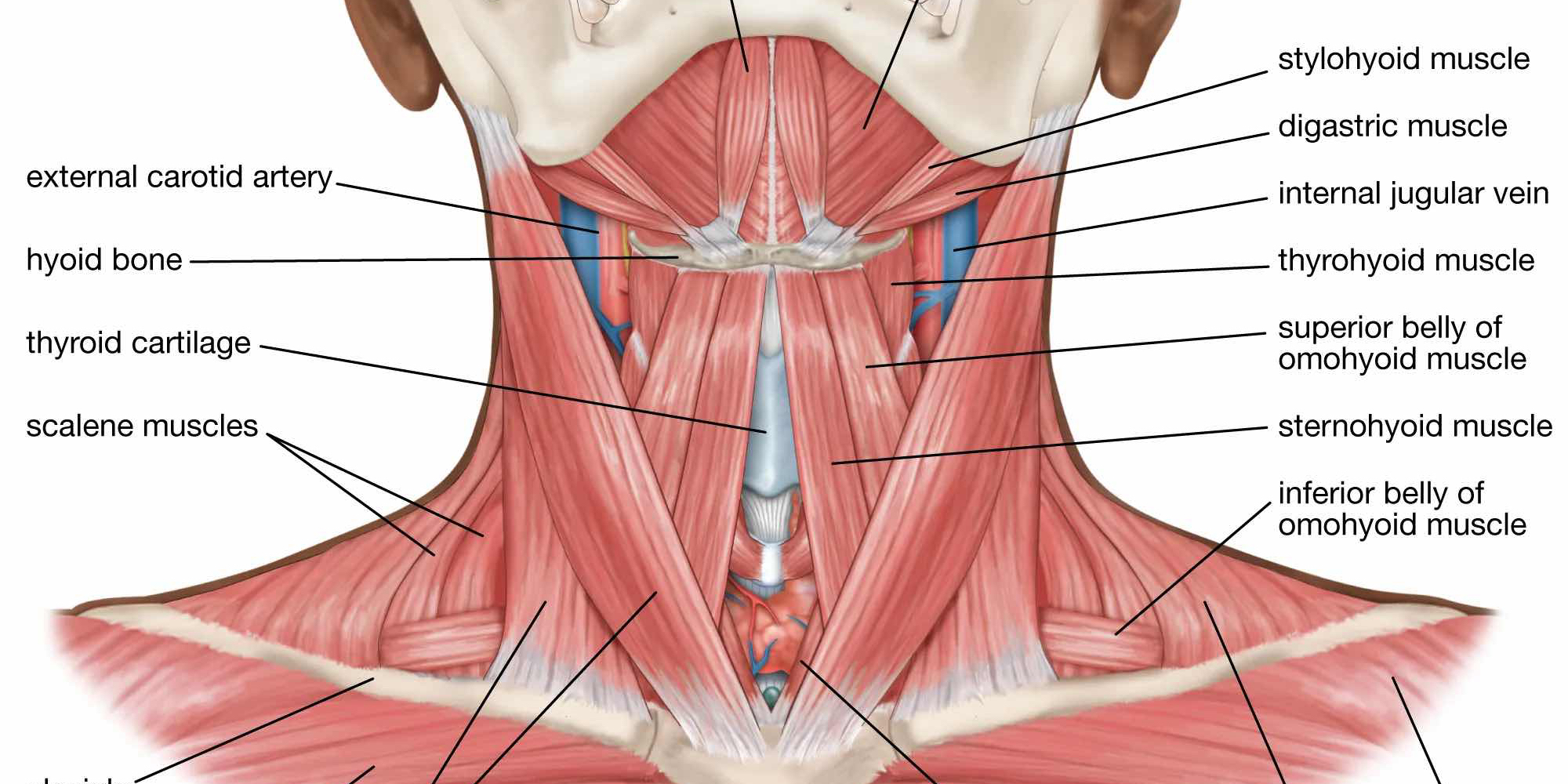
The image shows an anatomical illustration of the human neck, specifically detailing various muscles, arteries, veins, and cartilages. The question is likely asking to identify or understand the relationships between these labeled structures.
Answer
The diagram identifies neck structures including arteries, bones, cartilages, and an assortment of muscles.
The labels in the neck diagram identify the following structures: external carotid artery, hyoid bone, thyroid cartilage, scalene muscles, stylohyoid muscle, digastric muscle, internal jugular vein, thyrohyoid muscle, superior belly of omohyoid muscle, sternohyoid muscle, and inferior belly of omohyoid muscle.
Answer for screen readers
The labels in the neck diagram identify the following structures: external carotid artery, hyoid bone, thyroid cartilage, scalene muscles, stylohyoid muscle, digastric muscle, internal jugular vein, thyrohyoid muscle, superior belly of omohyoid muscle, sternohyoid muscle, and inferior belly of omohyoid muscle.
More Information
The neck contains essential structures such as muscles, glands, vessels, and vertebrae crucial for movement, support, and various physiological functions.
Tips
Carefully observe the direction of the labels and match them to the corresponding structure in the image. Pay attention to the different muscle layers and their names.
Sources
- Neck Anatomy: Muscles, glands, organs - Kenhub - kenhub.com
- Anatomy of the Throat and Neck | Dr. Larian - hyperparathyroidmd.com
- Neck Muscles: Labeled Diagrams and Pain Generators - verywellhealth.com
AI-generated content may contain errors. Please verify critical information