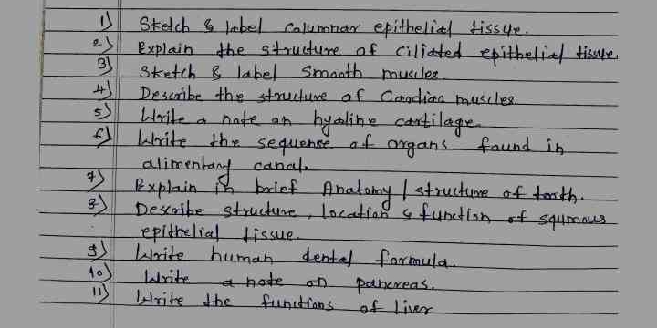
1. Sketch & label columnar epithelial tissue. 2. Explain the structure of ciliated epithelial tissue. 3. Sketch & label smooth muscles. 4. Describe the structure of cardiac muscles... 1. Sketch & label columnar epithelial tissue. 2. Explain the structure of ciliated epithelial tissue. 3. Sketch & label smooth muscles. 4. Describe the structure of cardiac muscles. 5. Write a note on hyaline cartilage. 6. Write the sequence of organs found in the alimentary canal. 7. Explain a brief anatomy/structure of teeth. 8. Describe structure, location & function of squamous epithelial tissue. 9. Write human dental formula. 10. Write a note on pancreases. 11. Write the functions of liver.

Understand the Problem
The question consists of multiple tasks related to anatomy and physiology, including sketches and explanations of various biological structures and functions.
Answer
Sketches and explanations for columnar and ciliated epithelial tissue, smooth and cardiac muscles, hyaline cartilage, alimentary sequence, teeth, squamous epithelial tissue, dental formula, pancreas note, and liver functions.
-
Sketch columnar epithelial tissue, showing tall, column-like cells. Label the nucleus, cytoplasm, and cell membrane.
-
Ciliated epithelial tissue has tall columnar cells with hair-like cilia on their free surfaces for movement of particles.
-
Draw elongated, spindle-shaped smooth muscle fibers. Label the nucleus and cytoplasm.
-
Cardiac muscles have striations and intercalated discs, located in the heart. They function to pump blood.
-
Hyaline cartilage is a firm tissue providing support and flexibility, found in joints.
-
Alimentary canal sequence: mouth, pharynx, esophagus, stomach, small intestine, large intestine, anus.
-
Teeth anatomy: crowns, roots, and enamel; function in food digestion.
-
Squamous epithelial tissue: flat, thin cells; located in areas like alveoli; function in protection and permeability.
-
Human dental formula: 2-1-2-3/2-1-2-3 (incisors, canines, premolars, molars).
-
Pancreas produces digestive enzymes and hormones like insulin.
-
Liver functions include detoxification, protein synthesis, and bile production.
Answer for screen readers
-
Sketch columnar epithelial tissue, showing tall, column-like cells. Label the nucleus, cytoplasm, and cell membrane.
-
Ciliated epithelial tissue has tall columnar cells with hair-like cilia on their free surfaces for movement of particles.
-
Draw elongated, spindle-shaped smooth muscle fibers. Label the nucleus and cytoplasm.
-
Cardiac muscles have striations and intercalated discs, located in the heart. They function to pump blood.
-
Hyaline cartilage is a firm tissue providing support and flexibility, found in joints.
-
Alimentary canal sequence: mouth, pharynx, esophagus, stomach, small intestine, large intestine, anus.
-
Teeth anatomy: crowns, roots, and enamel; function in food digestion.
-
Squamous epithelial tissue: flat, thin cells; located in areas like alveoli; function in protection and permeability.
-
Human dental formula: 2-1-2-3/2-1-2-3 (incisors, canines, premolars, molars).
-
Pancreas produces digestive enzymes and hormones like insulin.
-
Liver functions include detoxification, protein synthesis, and bile production.
More Information
Human dental formula indicates the arrangement of teeth types, useful for comparative anatomy.
Sources
- Epithelial Tissue | Anatomy and Physiology I - courses.lumenlearning.com
- Types of muscle tissue: MedlinePlus Medical Encyclopedia Image - medlineplus.gov
- Tissue Types | BIO103: Human Biology - Courses.lumenlearning.com. - courses.lumenlearning.com
AI-generated content may contain errors. Please verify critical information