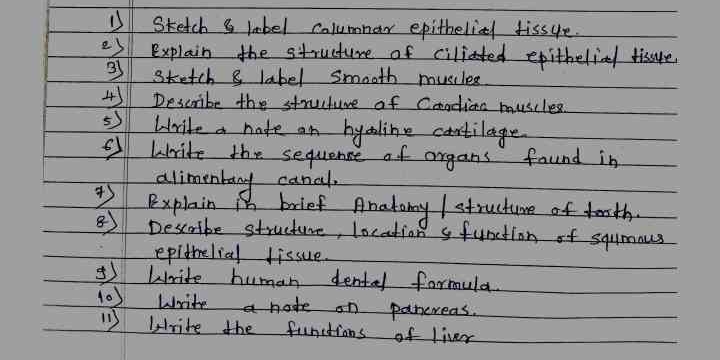
1. Sketch & label Columnar epithelial tissue. 2. Explain the structure of ciliated epithelial tissue. 3. Sketch & label Smooth muscle. 4. Describe the structure of Cardiac muscles.... 1. Sketch & label Columnar epithelial tissue. 2. Explain the structure of ciliated epithelial tissue. 3. Sketch & label Smooth muscle. 4. Describe the structure of Cardiac muscles. 5. Write a note on hyaline cartilage. 6. Write the sequence of organs found in alimentary canal. 7. Explain in brief Anatomy/structure of teeth. 8. Describe structure, location & function of squamous epithelial tissue. 9. Write human dental formula. 10. Write a note on pancreas. 11. Write the functions of liver.

Understand the Problem
The question consists of multiple tasks related to biology, including sketching, explaining structures, and describing functions of various tissues and organs.
Answer
1. Sketch: Columnar epithelial cells. 2. Cilia movement on tissue surface. 3. Sketch: Smooth muscle tissue. 4. Cardiac: Striated, branched, intercalated discs. 5. Hyaline: Joint-supportive cartilage. 6. Alimentary: Mouth to anus organs. 7. Teeth: Enamel, dentin, pulp. 8. Squamous: Flat protective layers. 9. Dental: 2-1-2-3. 10. Pancreas: Endocrine/exocrine functions. 11. Liver: Detox, bile, metabolism.
- Columnar epithelial tissue: tall, column-like cells with nuclei at the base; can be ciliated. 2. Ciliated epithelial tissue: columnar cells with cilia on the surface, found in respiratory tract. 3. Smooth muscle: spindle-shaped, non-striated, involuntary control. 4. Cardiac muscles: striated, branched, involuntary, interconnected by intercalated discs. 5. Hyaline cartilage: translucent cartilage found on joint surfaces, supportive tissue. 6. Alimentary canal: mouth, esophagus, stomach, small intestine, large intestine, anus. 7. Anatomy of teeth: crown, neck, root; made from enamel, dentin, pulp. 8. Squamous epithelial tissue: flat and thin cells, found in the lining of organs and vessels, protective barrier. 9. Human dental formula: 2-1-2-3/2-1-2-3 (incisors, canines, premolars, molars). 10. Pancreas note: dual-function gland, produces insulin and digestive enzymes. 11. Liver functions: detoxification, protein synthesis, bile production, metabolism of lipids and carbohydrates.
Answer for screen readers
- Columnar epithelial tissue: tall, column-like cells with nuclei at the base; can be ciliated. 2. Ciliated epithelial tissue: columnar cells with cilia on the surface, found in respiratory tract. 3. Smooth muscle: spindle-shaped, non-striated, involuntary control. 4. Cardiac muscles: striated, branched, involuntary, interconnected by intercalated discs. 5. Hyaline cartilage: translucent cartilage found on joint surfaces, supportive tissue. 6. Alimentary canal: mouth, esophagus, stomach, small intestine, large intestine, anus. 7. Anatomy of teeth: crown, neck, root; made from enamel, dentin, pulp. 8. Squamous epithelial tissue: flat and thin cells, found in the lining of organs and vessels, protective barrier. 9. Human dental formula: 2-1-2-3/2-1-2-3 (incisors, canines, premolars, molars). 10. Pancreas note: dual-function gland, produces insulin and digestive enzymes. 11. Liver functions: detoxification, protein synthesis, bile production, metabolism of lipids and carbohydrates.
More Information
Ciliated tissues help move particles out of the body, found in areas like the respiratory tract. Smooth muscles are crucial for involuntary body movements, such as in the digestive system. The liver performs numerous vital functions, including bile production essential for digestion.
Tips
Mixing up epithelial tissue types; each has specific locations and functions.
Sources
- Epithelial Tissue | Anatomy and Physiology I - courses.lumenlearning.com
- Types of tissue: Structure and function - Kenhub - kenhub.com
- Epithelium: What It Is, Function & Types - Cleveland Clinic - my.clevelandclinic.org
AI-generated content may contain errors. Please verify critical information