Podcast
Questions and Answers
What is the primary reason why animals have a wider peripheral vision than humans?
What is the primary reason why animals have a wider peripheral vision than humans?
- Because their visual fields of each eye completely overlap
- Because they have a more efficient pupillary light reflex
- Because they have more developed binocular vision
- Because their visual fields of each eye do not completely overlap (correct)
Which nerve controls the lateral rectus muscle of the eye?
Which nerve controls the lateral rectus muscle of the eye?
- Olfactory nerve (facial nerve 1)
- Trochlear nerve (facial nerve 4)
- Abducens nerve (facial nerve 6) (correct)
- Vestibulocochlear nerve (facial nerve 8)
What is the primary function of the area of centralis?
What is the primary function of the area of centralis?
- To regulate the pupillary light reflex
- To enable binocular vision
- To provide the greatest acuity (correct)
- To provide peripheral vision
What type of vision do dogs have only in the area directly in front of them?
What type of vision do dogs have only in the area directly in front of them?
Which of the following is a characteristic of prey species' eyes?
Which of the following is a characteristic of prey species' eyes?
Choose the correct statement.
Choose the correct statement.
Which nerve controls the superior oblique muscle of the eye?
Which nerve controls the superior oblique muscle of the eye?
What is the main function of horizontal cells in the retina?
What is the main function of horizontal cells in the retina?
Which type of cells primarily transmit signals vertically from the rods, cones, and horizontal cells to ganglion and amacrine cells?
Which type of cells primarily transmit signals vertically from the rods, cones, and horizontal cells to ganglion and amacrine cells?
What is the primary function of amacrine cells in the retina?
What is the primary function of amacrine cells in the retina?
What is the primary neurotransmitter released by ganglionic cells in the retina?
What is the primary neurotransmitter released by ganglionic cells in the retina?
What is the primary purpose of the fovea in the retina?
What is the primary purpose of the fovea in the retina?
What is unique about the structure of the fovea compared to the rest of the retina?
What is unique about the structure of the fovea compared to the rest of the retina?
What suspends the lens in the eye?
What suspends the lens in the eye?
Where is the lens located in the eye?
Where is the lens located in the eye?
What is the primary function of the ciliary body in the eye?
What is the primary function of the ciliary body in the eye?
What is the main purpose of the choroid in the eye?
What is the main purpose of the choroid in the eye?
What is the term for the enlargement of the pupil in the dark?
What is the term for the enlargement of the pupil in the dark?
What is the function of the tapetum lucidum in the eye?
What is the function of the tapetum lucidum in the eye?
What type of muscle cells are found in the pupillary dilator muscles?
What type of muscle cells are found in the pupillary dilator muscles?
What is the function of the iris in the eye?
What is the function of the iris in the eye?
What is the term for the reduction of the pupil size in bright light?
What is the term for the reduction of the pupil size in bright light?
What is the location of the tapetum lucidum in the eye?
What is the location of the tapetum lucidum in the eye?
What is the result of pupillary sphincter muscle contraction?
What is the result of pupillary sphincter muscle contraction?
What is the function of the optic nerve?
What is the function of the optic nerve?
What is the main function of rods in the retina?
What is the main function of rods in the retina?
What is the role of the outer segment in photoreceptors?
What is the role of the outer segment in photoreceptors?
What is the primary difference between rod cells and cone cells?
What is the primary difference between rod cells and cone cells?
What is the consequence of severe vitamin A deficiency?
What is the consequence of severe vitamin A deficiency?
How many types of cone cells are present in primates?
How many types of cone cells are present in primates?
What is the wavelength of the red sensitive pigment in cone cells?
What is the wavelength of the red sensitive pigment in cone cells?
What is the characteristic of most mammals in terms of color perception?
What is the characteristic of most mammals in terms of color perception?
What is the proportion of rod cells in the retina?
What is the proportion of rod cells in the retina?
Flashcards are hidden until you start studying
Study Notes
Visual System
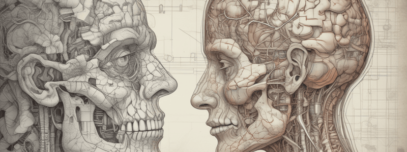
- Vision is an integral part of neural function, and the eyes are an extension of the brain.
- Animals have a wider peripheral vision than humans because their visual fields of each eye do not completely overlap.
Visual Field
- The visual field is the spatial area seen with one eye.
- The extent of overlap of the visual field depends on the anatomical placement of the eyes.
- Prey species have laterally oriented eyes, giving them a wider view.
- Dogs have binocular vision only in the area directly in front of them.
- Monocular vision does not provide good depth perception.
Eye Structure
Fovea
- The fovea is a minute area in the center of the retina, which is especially capable of acute and detailed vision.
- In animals, it is called the "area centralis".
- The fovea is composed almost entirely of cones, and their special long and slender bodies aid their detection in visual images.
- The more cones present, the more colors that can be interpreted.
Lens
- The lens is located behind the iris.
- The lens is suspended by the suspensory ligaments, and these fibers are attached to the ciliary body.
- The ciliary body is a muscular structure that helps with accommodation of the lens.
Choroid
- The choroid consists of loose connective tissue with numerous vasculature and pigmented cells, and it serves a nutritive function.
- Some diurnal animals have melanocytes that absorb light that has passed by the photoreceptors without stimulating them.
- Nocturnal and most domestic mammals have a patch of reflective material that is called the "tapetum lucidum".
Iris
- The iris is a diaphragm muscle that can contract/relax to control the rays of light that go into the eye via modification of the pupil diameter.
- In the dark, pupils will dilate (mydriasis).
- In the light, pupils will contract (miosis).
- The iris has dilator and sphincter muscles.
Pupillary Dilator Muscles
- The pupillary dilator muscles are radially arranged and oppose the action of the sphincter.
- The pupillary dilator muscles are apart of the pigmented anterior epithelial cells, known as myoepithelial cells, which are composed of smooth muscle.
- Its contraction results in pupillary dilation (mydriasis).
Pupillary Sphincter Muscles
- Pupillary sphincter muscles are circularly arranged near the pupillary margin that are innervated by parasympathetic fibers.
- Their contraction results in decreased pupillary size (miosis).
Eye Structure: Optic Nerve
- The axons leaving the eye at the optic disk give rise to the optic nerve (cranial nerve 2).
- There are more axons in both optic nerves than in all the dorsal roots of the spinal cord.
Photoreceptors (Rods and Cones)
- There are about 130 million photoreceptor cells in the retina, which can either be cones or rods.
- Rod and cone cells, each have an outer and inner segment.
- The outer segment is the photosensitive region.
- In cone cells, the outer segment is composed mainly of membranous invaginations.
- In rod cells, the outer segment contains numerous flattened membranous sacs that are arranged like a stack of coins.
- The membrane of these invaginations and sacs contains photopigments, which convert light stimulus to a receptor potential.
Rods
- Rods are photochemical neurotransmitters that are responsible for the perception of shades of grey (black and white vision), making them essential for night vision.
- Rod cells have rhodopsin, which has a low threshold of excitability.
- Rhodopsin is easy to stimulate by low-intensity light.
- Vitamin A is important for the formation of rhodopsin.
Cones
- Cones are photochemical neurotransmitters that are responsible for the perception of color.
- Cones have color pigments (or cone pigments), and they are less sensitive to light.
- This lack of sensitivity means that cones require relative high-intensity light than rhodopsin.
- Primates have the following 3 cone types: blue sensitive pigment (445 nanometers), green sensitive pigment (535 nanometers), and red sensitive pigment (570 nanometers).
- These 3 types make up polychromatic vision.
- The visual system mixes and contrasts the effects of each cone cell.
Studying That Suits You
Use AI to generate personalized quizzes and flashcards to suit your learning preferences.


