Podcast
Questions and Answers
What is a visual field?
What is a visual field?
- The area visible when both eyes are closed.
- The area visible only in darkness.
- Only the area visible on the left side.
- The area visible when one eye is fixed. (correct)
What is a hemifield?
What is a hemifield?
Half the visual field defined by quadrant created by vertical and horizontal meridian lines.
What is a quarterfield?
What is a quarterfield?
One quarter or quadrant of the visual field defined by the quadrant created by vertical and horizontal meridian lines.
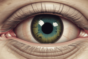
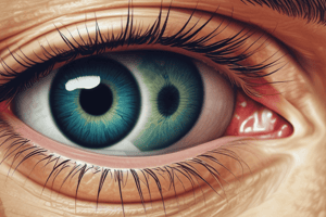
What is the anatomy of the eye?
What is the anatomy of the eye?
What is the function of the cornea?
What is the function of the cornea?
What does the pupil do?
What does the pupil do?
What is the lens and its purpose?
What is the lens and its purpose?
What is the blind spot?
What is the blind spot?
What are retinal ganglion cells?
What are retinal ganglion cells?
What are the types of photoreceptor cells?
What are the types of photoreceptor cells?
Rods are primarily responsible for color vision.
Rods are primarily responsible for color vision.
How do photoreceptor cells distribute across the retina?
How do photoreceptor cells distribute across the retina?
What happens when opsin absorbs light?
What happens when opsin absorbs light?
What are the recovery rates for photopigments?
What are the recovery rates for photopigments?
Rods are most active under ________ conditions.
Rods are most active under ________ conditions.
What is the Troxler effect?
What is the Troxler effect?
What are ipRGCs?
What are ipRGCs?
What are the three subtypes of cone photoreceptors?
What are the three subtypes of cone photoreceptors?
Flashcards are hidden until you start studying
Study Notes
Visual Field
- The visual field is defined as the entire area visible when an eye is fixated on a point in space.
- Vertical and horizontal meridians divide the visual field into left/right and top/bottom halves respectively.
- Hemifield refers to half of the visual field, while a quarterfield is a quadrant of the visual field.
Anatomy of the Eye
- The eye's anatomy includes various structures: iris (color), pupil (black center), and retina (light detection).
- The blind spot arises from a lack of light-sensitive photoreceptors in the optic disc, where the optic nerve exits the retina.
Key Structures of the Eye
- The cornea is a transparent dome-shaped covering affecting light entry.
- The fovea is part of the retina where color vision is most acute, containing only cone photoreceptors.
- Retinal ganglion cells serve as the output nodes to the brain.
Cornea's Role
- The cornea affects light waves by bending or refracting them, influenced by its thickness.
Pupil and Iris
- The pupil is the central hole through which light enters the eye, while the iris is the colored muscular surrounding structure regulating pupil size.
Lens Function
- The lens, located behind the iris, focuses incoming light on the retina and is designed to be transparent.
Sclera and Retina
- The sclera is the white outer layer providing protection, contrasted by the retina, part of the CNS where visual processing begins.
Blind Spot
- The blind spot corresponds to the location of the optic disc, lacking photoreceptors, creating an area of the visual field that goes undetected by the eye.
Optic Nerve
- Composed of retinal ganglion cell axons, the optic nerve transmits visual information to the brain and creates a blind spot due to absent photoreceptors.
Light Pathway to Retina
- The retina contains a layer of photoreceptors that absorb light, with additional modulatory neurons such as amacrine, horizontal, and bipolar cells aiding in visual information processing.
Photoreceptor Cells
- These cells change shape upon absorbing light, allowing for light detection; they come in two main types: rods (low-light) and cones (color).
Function and Process of Photoreceptors
- Photoreceptor cells convert light energy into a nervous system communicable form, known as signal transduction.
Types of Photoreceptors
- Rods are sensitive to low light, contributing to night vision without color differentiation.
- Cones dominate in the fovea, responsible for high visual acuity and color discrimination under bright conditions.
Distribution of Photoreceptors
- Rod density decreases towards the fovea, which consists solely of cones.
- Conversely, the peripheral retina has more rods than cones.
Cell Size and Information Gathering
- Larger cells capture more light but have lower resolution.
- Peripheral cells have larger receptive fields, enabling broader information gathering compared to central cells.
Cone Subtypes
- Cone photoreceptors are classified into three subtypes: short (blue), medium (green), and long (red).
Photoreceptor Proteins
- Photoreceptor proteins undergo a chemical change upon light absorption, facilitating light response.
- Rods use rhodopsin, while cones utilize photopsin; melanopsin is found in certain retinal ganglion cells.
Opponent-Processing Color Vision Theory
- Color perception operates through opponency channels, contrasting different cones (L vs M, S vs L+M, and dark vs bright).
Opsin Functionality
- Absorbing light requires opsins to regenerate, with rods (rhodopsin) taking longer to recover versus faster-recovering cones (photopsin).
Rod Activity Conditions
- Rods are most efficient in low-light or starlight conditions, requiring minimal light activation.
Troxler Effect
- This phenomenon occurs when prolonged focus on a stimulus leads to visual adaptation, causing the stimulus to fade as the eye becomes accustomed.
Cone Distribution in the Fovea
- The fovea contains short, medium, and long cones, with the very center consisting exclusively of medium and long cones.
Retinal Ganglion Cells (RGC)
- RGCs receive input from modulatory neurons, transmitting visual signals via the optic nerve.
Types of Retinal Ganglion Cells
- Classes include midget (parvocellular), parasol (magnocellular), and small bi-stratified cells (koniocellular), differentiated by size, function, and response characteristics.
Parvocellular and Magnocellular Pathways
- Parvocellular pathway: Midget RGCs process color, detail, and texture with a sustained response.
- Magnocellular pathway: Parasol RGCs detect motion and have larger cell pools over many receptors.
Koniocellular Pathway
- Small bistratified RGCs, which utilize S-cone input, project to visual areas with lower acuity information.
Melanopsin-Containing RGCs
- Known as intrinsically photosensitive RGCs, these cells can absorb light directly and manage circadian rhythms due to their unique photopigment.
Optic Chiasm
- The optic chiasm is a critical crossover point for optic nerves in the brain, directing visual field information to the opposite hemispheres.
RGC Proximity to the Fovea
- RGCs located closer to the fovea are fewer in number and smaller in size.
Size and Spatial Resolution Relations
- Midget RGCs located further from the fovea can match the spatial resolution of parasol RGCs positioned closer, indicating broader resolution in parasol cells.
ipRGCs
- Intrinsically photosensitive retinal ganglion cells (ipRGCs) function independently of traditional photoreceptors and play roles in signal modulation and circadian regulation.
Studying That Suits You
Use AI to generate personalized quizzes and flashcards to suit your learning preferences.