Podcast
Questions and Answers
Which part of the primary visual cortex represents the upper left quadrant of the visual field?
Which part of the primary visual cortex represents the upper left quadrant of the visual field?
- Lower left quadrant of the cortex
- Upper left quadrant of the cortex
- Upper right quadrant of the cortex
- Lower right quadrant of the cortex (correct)
Where is the calcarine sulcus located in relation to the primary visual cortex?
Where is the calcarine sulcus located in relation to the primary visual cortex?
- Does not relate to the visual cortex
- Above the visual cortex
- Below the visual cortex (correct)
- In between the two hemispheres
What causes lateral and vertical inversion in the projection of the visual field upon the visual cortex?
What causes lateral and vertical inversion in the projection of the visual field upon the visual cortex?
- Normal representation of visual input (correct)
- Retinal nerve damage
- Optic nerve misalignment
- Photoreceptor cell malfunction
Which neurological condition might affect the projection of the visual field onto the primary visual cortex?
Which neurological condition might affect the projection of the visual field onto the primary visual cortex?
How many quadrants does the visual field project to in terms of the primary visual cortex?
How many quadrants does the visual field project to in terms of the primary visual cortex?
Where are the cell bodies of both the 1st and 2nd order neurons of the central visual pathway located?
Where are the cell bodies of both the 1st and 2nd order neurons of the central visual pathway located?
Which type of neuron lies entirely within the retina?
Which type of neuron lies entirely within the retina?
What is the role of horizontal cells in the retina?
What is the role of horizontal cells in the retina?
Where do the axons of ganglion cells assemble before passing into the optic nerve?
Where do the axons of ganglion cells assemble before passing into the optic nerve?
What guarantees each hemisphere receives binocular input from the contralateral visual hemifield?
What guarantees each hemisphere receives binocular input from the contralateral visual hemifield?
Where does visual perception occur in the brain?
Where does visual perception occur in the brain?
What happens to the axons derived from the nasal halves of the retinae at the optic chiasma?
What happens to the axons derived from the nasal halves of the retinae at the optic chiasma?
Where do the 3rd order thalamocortical neurons project from the lateral geniculate nucleus?
Where do the 3rd order thalamocortical neurons project from the lateral geniculate nucleus?
Which structure lies immediately rostral to the optic chiasma?
Which structure lies immediately rostral to the optic chiasma?
Where do relatively small number of fibres leave before reaching the lateral geniculate nucleus?
Where do relatively small number of fibres leave before reaching the lateral geniculate nucleus?
What is the primary function of the visual association cortex?
What is the primary function of the visual association cortex?
Where do the axons that make up the optic nerve originate?
Where do the axons that make up the optic nerve originate?
What is the total length of the intraorbital portion of the optic nerve?
What is the total length of the intraorbital portion of the optic nerve?
Which part of the brain does the optic nerve originate from?
Which part of the brain does the optic nerve originate from?
What is the function of the fibres that leave the optic nerve before reaching the lateral geniculate nucleus?
What is the function of the fibres that leave the optic nerve before reaching the lateral geniculate nucleus?
Where does vision commence?
Where does vision commence?
Which part of the eye allows light to enter?
Which part of the eye allows light to enter?
Where do the fibers of the optic nerve project to?
Where do the fibers of the optic nerve project to?
Which region of the brain is responsible for visual perception?
Which region of the brain is responsible for visual perception?
What controls the muscle fibers of the iris?
What controls the muscle fibers of the iris?
Where are the cell bodies of the 1st and 2nd order neurons of the central visual pathway located?
Where are the cell bodies of the 1st and 2nd order neurons of the central visual pathway located?
What is the function of horizontal cells in the retina?
What is the function of horizontal cells in the retina?
Which cells are responsible for transferring information from photoreceptors to ganglion cells?
Which cells are responsible for transferring information from photoreceptors to ganglion cells?
Where do the fibers of the optic nerve undergo hemidecussation?
Where do the fibers of the optic nerve undergo hemidecussation?
What guarantees binocular input from the contralateral visual hemifield to each hemisphere?
What guarantees binocular input from the contralateral visual hemifield to each hemisphere?
Which type of neuron lies entirely within the retina?
Which type of neuron lies entirely within the retina?
What is the primary function of ganglion cells in the visual pathway?
What is the primary function of ganglion cells in the visual pathway?
What modulates transmission between bipolar cells and ganglion cells in the retina?
What modulates transmission between bipolar cells and ganglion cells in the retina?
Flashcards are hidden until you start studying
Study Notes
Innervation and Pupil Function
- Circular fibers are controlled by postganglionic parasympathetic neurons, causing pupil constriction and reducing light exposure to the retina.
- Radial fibers receive innervation from postganglionic sympathetic neurons, resulting in pupil dilation.
Structure of the Eye
- The ciliary body, located behind the iris, contains ciliary muscle innervated by the parasympathetic nervous system.
- The lens, situated within the ciliary body, focuses light onto the retina and is maintained in position by a suspensory ligament.
- Ciliary muscle contraction alters lens shape for focusing (known as accommodation).
Eye Compartments
- The lens and suspensory ligament divide the eyeball into two parts: anterior and posterior.
- The anterior segment contains aqueous humor, a thin fluid continuously secreted and reabsorbed by the ciliary body, draining into the canal of Schlemm to return to the venous system.
- The posterior segment is filled with vitreous humor, a gelatinous substance.
Choroid and Retina
- The choroid lines the inside of the sclera, containing dark pigment cells that absorb light, minimizing internal reflection.
- The inner surface of the choroid is lined by the photoreceptive retina, responsible for light perception.
Visual Processing Pathway
- Vision begins when an image is formed on the photoreceptive retina, encoding visual information through neuronal discharge.
- Neurons project visual information via the optic nerve, which undergoes hemidecussation at the optic chiasma before reaching the lateral geniculate nucleus of the thalamus.
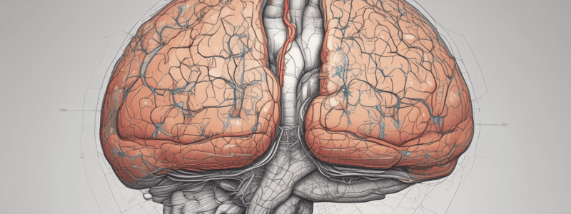
- Thalamocortical neurons transmit signals to the primary visual cortex located in the occipital lobe for visual perception.
Layers of the Eye
- The eyeball comprises three tissue layers; the outermost layer is tough, fibrous, and protective.
- The anterior pole of the eye features a transparent cornea that allows light entry.
- The anterior margin of the sclera includes two rings of smooth muscle, with the iris being the most anterior, containing the pupil that regulates light intake.
- Iris muscle fibers are arranged circularly and radially, controlled by the autonomic nervous system.
Studying That Suits You
Use AI to generate personalized quizzes and flashcards to suit your learning preferences.


