Podcast
Questions and Answers
What is the consequence of a volume overload in the heart due to a large amount of blood being ejected into the right ventricle?
What is the consequence of a volume overload in the heart due to a large amount of blood being ejected into the right ventricle?
- It leads to a permanent left to right shunt.
- Only the right ventricle experiences overload.
- The left ventricle compensates adequately without overload.
- Both ventricles experience an overload. (correct)
Which condition can arise from a ventricular septal defect (VSD) due to increased pulmonary vascular resistance?
Which condition can arise from a ventricular septal defect (VSD) due to increased pulmonary vascular resistance?
- Pulmonary artery dilation.
- Eisenmenger syndrome. (correct)
- Left ventricular hypertrophy only.
- Decreased pulmonary hypertension.
What are some common symptoms indicating heart failure in patients with a significant left to right shunt?
What are some common symptoms indicating heart failure in patients with a significant left to right shunt?
- Dyspnea, orthopnea, and weight gain. (correct)
- Syncope and temporary blindness.
- Chronic headaches and high blood pressure.
- Increased appetite and excessive thirst.
What type of cyanosis can result from the progression of Eisenmenger syndrome?
What type of cyanosis can result from the progression of Eisenmenger syndrome?
What imaging technique is recommended to visualize the interventricular septum in cases suspected of VSD?
What imaging technique is recommended to visualize the interventricular septum in cases suspected of VSD?
Which of the following is NOT typically a complication associated with a significant ventricular septal defect (VSD)?
Which of the following is NOT typically a complication associated with a significant ventricular septal defect (VSD)?
What is the primary issue causing a Ventricular Septal Defect (VSD)?
What is the primary issue causing a Ventricular Septal Defect (VSD)?
Which type of VSD is primarily located between the left and right ventricles and is often associated with multiple defects?
Which type of VSD is primarily located between the left and right ventricles and is often associated with multiple defects?
Which condition is often associated with an endocardial cushion defect?
Which condition is often associated with an endocardial cushion defect?
What range of percentages represents the occurrence of inlet septal VSDs?
What range of percentages represents the occurrence of inlet septal VSDs?
Which anatomical structures are typically involved in the formation of the atrioventricular defect?
Which anatomical structures are typically involved in the formation of the atrioventricular defect?
What is a common characteristic of medium-sized VSDs?
What is a common characteristic of medium-sized VSDs?
Which of the following statements accurately describes VSD types?
Which of the following statements accurately describes VSD types?
What characterizes the degree of VSD, where there is a complete absence of the septum?
What characterizes the degree of VSD, where there is a complete absence of the septum?
What is a significant consequence of untreated VSD in terms of pulmonary health?
What is a significant consequence of untreated VSD in terms of pulmonary health?
What does the Qp/Qs shunt ratio evaluate in patients with VSD?
What does the Qp/Qs shunt ratio evaluate in patients with VSD?
Which anatomical feature is indicative of Eisenmenger’s syndrome?
Which anatomical feature is indicative of Eisenmenger’s syndrome?
What is the primary pathophysiological change that occurs due to a left-to-right shunt in VSD?
What is the primary pathophysiological change that occurs due to a left-to-right shunt in VSD?
In the context of endocardial cushion defects, what is the primary consequence of the combined blood flow?
In the context of endocardial cushion defects, what is the primary consequence of the combined blood flow?
What is the classic physical finding associated with Eisenmenger syndrome?
What is the classic physical finding associated with Eisenmenger syndrome?
What is the primary anatomical location of a membranous septal VSD?
What is the primary anatomical location of a membranous septal VSD?
Which condition is strongly associated with outlet septal VSD?
Which condition is strongly associated with outlet septal VSD?
What is a characteristic feature of perimembranous VSDs?
What is a characteristic feature of perimembranous VSDs?
In what population is the prevalence of outlet septal VSD typically higher?
In what population is the prevalence of outlet septal VSD typically higher?
What is a significant consequence of a large VSD on heart function?
What is a significant consequence of a large VSD on heart function?
What congenital heart condition involves a failure to properly align the interventricular septum?
What congenital heart condition involves a failure to properly align the interventricular septum?
What major vessels can arise from a common trunk in Truncus Arteriosus?
What major vessels can arise from a common trunk in Truncus Arteriosus?
Which of the following is NOT characteristic of perimembranous VSDs?
Which of the following is NOT characteristic of perimembranous VSDs?
Which condition is an example of malalignment septal VSD?
Which condition is an example of malalignment septal VSD?
What is the prevalence percentage of outlet septal VSD among total VSD cases?
What is the prevalence percentage of outlet septal VSD among total VSD cases?
Flashcards are hidden until you start studying
Study Notes
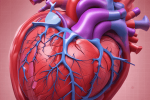
Ventricular Septal Defect (VSD)
- A hole between the left and right ventricle during fetal development
- Causes blood to shunt from the left ventricle to the right ventricle
- Can be asymptomatic in young patients with small shunts
- Can lead to pulmonary hypertension which increases resistance in the pulmonary arteries
- Can cause heart failure, dyspnea, orthopnea, paroxysmal nocturnal dyspnea, fatigue, cough, weight gain
- Can lead to Eisenmenger syndrome, where the shunt reverses to a right to left shunt and causes systemic cyanosis
- Can cause aortic insufficiency
- Can lead to infective endocarditis
VSD Types
- Inlet septal VSD: Borders the mitral valve, tricuspid valve, and muscle; often associated with an atrioventricular septal defect
- Outlet septal VSD: Borders the left ventricular outflow tract and right ventricular outflow tract; borders the aortic valve, pulmonic valve, and muscle; strongly associated with aortic valve prolapse and aortic regurgitation; prevalent in Asian populations
- Trabecular or muscular septal VSD: Located between the left and right ventricle bodies; usually low on the septal wall, in the thicker portion of the septum, close to the apex; surrounded by muscle
- Membranous septal or perimembranous septal VSD: Most common type; borders the tricuspid valve, aortic valve, and muscle; usually high in septal wall, in thinner, more flexible portion of the septum, closer to the valves and great vessels; frequently aneurysmal
- Malalignment septal VSD: The two portions of the interventricular septum fail to align properly; affects other structures within the heart, including tetralogy of Fallot and truncus arteriosus
VSD Echo Assessment
- VSD usually presents as an echo-free space along the interventricular septum
- Turbulent color flow Doppler (CFD) travels through the VSD between the left and right ventricles
- Document the VSD location, size, velocity, and direction
- Evaluate associated findings, including left ventricular volume overload, pulmonary hypertension, aortic valve prolapse, aortic insufficiency
- Calculate the Qp/Qs shunt ratio
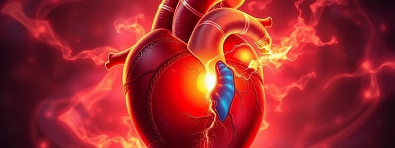
VSD and Pulmonary Distress
- Left-to-right shunt causes excess blood volume on the right side and in the lungs
- Increased blood volume in the lungs causes increased pressure
- Pressure backs up to the right heart
- Can result in Eisenmenger syndrome, which causes poor oxygenated blood to systemic circulation
Endocardial Cushion Defect
- aka atrioventricular canal defect or atrioventricular septal defect
- Combination of congenital heart anomalies creating a butterfly appearance
- Hole in the center of the heart where the upper chambers meet the lower chambers allows oxygenated blood to mix with deoxygenated blood
- Common valve in place of the mitral and tricuspid valves
- Frequently associated with trisomy 21 (Down Syndrome)
- Can be present in utero and anytime from birth to several months
- Partial endocardial cushion defects may not present until 20-30 years of age
- Requires surgery
- Symptoms in infant: coughing, cyanosis, dyspnea, fatigue, irregular or rapid heart rate, murmur, poor appetite, poor weight gain, swelling, wheezing
Cleft Mitral Valve
- Slit-like hole or division of the mitral valve leaflets, most commonly the anterior mitral valve leaflet
- Isolated cleft posterior leaflet is rare
- Evaluate valve anatomy, degree of cleft edge fibrosis, and retraction
- Regurgitation is common and should be addressed
- Surgical repair is typically required
- Associated cardiac lesions: Atrial Septal Defect (Primum)
Studying That Suits You
Use AI to generate personalized quizzes and flashcards to suit your learning preferences.