Podcast
Questions and Answers
What forms the superior vena cava?
What forms the superior vena cava?
The right and left brachiocephalic veins merge.
What is the primary function of the superior vena cava?
What is the primary function of the superior vena cava?
- Pumps blood to the heart
- Transports blood to the lungs
- Collects blood from the head and neck (correct)
- Filters blood in the kidneys
The superior vena cava has valves.
The superior vena cava has valves.
False (B)
Which group of veins does the occipital vein terminate in?
Which group of veins does the occipital vein terminate in?
What is the primary feature of cerebral veins?
What is the primary feature of cerebral veins?
Cerebral veins open into venous sinuses in the direction of blood flow.
Cerebral veins open into venous sinuses in the direction of blood flow.
What is the role of the superior anastomotic vein?
What is the role of the superior anastomotic vein?
Match the following veins with their regions of drainage:
Match the following veins with their regions of drainage:
What is the length of the superior vena cava?
What is the length of the superior vena cava?
What are the two groups of veins of the brain?
What are the two groups of veins of the brain?
Which vein is formed by the union of the supratrochlear and supraorbital veins?
Which vein is formed by the union of the supratrochlear and supraorbital veins?
The superior vena cava has multiple valves.
The superior vena cava has multiple valves.
What is a significant feature of the cerebral veins regarding their structure?
What is a significant feature of the cerebral veins regarding their structure?
Match the following veins with their corresponding regions of drainage:
Match the following veins with their corresponding regions of drainage:
The anterior cerebral veins drain into the ______.
The anterior cerebral veins drain into the ______.
Which veins connect extracranial and intracranial veins?
Which veins connect extracranial and intracranial veins?
What role do the diploic veins play?
What role do the diploic veins play?
Flashcards are hidden until you start studying
Study Notes
Veins of the Head and Neck
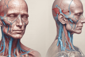
- Blood from the head and neck flows into the superior vena cava system.
- The superior vena cava is a thick, short trunk that is 5-6 cm long and 2-2.5 cm in diameter.
- It has no valves.
- It is formed by the merging of the right and left brachiocephalic veins behind the junction of the cartilage of the first right rib and the sternum.
- It descends down and opens into the right atrium.
- The superior vena cava collects blood from the head, neck, upper limbs, and the walls and organs of the chest cavity.
Veins of the Brain
- Cerebral veins form anastomoses with the diploic and extracranial veins.
- Cerebral veins have multiple ways of drainage.
- Cerebral veins have no valves.
- Cerebral veins have no muscles in their walls.
- Cerebral veins have specific features that maintain constant blood pressure and pressure of the cerebrospinal fluid.
- Some veins open into the venous sinuses against the direction of blood flow.
- The pachimeningeal sinuses do not collapse and cause hemivacuum.
- The veins of the brain are subdivided into superficial and deep veins.
Superficial Veins of the Brain
- The veins of the scalp accompany the arteries and have similar names.
- The supratrochlear and supraorbital veins unite at the medial angle of the eye, forming the angular vein.
- The angular vein continues as the facial vein.
- The superficial temporal vein descends in front of the tragus, enters the parotid gland, and joins the maxillary vein to form the retromandibular vein.
- The anterior part of the retromandibular vein unites with the facial vein to form the common facial vein, which drains into the internal jugular vein and ultimately the subclavian vein.
- The occipital vein terminates in the suboccipital plexus.
- Other veins, like the emissary veins and frontal diploic veins, also contribute to the venous drainage of the scalp.
Deep Veins of the Brain
- The outflow of blood through the deep veins is carried out into the venous sinuses.
- The superior anastomotic vein connects the superior sagittal and cavernous sinuses and parietal veins with the temporal veins.
- The inferior anastomotic vein connects the transverse venous sinus with the cavernous or sphenoid-parietal temporal and parietal veins with the occipital veins.
Venous Drainage of the Brain
- Due to the special function of the brain, multidirectional blood flow paths are formed on the head.
- This ensures high reliability of blood flow.
- Inside the skull, venous sinuses of the dura mater act as non-decaying venous collectors.
- Multilateral and multitiered connections of various outflow paths exist.
- The venous drainage of the head includes:
- Diploic veins
- Sinuses of the dura mater
- Emissary veins
- Cerebral venous bed and its outflow paths
- Veins of the orbit and eyeball
- Venous bed of the face
Veins of the Head and Neck
- Blood flow from the head and neck flows into the superior vena cava system
- The superior vena cava is about 5-6 cm long and 2-2.5 cm in diameter
- The superior vena cava has no valves
- The superior vena cava is formed by the merging of the right and left brachiocephalic veins behind the junction of the cartilage of the first right rib and the sternum
- The superior vena cava descends and empties into the right atrium
- The superior vena cava collects blood from the head, neck, upper limbs, and the walls and organs of the chest cavity.
Cerebral Veins
- Cerebral veins form anastomoses with the diploic and extracranial veins
- Cerebral veins have multiple drainage pathways
- Cerebral vein walls lack muscle
- Cerebral veins lack valves
- Some cerebral veins open into the venous sinuses against the direction of blood flow to maintain patency and prevent hemivacuum
- There are two groups of cerebral veins: superficial and deep
- The outflow of blood from these veins is carried to the venous sinuses
Superficial Cerebral Veins
- The superficial veins form a venous network on the surface of the brain
- This network allows for collateral blood flow in different directions
- The superior anastomotic vein connects the superior sagittal and cavernous sinuses, parietal veins, and temporal veins
- The inferior anastomotic vein connects the transverse venous sinus with the cavernous or sphenoid-parietal temporal and parietal veins with the occipital veins
Superficial Cerebral Vein Drainage
- Veins | Region of Drainage | Termination
- Superior Cerebral Veins | Superolateral surface of the hemisphere | The superior sagittal sinus
- Superficial Middle Cerebral Veins | The area around the posterior branch of the lateral groove | The cavernous sinus or the sphenoparietal sinus
- Deep Middle Cerebral Vein | Surface of the insula | The basal vein
- Inferior Cerebral Veins | The orbit | The superior cerebral veins
- Temporal Lobe Veins | The temporal lobe | The superior sagittal sinus, the cavernous sinus, or neighboring sinuses.
- Anterior Cerebral Veins | The corpus callosum, the anterior part of the medial surface of the hemisphere | The basal vein
Drainage Pathways of the Brain
- The brain has a multidirectional blood flow system due to its high energy needs and continuous function
- This ensures reliable blood supply to maintain body integrity
- Venous sinuses of the dura mater are specially arranged non-decaying venous collectors, providing multilateral and multitiered connections for outflow pathways
- The head region is comprised of:
- Diploic veins (veins of the skull bones)
- Sinuses of the dura mater
- Emissary veins (venous anastomoses connecting extra and intracranial veins)
- Cerebral venous bed and outflow (cerebral veins)
- Veins of the orbit and eyeball
- Venous bed of the face, carrying blood into the facial, retromandibular, and partially into the ...
Studying That Suits You
Use AI to generate personalized quizzes and flashcards to suit your learning preferences.


