Podcast
Questions and Answers
What is the role of microfilaments in cell shape and movement?
What is the role of microfilaments in cell shape and movement?
Microfilaments are involved in cell shape and movement, interact with myosin for muscle contraction, and are involved in cell migration, amoeboid movement, and cytoplasmic streaming.
What is the function of actin in microfilaments?
What is the function of actin in microfilaments?
Actin is the protein building block of microfilaments, folding into a globular-shaped molecule that can bind ATP or ADP.
How do G-actin monomers assemble into microfilaments?
How do G-actin monomers assemble into microfilaments?
G-actin monomers self-assemble into microfilaments through a lag phase and elongation phase, similar to tubulin assembly, forming F-actin filaments composed of two linear strands of polymerized G-actin wound into a helix.
What is the polarity of actin monomers in a microfilament?
What is the polarity of actin monomers in a microfilament?
What is the role of myosin in microfilaments?
What is the role of myosin in microfilaments?
What is cytoplasmic streaming, and how does it relate to microfilaments?
What is cytoplasmic streaming, and how does it relate to microfilaments?
How do microfilaments contribute to cell crawling and chemotaxis?
How do microfilaments contribute to cell crawling and chemotaxis?
What is the structural core of microvilli, and how does it relate to microfilaments?
What is the structural core of microvilli, and how does it relate to microfilaments?
What is the primary function of myosin II in cells?
What is the primary function of myosin II in cells?
How do myosin II molecules move in relation to actin filaments during muscle contraction?
How do myosin II molecules move in relation to actin filaments during muscle contraction?
What is the significant difference between kinesins and myosin II in terms of their function?
What is the significant difference between kinesins and myosin II in terms of their function?
What is the sliding filament model, and how does it explain muscle contraction?
What is the sliding filament model, and how does it explain muscle contraction?
What is the role of cross-bridges in muscle contraction?
What is the role of cross-bridges in muscle contraction?
How do myosin II molecules operate during muscle contraction?
How do myosin II molecules operate during muscle contraction?
What is the structure of a muscle fiber, and how does it relate to muscle contraction?
What is the structure of a muscle fiber, and how does it relate to muscle contraction?
How much does a single myosin II molecule slide an actin filament during muscle contraction?
How much does a single myosin II molecule slide an actin filament during muscle contraction?
What is the function of primary cilia in sensing and responding to external stimuli?
What is the function of primary cilia in sensing and responding to external stimuli?
What is the structure of the axoneme in primary cilia?
What is the structure of the axoneme in primary cilia?
What is the result of defects in primary cilia development?
What is the result of defects in primary cilia development?
What is the role of myosins in cellular events?
What is the role of myosins in cellular events?
What is the result of a loss of dynein in primary cilia?
What is the result of a loss of dynein in primary cilia?
What is the role of genes associated with ciliogenesis?
What is the role of genes associated with ciliogenesis?
What is the function of RPE65 in primary cilia?
What is the function of RPE65 in primary cilia?
What is the characteristic of primary cilia that distinguishes them from other cilia?
What is the characteristic of primary cilia that distinguishes them from other cilia?
What is the primary function of myosin II in cells, and how does it differ from kinesins?
What is the primary function of myosin II in cells, and how does it differ from kinesins?
Describe the sliding filament model and its role in muscle contraction.
Describe the sliding filament model and its role in muscle contraction.
How do myosin II molecules move along actin filaments during muscle contraction?
How do myosin II molecules move along actin filaments during muscle contraction?
What is the significance of cross-bridges in muscle contraction?
What is the significance of cross-bridges in muscle contraction?
What is the structural organization of a muscle fiber, and how does it relate to muscle contraction?
What is the structural organization of a muscle fiber, and how does it relate to muscle contraction?
How do the characteristics of myosin II molecules contribute to their function in muscle contraction?
How do the characteristics of myosin II molecules contribute to their function in muscle contraction?
What is the significance of the polarity of actin monomers in a microfilament, and how does it relate to muscle contraction?
What is the significance of the polarity of actin monomers in a microfilament, and how does it relate to muscle contraction?
How does the structure of actin filaments contribute to their function in muscle contraction?
How does the structure of actin filaments contribute to their function in muscle contraction?
What is the primary mechanism by which myosin II generates force during muscle contraction?
What is the primary mechanism by which myosin II generates force during muscle contraction?
How do actin filaments regulate the movement of myosin II motors during cell crawling and chemotaxis?
How do actin filaments regulate the movement of myosin II motors during cell crawling and chemotaxis?
What is the structural significance of the 'barbed end' and 'pointed end' of an actin filament?
What is the structural significance of the 'barbed end' and 'pointed end' of an actin filament?
How do myosin VI and myosin VII motors differ in their function and regulation?
How do myosin VI and myosin VII motors differ in their function and regulation?
What is the role of tropomyosin in regulating actin filament dynamics and myosin function?
What is the role of tropomyosin in regulating actin filament dynamics and myosin function?
How do kinesin motors coordinate with myosin motors to regulate organelle transport and cell migration?
How do kinesin motors coordinate with myosin motors to regulate organelle transport and cell migration?
What is the significance of the actin-binding domain in myosin II motors?
What is the significance of the actin-binding domain in myosin II motors?
How do post-translational modifications regulate myosin II motor activity?
How do post-translational modifications regulate myosin II motor activity?
What is the significance of the '9 + 0' axoneme structure in primary cilia?
What is the significance of the '9 + 0' axoneme structure in primary cilia?
How do myosins interact with actin microfilaments to facilitate cellular events?
How do myosins interact with actin microfilaments to facilitate cellular events?
What is the consequence of defects in primary cilia development, and what is the resulting disorder?
What is the consequence of defects in primary cilia development, and what is the resulting disorder?
What is the function of dynein in primary cilia, and what is the result of its loss?
What is the function of dynein in primary cilia, and what is the result of its loss?
What is the role of primary cilia in embryonic development, and what is the significance of their dysfunction?
What is the role of primary cilia in embryonic development, and what is the significance of their dysfunction?
How do myosins contribute to the process of phagocytosis, and what is the significance of this process?
How do myosins contribute to the process of phagocytosis, and what is the significance of this process?
What is the relationship between primary cilia and the apical surface of epithelial cells, and what is the significance of this relationship?
What is the relationship between primary cilia and the apical surface of epithelial cells, and what is the significance of this relationship?
What is the role of genes associated with ciliogenesis, and how do they contribute to primary cilia development?
What is the role of genes associated with ciliogenesis, and how do they contribute to primary cilia development?
What is the primary function of myosin II, and how does it differ from kinesins in terms of their cellular function?
What is the primary function of myosin II, and how does it differ from kinesins in terms of their cellular function?
Describe the sliding filament model, and how does it explain muscle contraction?
Describe the sliding filament model, and how does it explain muscle contraction?
What is the significance of the polarity of actin monomers in a microfilament, and how does it relate to muscle contraction?
What is the significance of the polarity of actin monomers in a microfilament, and how does it relate to muscle contraction?
How do myosin II molecules operate during muscle contraction, and what is the significance of cross-bridges in this process?
How do myosin II molecules operate during muscle contraction, and what is the significance of cross-bridges in this process?
What is the structural organization of a muscle fiber, and how does it relate to muscle contraction?
What is the structural organization of a muscle fiber, and how does it relate to muscle contraction?
How do the characteristics of myosin II molecules contribute to their function in muscle contraction?
How do the characteristics of myosin II molecules contribute to their function in muscle contraction?
What is the primary mechanism by which myosin II generates force during muscle contraction?
What is the primary mechanism by which myosin II generates force during muscle contraction?
How does the structure of actin filaments contribute to their function in muscle contraction?
How does the structure of actin filaments contribute to their function in muscle contraction?
What is the primary function of primary cilia, and what is their characteristic structure that distinguishes them from other cilia?
What is the primary function of primary cilia, and what is their characteristic structure that distinguishes them from other cilia?
What is the role of myosins in cellular events, and how do they interact with actin microfilaments?
What is the role of myosins in cellular events, and how do they interact with actin microfilaments?
What is the consequence of defects in primary cilia development, and what is the resulting disorder?
What is the consequence of defects in primary cilia development, and what is the resulting disorder?
What is the role of dynein in primary cilia, and what is the result of its loss?
What is the role of dynein in primary cilia, and what is the result of its loss?
What is the function of RPE65 in primary cilia, and what is its significance?
What is the function of RPE65 in primary cilia, and what is its significance?
How do myosins contribute to the process of phagocytosis, and what is the significance of this process?
How do myosins contribute to the process of phagocytosis, and what is the significance of this process?
What is the relationship between primary cilia and the apical surface of epithelial cells, and what is the significance of this relationship?
What is the relationship between primary cilia and the apical surface of epithelial cells, and what is the significance of this relationship?
What is the significance of the '9 + 0' axoneme structure in primary cilia, and how does it differ from other cilia?
What is the significance of the '9 + 0' axoneme structure in primary cilia, and how does it differ from other cilia?
What is the underlying mechanism by which myosin II generates force during muscle contraction, and how does it relate to the polarity of actin monomers in a microfilament?
What is the underlying mechanism by which myosin II generates force during muscle contraction, and how does it relate to the polarity of actin monomers in a microfilament?
Compare and contrast the structure and function of kinesins and myosin II in cellular events, highlighting their differences in motor protein activity.
Compare and contrast the structure and function of kinesins and myosin II in cellular events, highlighting their differences in motor protein activity.
Describe the role of actin-binding proteins in regulating actin filament dynamics and myosin function, highlighting their impact on cell migration and chemotaxis.
Describe the role of actin-binding proteins in regulating actin filament dynamics and myosin function, highlighting their impact on cell migration and chemotaxis.
What is the significance of the 'barbed end' and 'pointed end' of an actin filament in terms of myosin motor activity, and how do these ends influence the direction of movement?
What is the significance of the 'barbed end' and 'pointed end' of an actin filament in terms of myosin motor activity, and how do these ends influence the direction of movement?
How do myosin VI and myosin VII motors differ in their function and regulation, and what are the implications for cellular events?
How do myosin VI and myosin VII motors differ in their function and regulation, and what are the implications for cellular events?
What is the significance of the sliding filament model in explaining muscle contraction, and how does it relate to the structure of actin filaments and myosin motors?
What is the significance of the sliding filament model in explaining muscle contraction, and how does it relate to the structure of actin filaments and myosin motors?
How do post-translational modifications, such as phosphorylation, regulate myosin II motor activity, and what are the implications for cellular events?
How do post-translational modifications, such as phosphorylation, regulate myosin II motor activity, and what are the implications for cellular events?
What is the role of tropomyosin in regulating actin filament dynamics and myosin function, and how does it influence cell migration and chemotaxis?
What is the role of tropomyosin in regulating actin filament dynamics and myosin function, and how does it influence cell migration and chemotaxis?
Flashcards are hidden until you start studying
Study Notes
Myosin II Function
- Myosin II pulls arrays of actin filaments together, resulting in contraction of a cell or group of cells.
- Resembles kinesin, with globular domains that walk along a protein filament, using ATP hydrolysis to change shape.
Myosin II vs. Kinesin
- Myosin II molecules move short distances but operate in large arrays, in some cases billions of motors working together to mediate muscle contraction.
- Kinesins operate alone or in small numbers to transport vesicles over large distances.
Muscle Contraction
- Muscle contraction is the most familiar example of mechanical work mediated by intracellular filaments.
- A muscle consists of parallel muscle fibers, each of which is a long, thin, highly specialized, multinucleate cell.
- Each muscle fiber contains numerous myofibrils, each of which is divided along its length into repeating units called sarcomeres.
The Sliding-Filament Model
- The sliding filament model explains muscle contraction, where thin filaments slide past thick filaments, with no change in length of either.
- Cross-bridges are formed between F-actin of thin filaments and myosin heads of thick filaments, dissociating rapidly and requiring lots of ATP.
- The result is shortening of sarcomeres and muscle fiber contraction.
Cytoskeleton and Cellular Movement
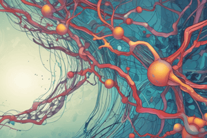
- Microfilaments are involved in cell shape and movement, interacting with myosin for muscle contraction.
- They are also involved in cell migration, amoeboid movement, and cytoplasmic streaming.
- Microfilaments contribute to development and maintenance of cell shape, forming the structural core of microvilli.
Actin and Microfilaments
- Actin is the protein building block of microfilaments, found in all eukaryotic cells.
- G-actin monomers self-assemble into microfilaments, with a lag phase and elongation phase, similar to tubulin assembly.
- F-actin filaments are composed of two linear strands of polymerized G-actin wound into a helix, with all actin monomers in the filament having the same orientation (polarity).
Primary Cilia
- Primary cilium is a long, thin organelle found on nearly all cells, common on apical surface of epithelial cells.
- Primary cilia are important in development, with a role in embryonic patterning and organogenesis, and defects can result in disorders such as deafness and left-right asymmetry reversals (ciliopathies).
- Primary cilia have a "9 + 0" axoneme structure, lacking the central pair, and do not move.
Myosins
- Myosins are ATP-dependent motors that interact with and exert force on actin microfilaments.
- Myosins function in a wide range of cellular events, including muscle contraction, cell movement, phagocytosis, and vesicle transport.
Myosin II Function
- Myosin II pulls arrays of actin filaments together, resulting in contraction of a cell or group of cells.
- Resembles kinesin, with globular domains that walk along a protein filament, using ATP hydrolysis to change shape.
Myosin II vs. Kinesin
- Myosin II molecules move short distances but operate in large arrays, in some cases billions of motors working together to mediate muscle contraction.
- Kinesins operate alone or in small numbers to transport vesicles over large distances.
Muscle Contraction
- Muscle contraction is the most familiar example of mechanical work mediated by intracellular filaments.
- A muscle consists of parallel muscle fibers, each of which is a long, thin, highly specialized, multinucleate cell.
- Each muscle fiber contains numerous myofibrils, each of which is divided along its length into repeating units called sarcomeres.
The Sliding-Filament Model
- The sliding filament model explains muscle contraction, where thin filaments slide past thick filaments, with no change in length of either.
- Cross-bridges are formed between F-actin of thin filaments and myosin heads of thick filaments, dissociating rapidly and requiring lots of ATP.
- The result is shortening of sarcomeres and muscle fiber contraction.
Cytoskeleton and Cellular Movement
- Microfilaments are involved in cell shape and movement, interacting with myosin for muscle contraction.
- They are also involved in cell migration, amoeboid movement, and cytoplasmic streaming.
- Microfilaments contribute to development and maintenance of cell shape, forming the structural core of microvilli.
Actin and Microfilaments
- Actin is the protein building block of microfilaments, found in all eukaryotic cells.
- G-actin monomers self-assemble into microfilaments, with a lag phase and elongation phase, similar to tubulin assembly.
- F-actin filaments are composed of two linear strands of polymerized G-actin wound into a helix, with all actin monomers in the filament having the same orientation (polarity).
Primary Cilia
- Primary cilium is a long, thin organelle found on nearly all cells, common on apical surface of epithelial cells.
- Primary cilia are important in development, with a role in embryonic patterning and organogenesis, and defects can result in disorders such as deafness and left-right asymmetry reversals (ciliopathies).
- Primary cilia have a "9 + 0" axoneme structure, lacking the central pair, and do not move.
Myosins
- Myosins are ATP-dependent motors that interact with and exert force on actin microfilaments.
- Myosins function in a wide range of cellular events, including muscle contraction, cell movement, phagocytosis, and vesicle transport.
Studying That Suits You
Use AI to generate personalized quizzes and flashcards to suit your learning preferences.