Podcast
Questions and Answers
What event signifies the end of the atrial diastole phase in the cardiac cycle?
What event signifies the end of the atrial diastole phase in the cardiac cycle?
- Opening of the atrioventricular valves.
- Closure of the atrioventricular valves.
- Opening of the aortic valve.
- Closure of the aortic and pulmonary valves. (correct)
During which phase of the cardiac cycle does the atrial contraction occur?
During which phase of the cardiac cycle does the atrial contraction occur?
- Ventricular Ejection
- Ventricular Filling Stage
- Isovolumic Relaxation
- Atrial Systole (correct)
What is the duration of one cardiac cycle in a healthy human heart?
What is the duration of one cardiac cycle in a healthy human heart?
- 1.0 seconds
- 0.6 seconds
- 1.2 seconds
- 0.8 seconds (correct)
Which event occurs during the isovolumic contraction phase of the cardiac cycle?
Which event occurs during the isovolumic contraction phase of the cardiac cycle?
Which activity is associated with the T wave in an electrocardiogram?
Which activity is associated with the T wave in an electrocardiogram?
What is the clinical significance of the QRS complex in an ECG tracing?
What is the clinical significance of the QRS complex in an ECG tracing?
What activity does the P wave on an ECG tracing represent?
What activity does the P wave on an ECG tracing represent?
If the cardiac cycle is analogous to an engine, which event would be considered the 'power stroke' that propels blood into circulation?
If the cardiac cycle is analogous to an engine, which event would be considered the 'power stroke' that propels blood into circulation?
In what manner do the tissues and fluids surrounding the heart contribute during the cardiac cycle?
In what manner do the tissues and fluids surrounding the heart contribute during the cardiac cycle?
What is the main purpose of placing electrodes on the skin for electrocardiography?
What is the main purpose of placing electrodes on the skin for electrocardiography?
What is the standard number of electrodes used in a conventional 12-lead ECG?
What is the standard number of electrodes used in a conventional 12-lead ECG?
What information does an ECG lead provide?
What information does an ECG lead provide?
What is the significance of the electrode position in electrocardiography?
What is the significance of the electrode position in electrocardiography?
How is ''electrical potential difference'' defined in the context of electrocardiography?
How is ''electrical potential difference'' defined in the context of electrocardiography?
What concept is described by 'a combination of electrodes for recording' during electrocardiography?
What concept is described by 'a combination of electrodes for recording' during electrocardiography?
How should leads be angled in relation to the plane of interest to optimally detect electrical vectors?
How should leads be angled in relation to the plane of interest to optimally detect electrical vectors?
Which ECG leads are categorized as augmented limb leads?
Which ECG leads are categorized as augmented limb leads?
In the context of ECG limb leads, what do leads I, II, and III primarily compare?
In the context of ECG limb leads, what do leads I, II, and III primarily compare?
What is the significance of inverting lead aVR in ECG interpretation?
What is the significance of inverting lead aVR in ECG interpretation?
Which electrode does Lead I compare to the electrode on the right arm?
Which electrode does Lead I compare to the electrode on the right arm?
What formula accurately represents Einthoven's Law?
What formula accurately represents Einthoven's Law?
What describes the function of a central terminal in Goldberger's augmented limb leads?
What describes the function of a central terminal in Goldberger's augmented limb leads?
In lead aVF, where is the exploring electrode placed?
In lead aVF, where is the exploring electrode placed?
What is the angle by which lead aVF views the heart's electrical activity?
What is the angle by which lead aVF views the heart's electrical activity?
Which leads are referred to as inferior limb leads, providing insight into the inferior aspect of the left ventricle?
Which leads are referred to as inferior limb leads, providing insight into the inferior aspect of the left ventricle?
What anatomical plane do precordial leads lie in?
What anatomical plane do precordial leads lie in?
With reference to precordial leads, what acts as the positive poles for the six corresponding leads?
With reference to precordial leads, what acts as the positive poles for the six corresponding leads?
In the Cabrera system for ECG presentation, how are the leads arranged?
In the Cabrera system for ECG presentation, how are the leads arranged?
What does the standard grid on ECG paper represent on its horizontal axis?
What does the standard grid on ECG paper represent on its horizontal axis?
Of the conditions listed, which one is least likely to lead to the requirement of an electrocardiogram?
Of the conditions listed, which one is least likely to lead to the requirement of an electrocardiogram?
What does the term 'cardiac cycle' refer to?
What does the term 'cardiac cycle' refer to?
Considering the chambers of the human heart, what is the key role of the atria?
Considering the chambers of the human heart, what is the key role of the atria?
During which stage of the cardiac cycle does the closure of the aortic valve and pulmonary artery occur?
During which stage of the cardiac cycle does the closure of the aortic valve and pulmonary artery occur?
How does atrial contraction contribute to the flow of blood in the heart?
How does atrial contraction contribute to the flow of blood in the heart?
During isovolumic relaxation, what pressure changes occur in the ventricles?
During isovolumic relaxation, what pressure changes occur in the ventricles?
Following ventricular systole/contraction on an ECG, which wave would you expect to see?
Following ventricular systole/contraction on an ECG, which wave would you expect to see?
What term defines the graph representing voltage versus time of the electrical activity of the heart?
What term defines the graph representing voltage versus time of the electrical activity of the heart?
Upon which body parts are the electrodes attached to, during electrocardiography?
Upon which body parts are the electrodes attached to, during electrocardiography?
During the isovolumic contraction phase, what key characteristic defines the ventricular volume?
During the isovolumic contraction phase, what key characteristic defines the ventricular volume?
How does the direction of an electrical vector influence the appearance of a wave on an ECG?
How does the direction of an electrical vector influence the appearance of a wave on an ECG?
In the context of electrocardiography, what is the key distinction between an electrode and a lead?
In the context of electrocardiography, what is the key distinction between an electrode and a lead?
If a cardiologist observes an inverted T wave in multiple leads of an ECG, what condition should they consider?
If a cardiologist observes an inverted T wave in multiple leads of an ECG, what condition should they consider?
How does the spatial organization of Einthoven's leads (I, II, and III) relate to Kirchhoff's Law?
How does the spatial organization of Einthoven's leads (I, II, and III) relate to Kirchhoff's Law?
Flashcards
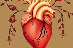
Cardiac cycle
Cardiac cycle
The sequence of events that take place when the heart beats.
Systole
Systole
The period of contraction the heart undergoes while pumping blood into circulation.
Diastole
Diastole
The period of relaxation that occurs as the heart chambers fill with blood.
Atrium
Atrium
Signup and view all the flashcards
Ventricles
Ventricles
Signup and view all the flashcards
Atrial Diastole
Atrial Diastole
Signup and view all the flashcards
Atrial Systole
Atrial Systole
Signup and view all the flashcards
Isovolumic Contraction
Isovolumic Contraction
Signup and view all the flashcards
Ventricular Ejection
Ventricular Ejection
Signup and view all the flashcards
Isovolumic Relaxation
Isovolumic Relaxation
Signup and view all the flashcards
Ventricular Filling
Ventricular Filling
Signup and view all the flashcards
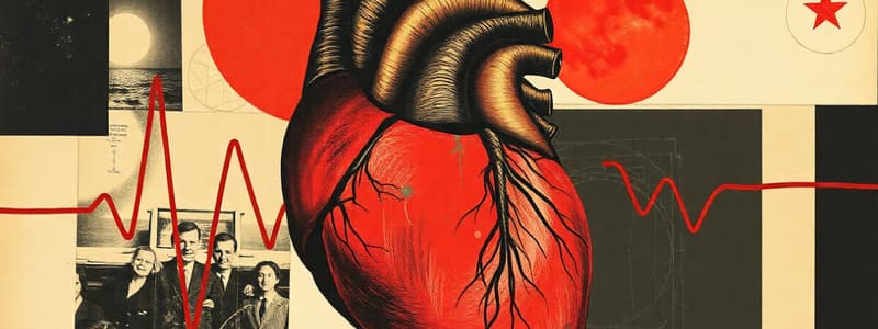
Electrocardiogram (ECG/EKG)
Electrocardiogram (ECG/EKG)
Signup and view all the flashcards
Electrode
Electrode
Signup and view all the flashcards
ECG Lead
ECG Lead
Signup and view all the flashcards
12-Lead ECG
12-Lead ECG
Signup and view all the flashcards
ECG Lead Derivation
ECG Lead Derivation
Signup and view all the flashcards
Anatomical Planes
Anatomical Planes
Signup and view all the flashcards
Types of ECG Leads
Types of ECG Leads
Signup and view all the flashcards
Limb Leads
Limb Leads
Signup and view all the flashcards
Lead aVR
Lead aVR
Signup and view all the flashcards
Lead aVL
Lead aVL
Signup and view all the flashcards
Lead aVF
Lead aVF
Signup and view all the flashcards
Precordial Leads
Precordial Leads
Signup and view all the flashcards
V1-V2 septal chest leads
V1-V2 septal chest leads
Signup and view all the flashcards
V3-V4 anterior leads
V3-V4 anterior leads
Signup and view all the flashcards
V5-V6 anterolateral leads
V5-V6 anterolateral leads
Signup and view all the flashcards
Background Grid
Background Grid
Signup and view all the flashcards
Why ECG?
Why ECG?
Signup and view all the flashcards
Heart rate
Heart rate
Signup and view all the flashcards
Healthy Heart Beats
Healthy Heart Beats
Signup and view all the flashcards
Cardiac Cycle Composition
Cardiac Cycle Composition
Signup and view all the flashcards
Atriums Function
Atriums Function
Signup and view all the flashcards
Ventricles Function
Ventricles Function
Signup and view all the flashcards
ECG purpose
ECG purpose
Signup and view all the flashcards
SA node origin
SA node origin
Signup and view all the flashcards
Purkinje fibers.
Purkinje fibers.
Signup and view all the flashcards
P wave
P wave
Signup and view all the flashcards
T wave
T wave
Signup and view all the flashcards
QRS complex
QRS complex
Signup and view all the flashcards
Electrodes
Electrodes
Signup and view all the flashcards
electrode
electrode
Signup and view all the flashcards
Electrocardiograph function
Electrocardiograph function
Signup and view all the flashcards
3 types of the leads
3 types of the leads
Signup and view all the flashcards
Study Notes
- Cardiac cycle refers to the sequence of events that take place when the heart beats.
Cardiac Cycle
- The occurrence of a cardiac cycle is illustrated by a heart rate, indicated as beats per minute.
- A healthy human heart beats 72 times per minute and completes 72 cardiac cycles per minute.
- The cardiac cycle comprises the diastole, the systole, and the intervening pause.
- It includes a complete contraction and relaxation of both the atria and ventricles, lasting about 0.8 seconds.
- Systole involves the period of contraction when the heart pumps blood into circulation.
- Diastole involves the period of relaxation as the heart chambers fill with blood.
- The human heart consists of four chambers.
- Two upper chambers are the left and right atria, while the lower two are the right and left ventricles.
- Atria are structures where blood comes to the heart, and ventricles are where blood leaves.
Cardiac Cycle Phases
- Atrial Diastole: Chambers of the heart are calm. The aortic valve and pulmonary artery close while atrioventricular valves open, allowing the chambers to relax.
- Atrial Systole: Blood cells flow from the atrium to the ventricle as the atrium contracts.
- As the atria contract, pressure increases, forcing more blood flow across open atrioventricular (AV) valves, leading to a rapid flow of blood into the ventricles.
- Isovolumic Contraction: Ventricles begin to contract, but ventricular volume does not change because all valves are closed.
- Ventricular Ejection: Ventricles contract and empty, causing the pulmonary artery and aortic valve close.
- Isovolumic Relaxation: No blood enters the ventricles, causing pressure to decrease, and ventricles stop contracting and relax. Due to the pressure in the aorta, the pulmonary artery and aortic valve close.
- Ventricular Filling Stage: Ventricles relax, and the pressure difference between atria and ventricles causes blood to flow back into the ventricles through open A-V valves.
Electrocardiography (ECG or EKG)
-
Electrical currents are detected during a routine physical via electrodes attached to the skin, typically at the wrist, ankles, and six separate locations on the chest.
-
Electrocardiogram is a graph of voltage versus time representing electrical activity of the heart.
-
Electrodes (small, plastic patches that stick to the skin) are put on spots on the chest, arms, and legs, connected to an ECG machine by lead wires.
-
Comparisons of readings from the different electrodes provide an accurate and comprehensive assessment of the heart's electrical activity.
-
An ECG provides a composite tracing of all muscle impulses formed by myocardial cells.
-
It records changes in voltage (potential) across the sarcolemma that:
- Originates in the SA node.
- Radiates through both atria to the AV node.
- Passes through the AV node and interventricular septum to the heart apex.
- Stimulates Purkinje fibers in ventricular myocardium.
-
An typical ECG tracing features 3 principal deflections for one heart cycle.
- A P wave above the baseline
- A QRS complex: it begins (Q) and ends (S) with small downward deflection from the baseline and a large deflection (R) above the baseline
- A T wave above the baseline
Electrocardiogram Waves
- These waves are indicators of depolarization and repolarization in specific heart regions.
- I. P wave: Generated when impulse starts in the SA node and depolarizes the cells of atria.
- II. QRS complex: Identifies start of ventricles' depolarization; Atria repolarize at the same time
- III. T wave: small, rounded peak denoting ventricular repolarization.
- P-wave shows depolarization of atria that begins contracting about 25 msec after the start of the P-wave
- QRS-complex is ventricular depolarization where ventricles start contracting shortly after the peak of the R wave
- T-wave is ventricular repolarization
ECG Leads and Electrical Potentials
- Electrode: A conductive pad attached to the skin to record electrical currents.
- ECG lead: A graphical description of the electrical activity of the heart, analyzed with multiple electrodes.
- Standard ECG: Also known as a 12-lead ECG since it includes 12 leads - it's obtained using 10 electrodes.
- A combination of electrodes for recording is called a LEAD.
- Electrical potential differences arise as the electrical impulse travels through the heart.
- Electric potential difference is defined as a difference in electric potential between two measurement points.
- This means that the electrical potential difference is the difference in the electrical potential detected by two (or more) electrodes.
- Depolarization and repolarization generate electrical currents.
- Tissues and fluids surrounding the heart act as electrical conductors.
- Electrodes placed on the skin detect these electrical currents.
- The electrocardiograph (ECG machine) compares, amplifies, and filters electrical potential differences recorded by the electrodes.
- In a conventional 12-lead ECG, ten electrodes are placed on the limbs and chest surface; The heart's electrical potential is measured from twelve angles ("leads").
- Every lead represents differences in electrical potentials measured in two points in space. The simplest leads are composed using only two electrodes.
- The electrocardiograph defines one electrode as an exploring (positive) and the other as a reference (negative) electrode.
Anatomical Planes and Lead Placements
- Electrical activity of the heart is observed from the horizontal and frontal planes.
- Detection of vectors in a certain plane depends on the angle of the lead.
- This angle depends on placement of the exploring lead and the reference point.
- A lead with leads on the head and the left foot would be angled in the frontal plane and detect vertical vectors.
- A lead with leads on the sternum and on the back. This lead will be angled from the back to the anterior chest wall, which is the horizontal plane.
- Leads are broken down into three types: limb, augmented limb, and precordial or chest.
- The 12-lead ECG has:
- Three limb leads
- Three augmented limb leads arranged like spokes of a wheel in the coronal plane (vertical).
- Six precordial or chest leads on the perpendicular transverse plane (horizontal).
- Principles of the Limb Leads.Leads I, II, III, aVF, aVL and aVR uses three electrodes placed on the right arm, the left arm and the left leg in order to detect electrical activity in the frontal plane.
- Leads I, II and III show compare electrical potential differences between two leads, for example,
- Lead I : compares electrode of the left are versus that of right arm
- Lead II: Left leg compares to that of right arm
- Lead III: The left leg Electrode compares to that of the left arm
Wilhelm Einthoven, Kirchhoff's Law, Einthoven's Law, and Lead Voltage
- Leads I, II and III are original leads constructed by Wilhelm Einthoven. Spatial organization of these leads forms a triangle in the chest.
- According to Kirchhoff's law, the sum of all currents in a closed circuit must be zero. Since Einthoven's triangle can be viewed as a circuit applies to it. Thus emerges Einthoven's law :
- Lead I + Lead III = Lead II
- Voltage is calculated as:
- I = LA - RA
- II = LL - RA
- III = LL - LA
Augmented limb leads (aVR, aVL, and aVF)
- Derived from same three electrodes as leads I, II, and III using Goldberger's central terminal as their negative pole.
- Letter 'a' stands for augmented, V for voltage, R for right arm, L for left arm, and F for foot.
- In aVR, the right arm is the exploring electrode, with the reference composed by averaging the left arm and left leg.
- In lead aVL, the left arm electrode is exploring, viewing the heart ≈ −30°.
- In lead aVF, the exploring electrode is the left leg.
- Since Goldberger's leads are composed of the same electrodes that Einthoven's display Goldberger's mathematical relationship:
- aVL: (Lead I – Lead III)/2
- (-aVR): (Lead I + Lead II)/2
- aVF: (Lead II + Lead III)/2
- The electrode on the left leg serves as the exploring electrode while the reference is composed of the average of the arm electrodes. The angle by which aVF views the heart’s electrical activity is 90°.
- Clinically, the aVF viewed as "views the inferior wall of the left ventricle". The principals apply to lead aVR/ aVL:
- II, aVF and III are known as interior or diaphragmal limb leads that are located on the left ventricle.
- aVL., I, and aVR are lateral limb leads located on the lateral side of the ventricles.
Hexaxial references
- In conjunction with leads I, II, and III, leads aVR, aVL, and aVF complete the hexaxial reference system. The system helps calculate the heart's electrical axis in the frontal plane.
Precordial Leads and the Background Grid
- Precordial leads lie in the transverse (horizontal) plane, which is perpendicular to the other six leads.
- The six precordial electrodes act as the positive poles for the six corresponding precordial leads: (V1, V2, V3, V4, V5, and V6). Wilson's central terminal is used as the negative pole.
- Precordial and Unipolar leads can be used to generate bipolar precordial leads that explore the horizontal plane (left to right axis).
- In the Cabrera system, the leads are placed in anatomical order, i.e.,
- V1-V2 ("septal leads"): primarily observe the ventricular septum but may display ECG originating from the right ventricle.
- V3-V4 ("anterior leads"): observe the anterior wall of the left ventricle.
- V5-V6 ("anterolateral leads"): observe the lateral wall of the left ventricle.
- Note that none of the leads in the 12-lead ECG can precisely detect vectors in the ventricals
- ECGs are normally printed on a grid, with the horizontal axis representing time and the vertical axis representing voltage.
Background Grid
- ECGs are normally printed on a grid. The horizontal axis represents time, and the vertical axis represents voltage. The standard values on this grid are shown in the adjacent image at 25mm/sec:
- A small box is 1 mm × 1 mm and represents 0.1 mV × 0.04 seconds.
- A large box is 5 mm x 5 mm and represents 0.5 mV × 0.20 seconds.
Application
- Why ECG Needed:
- Chest pain
- Severe tiredness (fatigue), shortness of breath, dizziness, or fainting
- Irregular heartbeats
- Overall health of the heart before procedures, such as surgery;
- After treatment for a heart attack (myocardial infarction)
- How implanted pacemaker is working
- How certain heart medicines are working
- Baseline tracing of the heart's function during a physical exam, which can be compared with future ECGs
Studying That Suits You
Use AI to generate personalized quizzes and flashcards to suit your learning preferences.