Podcast
Questions and Answers
What physiological change primarily characterizes anemia?
What physiological change primarily characterizes anemia?
- Reduced oxygen-carrying capacity (correct)
- Increased oxygen-carrying capacity
- Elevated red blood cell production
- Enhanced blood clotting ability
What two classifications relate to the pathophysiology of anemia?
What two classifications relate to the pathophysiology of anemia?
- Size and Hemoglobin content (correct)
- Shape and Blood Volume
- Size and Erythrocyte count
- Shape and Hemoglobin content
What distinguishes microcytic anemia from other types of anemia?
What distinguishes microcytic anemia from other types of anemia?
- Small red blood cell size (correct)
- Normal red blood cell size
- Increased hemoglobin concentration
- Large red blood cell size
In normocytic anemia, what does an increase in reticulocytes typically indicate?
In normocytic anemia, what does an increase in reticulocytes typically indicate?
What is the primary cause of macrocytic anemia?
What is the primary cause of macrocytic anemia?
Why does iron deficiency anemia occur in the context of blood loss?
Why does iron deficiency anemia occur in the context of blood loss?
What is a key characteristic of aplastic anemia?
What is a key characteristic of aplastic anemia?
In hemolytic anemia, what process leads to the characteristic signs and symptoms?
In hemolytic anemia, what process leads to the characteristic signs and symptoms?
What causes relative polycythemia?
What causes relative polycythemia?
Which genetic defect is associated with hereditary hemochromatosis?
Which genetic defect is associated with hereditary hemochromatosis?
What is the primary distinction between quantitative and qualitative leukocyte disorders?
What is the primary distinction between quantitative and qualitative leukocyte disorders?
How is leukocytosis best described?
How is leukocytosis best described?
What is indicated when immature neutrophils are released into the blood?
What is indicated when immature neutrophils are released into the blood?
What is Granulocytopenia?
What is Granulocytopenia?
Which conditions are associated with eosinophilia?
Which conditions are associated with eosinophilia?
What typically triggers monocytosis?
What typically triggers monocytosis?
What is the primary cause of lymphocytosis?
What is the primary cause of lymphocytosis?
What distinguishes localized from generalized lymphadenopathy?
What distinguishes localized from generalized lymphadenopathy?
What is a key characteristic of multiple myeloma (MM)?
What is a key characteristic of multiple myeloma (MM)?
What is the primary cause of hypersplenism?
What is the primary cause of hypersplenism?
In the blood clotting process, what is the role of thrombin?
In the blood clotting process, what is the role of thrombin?
Which condition is characterized by a decrease in the number of circulating platelets?
Which condition is characterized by a decrease in the number of circulating platelets?
Why is Vitamin K deficiency associated with impaired hemostasis?
Why is Vitamin K deficiency associated with impaired hemostasis?
Which of the following describes an embolus?
Which of the following describes an embolus?
What is the primary underlying cause of iron deficiency anemia (IDA) in infants and children?
What is the primary underlying cause of iron deficiency anemia (IDA) in infants and children?
What is a primary mechanism behind hemolytic disease of the fetus and newborn?
What is a primary mechanism behind hemolytic disease of the fetus and newborn?
What causes Hb S to stretch and elongate forming sickle shape?
What causes Hb S to stretch and elongate forming sickle shape?
What is an aplastic crisis?
What is an aplastic crisis?
Which factor is deficient in Hemophilia B?
Which factor is deficient in Hemophilia B?
What is the primary mechanism behind primary immune thrombocytopenia (ITP)?
What is the primary mechanism behind primary immune thrombocytopenia (ITP)?
What cell population is affected by Acute lymphoblastic leukemia (ALL)?
What cell population is affected by Acute lymphoblastic leukemia (ALL)?
What change in blood cells defines leukemia?
What change in blood cells defines leukemia?
What is the relationship between Von Willebrand factor (vWF) and hemostasis?
What is the relationship between Von Willebrand factor (vWF) and hemostasis?
What change is related to thrombocythemia?
What change is related to thrombocythemia?
In the coagulation cascade, what is the role of Factor Xa?
In the coagulation cascade, what is the role of Factor Xa?
What process is initiated by tissue factor?
What process is initiated by tissue factor?
Flashcards
What is Anemia?
What is Anemia?
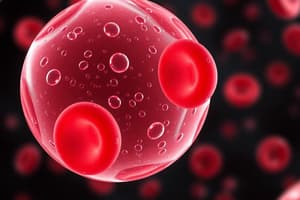
Reduced oxygen-carrying capacity leading to hypoxemia due to reduced erythrocytes or hemoglobin.
What is MCV in Anemia?
What is MCV in Anemia?
Classifies anemia based on the average size of red blood cells (measured in femtoliters).
What is Microcytic Anemia?
What is Microcytic Anemia?
Small red blood cell size caused by lack of iron or hemoglobin production.
What is Normocytic Anemia?
What is Normocytic Anemia?
Signup and view all the flashcards
Macrocytic Anemia?
Macrocytic Anemia?
Signup and view all the flashcards
Anemia of Blood Loss
Anemia of Blood Loss
Signup and view all the flashcards
Aplastic/Hemolytic Anemia
Aplastic/Hemolytic Anemia
Signup and view all the flashcards
Polycythemia
Polycythemia
Signup and view all the flashcards
Hereditary Hemochromatosis
Hereditary Hemochromatosis
Signup and view all the flashcards
Alterations in Leukocyte
Alterations in Leukocyte
Signup and view all the flashcards
What is Leukocytosis?
What is Leukocytosis?
Signup and view all the flashcards
What is Leukopenia?
What is Leukopenia?
Signup and view all the flashcards
Granulocytosis
Granulocytosis
Signup and view all the flashcards
Neutropenia
Neutropenia
Signup and view all the flashcards
Eosinophilia
Eosinophilia
Signup and view all the flashcards
Basophilia
Basophilia
Signup and view all the flashcards
Monocytosis
Monocytosis
Signup and view all the flashcards
Lymphocytosis
Lymphocytosis
Signup and view all the flashcards
Lymphadenopathy
Lymphadenopathy
Signup and view all the flashcards
Multiple Myeloma (MM)
Multiple Myeloma (MM)
Signup and view all the flashcards
Splenomegaly/Hypersplenism
Splenomegaly/Hypersplenism
Signup and view all the flashcards
Thrombocytopenia
Thrombocytopenia
Signup and view all the flashcards
Thrombocythemia
Thrombocythemia
Signup and view all the flashcards
Impaired Hemostasis
Impaired Hemostasis
Signup and view all the flashcards
Thrombus/Embolus
Thrombus/Embolus
Signup and view all the flashcards
Iron Deficiency Anemia (IDA)
Iron Deficiency Anemia (IDA)
Signup and view all the flashcards
Hemolytic Disease of Newborn
Hemolytic Disease of Newborn
Signup and view all the flashcards
Sickle Cell Disease
Sickle Cell Disease
Signup and view all the flashcards
Hemophilias
Hemophilias
Signup and view all the flashcards
Immune Thrombocytopenia (ITP)
Immune Thrombocytopenia (ITP)
Signup and view all the flashcards
Leukemia
Leukemia
Signup and view all the flashcards
Study Notes
Anemia
- Anemia is a physiologic manifestation
- It results in reduced oxygen-carrying capacity, leading to hypoxemia.
- Symptoms can vary based on the severity and the body’s ability to compensate.
- Common symptoms include fatigue, weakness, dyspnea, and pallor.
- It involves a reduction in the total number of erythrocytes in circulating blood
- It also concerns the quality or quantity of hemoglobin.
- Causes can include:
- Impaired erythrocyte production
- Acute or chronic blood loss
- Increased erythrocyte destruction
- A combination of all these factors
Anemia Classification Using MCV Value
- Helps classify different types of anemia
- Based on the mean corpuscular volume (MCV)
- MCV is a measure of the average size of red blood cells (measured in femtoliters)
- Size is identified by terms ending in "-cytic" (macrocytic, microcytic, normocytic)
- Hemoglobin content is identified by terms ending in "-chromic" (normochromic, hyperchromic, hypochromic)
Microcytic Anemia
- The MCV is less than 80 fl
- Involves small red blood cells
- Caused by a lack of iron in hemoglobin (hypochromic)
- It can also be caused by lack of hemoglobin production (normochromic)
- Examples: iron-deficiency anemia and thalassemia
Normocytic Anemia
- An MCV is between 80-100 fl
- Occurs with normal red blood cell size.
- Examples: anemia of chronic disease, aplastic anemia, and hemolytic anemia.
Macrocytic Anemia
- An MCV is greater than 100 fl.
- Involves large red blood cells
- Examples: Vitamin B12 deficiency anemia and folate deficiency anemia
- Macrocytic anemia can coexist with microcytic or normocytic anemia.
Normocytic Anemia
- Often grouped by whether reticulocytes are increased vs. decreased and hemolytic vs. non-hemolytic
- Increased reticulocytes means the body is trying to make new RBCs.
Macrocytic Anemia
- It involves large size caused by defects in RBC maturation or a disproportionate number of reticulocytes (reticulocytosis).
- Has 2 categories:
- Megaloblastic anemia refers to the large-sized cells that are caused by defects in DNA synthesis in RBCs.
- Non-megaloblastic anemia refers to the large size caused by RBC developmental issues.
Anemias of Blood Loss
- Acute blood loss: Posthemorrhagic anemia
- Chronic blood loss:
- Anemia can occur if the blood loss exceeds bone marrow replacement capacity.
- Iron deficiency anemia can occur.
Aplastic and Hemolytic Anemias
- Aplastic anemia: Body is not making enough RBCs due to hematopoietic failure or bone marrow aplasia, Reduced production of mature cells, Pancytopenia- reduction in RBC, WBC, platelets
- Hemolytic anemia: Premature accelerated destruction of erythrocytes
Myeloproliferative RBC Disorders
- Polycythemia involves the overproduction of red blood cells.
- Relative polycythemia (dehydration) occurs when fluid loss results in relative increases of red cell counts and Hgb and Hct values.
- Hereditary hemochromatosis:
- Involves iron overload disorder, an autosomal recessive disorder of iron metabolism
- Exhibits mutations in Hepcidin,the protein that governs iron regulation
- From excessive storage of iron in the liver, skin, pancreas, heart, joints, and testes
- It may or may not have polycythemia.
Alterations of Leukocyte Function
- Quantitative disorders involve increases or decreases in cell numbers due to bone marrow disorders or premature destruction of cells
- It is also caused by a response to infectious microorganism invasion.
- Qualitative disorders involve disruptions of leukocyte function, causing phagocytes or lymphocytes to lose their abilities.
Quantitative Alterations of Leukocytes
- Leukocytosis:
- High leukocyte count
- It is a normal protective physiologic response to physiologic stressors, such as infectious microorganisms.
- Leukopenia:
- Low leukocyte count that is not normal or beneficial, and predisposes a patient to infections.
Quantitative Alterations of Granulocytes
- Granulocytosis (neutrophilia):
- Increase in granulocytes
- Evident in the first stages of an infection or inflammation
- Immature neutrophils are released if the need for neutrophils increases beyond the supply.
- Neutropenia:
- Reduction in circulating neutrophils
- Caused by prolonged severe infection
- Decreased production
- Reduced survival
- Abnormal neutrophil distribution and sequestration Chemotherapy can cause granulocytopenia (severe neutropenia) or agranulocytosis (absence of granulocytes).
- Reduction in circulating neutrophils
Quantitative Alterations of Granulocytes
- Eosinophilia involves an increase in circulating eosinophils caused by hypersensitivity reactions
- Also allergic disorders and parasitic invasions
- Eosinopenia involves a decrease in circulation numbers of eosinophils, caused by migration of cells to inflammatory sites.
- Basophilia involves an increase in circulating basophils caused by response to inflammation and hypersensitivity reactions.
- Seen in myeloproliferative disorders
- Basopenia involves a decrease in circulating numbers of basophils
- Occurs in acute infections, hyperthyroidism, ovulation, pregnancy, and long-term steroid therapy.
Quantitative Alterations of Monocytes
- Monocytosis involves an increase in circulating monocytes
- It is often transient and unrelated to dysfunction of monocyte production.
- It usually occurs with neutropenia in later stages of infections
- This occurs when monocytes are needed to phagocytize organisms and debris.
- Monocytopenia involves a decrease in circulating monocytes, but is very rare.
Quantitative Alterations of Lymphocytes
- Lymphocytosis involves an increase in the number or proportion of lymphocytes
- Caused by acute viral infections, particularly caused by Epstein-Barr virus (EBV).
- Lymphocytopenia involves a decrease in the number of circulating lymphocytes
- Caused by abnormal production due to immune deficiencies or caused by destruction by drugs, viruses, or radiation.
Lymphadenopathy
- Involves enlarged lymph nodes that become palpable and tender.
- Can be localized, such as drainage of an inflammatory lesion located near the enlarged node.
- Can be generalized: occurs in the presence of infections, autoimmune diseases, or disseminated malignancy.
Plasma Cell Malignancy
- Multiple Myeloma (MM) is a malignant proliferation of plasma cells that infiltrate bone marrow and aggregate into tumor masses in the skeletal system, causing lytic bone lesions
- Manifestations include hypercalcemia, renal failure, anemia, and immune abnormalities, preceded by monoclonal gammopathy of undetermined significance.
Alterations of Splenic Function
- Splenomegaly: Enlargement of spleen, may or may not be pathologic.
- Congestive: Red pulp
- Infiltrative: White pulp
- Infectious: White pulp
- Hypersplenism: Overactivity of spleen that causes anemia.
Blood Clotting Process
- Hemostasis relies on major components to function normally
- These include the blood vessels, platelets and plasma coagulation factors.
Quantitative Disorders of Platelets
- Thrombocytopenia involves a decrease in the number of circulating platelets.
- Thrombocythemia involves an increase in the number of circulating platelets.
Impaired Hemostasis
- It is defined as an inability of coagulation and stable fibrin clot.
- Vitamin K deficiency:
- Vitamin K is necessary for synthesis and regulation of prothrombin
- Vitamin K is necessary for the procoagulant factors (VII, XI, X) and proteins C and S (anticoagulants).
- Liver disease:
- Hepatic cells produce most factors involved in hemostasis
- Damage causes production of fewer clotting factors
Thromboembolic Disorders
- Thrombus: stationary clot attached to vessel wall.
- Arterial or venous thrombi
- Virchow triad:
- Injury to vessel
- Abnormal blood flow
- Hypercoagulopathy
- Hereditary thrombophilias: mutations in coagulation
- Embolus: Unattached mass circulating within bloodstream.
Acquired Disorders of Erythrocytes (Children)
- Iron deficiency anemia (IDA)
- It is the most common nutritional disorder of infancy and childhood.
- It is caused by lack of iron intake, problems with iron absorption, blood loss, and increased iron requirements.
- Hemolytic disease of the fetus and newborn (erythroblastosis fetalis)
- Maternal antibody directed against fetal antigens
- Rh incompatibility (most common), ABO incompatibility, or other RBC antigen.
- It can be manifested by anemia, hyperbilirubinemia, neonatal jaundice, or bilirubin encephalopathy.
Inherited Disorders of Erythrocytes
- Sickle cell disease: Disorders characterized by the presence of hemoglobin S.
- Deoxygenation and dehydration cause Hb S to stretch and elongate, forming a sickle shape.
- It can result in vaso-occlusive crisis (thrombotic crisis).
- It may also result in aplastic crisis (not enough red blood cells) or sequestration crisis (red blood cells retained in spleen).
- Can also result in hyperhemolytic crisis(increased hemolysis).
Inherited Hemorrhagic Disease
- Hemophilias: Serious bleeding disorders that involve mutations in coagulation factors.
- Types:
- Hemophilia A: factor VIII deficiency
- Hemophilia B: factor IX deficiency
- Hemophilia C: factor XI deficiency
Antibody-Mediated Hemorrhagic Disease
- Primary immune thrombocytopenia is the most common platelet consumption disorder.
- It is defined by a platelet destruction rate that exceeds production.
- 70% of cases occur after a viral disease, with a typical onset at 1-6 years old.
- Manifested by bruising and petechial rash.
Leukemia
- Cancer of blood-forming tissues.
- Most common cancer in children.
- Acute lymphoblastic leukemia (ALL): acute myelogenous leukemia (AML).
- Most common cancer in children.
Studying That Suits You
Use AI to generate personalized quizzes and flashcards to suit your learning preferences.