Podcast
Questions and Answers
In the brachium, where is the ulnar nerve first palpable?
In the brachium, where is the ulnar nerve first palpable?
- Medial intermuscular septum (correct)
- Posterior intermuscular septum
- Anterior intermuscular septum
- Lateral intermuscular septum
What anatomical structures form the ulnar groove through which the ulnar nerve passes?
What anatomical structures form the ulnar groove through which the ulnar nerve passes?
- Olecranon and medial epicondyle (correct)
- Lateral epicondyle and olecranon
- Coracoid process and acromion
- Medial epicondyle and radial head
Which of the following is the MOST likely location for ulnar nerve compression due to sustained elbow flexion?
Which of the following is the MOST likely location for ulnar nerve compression due to sustained elbow flexion?
- Cubital tunnel (correct)
- Guyon's canal
- Carpal tunnel
- Axilla
Which nerve roots contribute to the formation of the ulnar nerve?
Which nerve roots contribute to the formation of the ulnar nerve?
Which ligament covers the ulnar groove, contributing to the potential for cubital tunnel syndrome?
Which ligament covers the ulnar groove, contributing to the potential for cubital tunnel syndrome?
In which direction should PROM be applied to joints affected by nerve injury?
In which direction should PROM be applied to joints affected by nerve injury?
What is the MOST appropriate initial treatment approach for flaccid tissue resulting from ulnar nerve injury?
What is the MOST appropriate initial treatment approach for flaccid tissue resulting from ulnar nerve injury?
Which of the following muscles is NOT innervated by the ulnar nerve?
Which of the following muscles is NOT innervated by the ulnar nerve?
Why is hydrotherapy contraindicated when autonomic dysfunction is present following an ulnar nerve injury?
Why is hydrotherapy contraindicated when autonomic dysfunction is present following an ulnar nerve injury?
Following an ulnar nerve injury, what sign would indicate loss of adductor pollicis function and compensatory recruitment of flexor pollicis longus?
Following an ulnar nerve injury, what sign would indicate loss of adductor pollicis function and compensatory recruitment of flexor pollicis longus?
Flashcards
Ulnar Nerve Anatomy
Ulnar Nerve Anatomy
Originates from the medial cord of the brachial plexus, deriving from nerve roots C8 and T1.
Ulnar Nerve Function
Ulnar Nerve Function
Motor: FCU, FDP(Flexor digitorum profundus), hypothenar muscles, interossei, adductor pollicis, 3rd & 4th lumbricals. Sensory: Palmar and dorsal surfaces of medial hand and digits.
Ulnar Nerve Injuries
Ulnar Nerve Injuries
The ulnar nerve is subject to neuropraxia, axonotmesis, and neurotmesis.
Ulnar Nerve Injury Causes
Ulnar Nerve Injury Causes
Signup and view all the flashcards
Ulnar Nerve Damage Symptoms
Ulnar Nerve Damage Symptoms
Signup and view all the flashcards
Froment's Sign
Froment's Sign
Signup and view all the flashcards
Ulnar Nerve Assessment
Ulnar Nerve Assessment
Signup and view all the flashcards
Ulnar Nerve Treatment Precautions
Ulnar Nerve Treatment Precautions
Signup and view all the flashcards
Ulnar Nerve Home Care
Ulnar Nerve Home Care
Signup and view all the flashcards
Cubital Tunnel Syndrome
Cubital Tunnel Syndrome
Signup and view all the flashcards
Study Notes
- Ulnar nerve
Anatomy
- A terminal branch of the medial cord of the brachial plexus, originating from nerve roots C8 and T1
Palpation
- Can be palpated within the medial intermuscular septum of the brachium
- It runs alongside the median nerve and brachial artery to the mid-point of the brachium near the coracobrachialis insertion
- The ulnar nerve then runs more medially while the median nerve continues alongside the brachial artery
- Symptoms of ulnar nerve compression may be elicited by palpating medial to lateral between the biceps and triceps
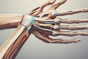
- The ulnar nerve passes through the ulnar groove between the olecranon and the medial epicondyle, known as the cubital tunnel
- This tunnel is covered by the arcuate ligament (ligament of Osborne)
- At the elbow, the ulnar nerve passes between the two heads of flexor carpi ulnaris (FCU) and deep to the muscle lying between FCU and flexor digitorum profundus
- Motor fibers proceed toward the wrist within this space
- At the forearm's halfway point, it branches into palmar and dorsal sensory nerves supplying the skin on the palm's ulnar side
- At the wrist, it travels over the flexor retinaculum, entering Guyon's canal, located between the pisiform and the hook of hamate, beneath the palmaris brevis and volar carpal ligament
- Gives off a motor branch to the intrinsic hand muscles, as well as sensory branches to the palmar and dorsal sides of digits 4 and 5
Palpation of affected tissue
- Focus on the tone and tissue health of the intrinsic hand muscles and FCU
- Assess sensation in the 4th and 5th digits, primarily the 5th digit
Function
- The motor functions include actions of the flexor carpi ulnaris (FCU) and flexor digitorum profundus (FDP)
- Innervates the hypothenar eminence (abductor digiti minimi (ADMM), flexor digiti minimi brevis (FDMM), opponens digiti minimi (ODM))
- Innervates the intrinsic compartment including lumbricals 3 & 4, palmar interossei muscles (PIM), dorsal interossei muscles (DIM), and adductor pollicis
- The flexor pollicis brevis is sometimes innervated, but is mainly innervated by the median nerve
- Responsible for sensation in the palmar and dorsal aspects of the medial hand and digits
Pathophysiology - Ulnar Nerve Injuries
- The ulnar nerve can be subject to neuropraxia, axonotmesis, and neurotmesis
- Determining the correct treatment depends on the the injury
Mechanism of Injury (MOI)
- Fractures in the medial epicondyle, mid-forearm, or wrist (Colles' fracture)
- Elbow dislocation
- Prolonged compression, such as leaning the elbow on a hard surface, handcuff use, or bicycling
- Repetitive actions leading to fibrosis and compression
- Trauma resulting in contusion or laceration
- Most vulnerable at the elbow and wrist
Signs & Symptoms (degeneration)
- A complete lesion results in claw hand, affecting the 5th digit with MCP extension, abduction, and IP flexion, and the 4th digit with MCP extension and IP flexion
- Loss of thumb adduction
- Muscle wasting in hypothenar eminence and interosseous spaces
- Anhidrosis (decreased sweating) and potential vasomotor changes
- Altered sensation in the 5th digit and half of the 4th digit, as well as the palm
- Anesthesia in the 5th digit
- Can lead to classic deformities
Ulnar Nerve Problems
- Bishop's Hand (Benediction Hand): Loss of ulnar lumbricals causes digits 4 & 5 to rest in a position opposite to the lumbricals' action
- Claw Hand: Same as Bishop's hand but with abduction of digits 4 & 5
- Froment's Sign: Loss of adductor pollicis leads to compensatory recruitment of flexor pollicis longus to grip
Palsy/Lesion and Description
- Tardy ulnar palsy may occur years after a fracture, related to callus formation or valgus deformity of the elbow, leading to adverse tension at the ulnar groove
- Cubital tunnel syndrome occurs when elbow flexion flattens the tunnel, putting pressure on the nerve and sustained elbow flexion can provoke ulnar nerve compression or lesions
- Ulnar tunnel/Guyon's tunnel/Guyon's canal syndrome/handlebar palsy is due to pressure on the hypothenar eminence from biking, jack hammering, keyboard use, overuse of the wrist from gripping or twisting, trauma to the wrist causing swelling and carpal fracture or arthritis
Ulnar Nerve Assessment
- Assess the degree of nerve regeneration at Tinel's sign at the ulnar groove, where gently tapping over the nerve will elicit tingling to the point of regeneration
- Use Froment's sign assessment by having someone grasp a piece of paper between the thumb & index finger (adductor pollicis), if the examiner pulls the paper away – and the patient flexes at the IP, is positive
- Assess the 4th & 5th digit strength for general weakness in RROM of the 4th and 5th digit (flexion, abduction, adduction)
Ulnar Nerve Treatment
- Context: managing a regenerating lesion post-fracture
- Do not traction the regenerating nerve or stretch denervated tissue
- Place the arm in a neutral position with pillows to reduce elbow, wrist, & finger flexion
- Apply segmental treatment proximal to the lesion with techniques applied perpendicular to the nerve
- Stabilize tissue just proximal to the lesion to prevent drag
- Treat flaccid tissue with light stroking and compression
- Treat unaffected tissue distal to the lesion site with strain toward (but not onto) flaccid tissue
- PROM to joints in direction that shortens affected tissue & nerve
Goals and Approach
- Promote relaxation
- Decrease edema
- Decrease tone and TrP in muscles proximal to the lesion
- Promote tissue health in denervated tissue through light stroking that shortens the nerve/bunches up tissue or is cross-fiber and/or gentle compressions
- Prevent contracture of unopposed antagonist muscles
- Apply segmental work (distal to lesion is okay on unaffected muscles/antagonists only)
- Promote joint health with PROM, if possible
- Handle limbs with care to avoid traction on the nerve
- Encourage returning motor function by facilitating ROODS techniques or similar or AAROM on the returned function (incorporate visualization)
- Promote tissue health in reinnervated tissue
- Use gentle Swedish techniques or sensory stimulation using different textures
Hydrotherapy
- If there is autonomic dysfunction, hydrotherapy is contraindicated anywhere on affected limb, even proximal to the lesion site
- If autonomics not affected, then use cool compress over acute injury, modified temperature over affected tissue, and mild contrast washes
Home Care
- If wearing a splint be vigilant for pressure sores
- PROM of joints that shortens affected tissue
- Visualize performing actions with the affected limb
- Elevate limb if edema present
Studying That Suits You
Use AI to generate personalized quizzes and flashcards to suit your learning preferences.