Podcast
Questions and Answers
What is the common name for Toxocara canis?
What is the common name for Toxocara canis?
- Heartworm
- Dog roundworm (correct)
- Cat worm
- Hookworm
Where do adult female Toxocara canis worms reside in an infected dog?
Where do adult female Toxocara canis worms reside in an infected dog?
- Large intestine
- Small intestine (correct)
- Liver
- Stomach
Which of the following routes does NOT contribute to Toxocara canis infection in dogs?
Which of the following routes does NOT contribute to Toxocara canis infection in dogs?
- Transplacental transmission
- Consumption of paratenic hosts
- Airborne transmission (correct)
- Ingestion of infective eggs
What is somatic migration in older dogs infected with Toxocara canis?
What is somatic migration in older dogs infected with Toxocara canis?
What clinical signs are commonly observed in young puppies with heavy Toxocara canis infections?
What clinical signs are commonly observed in young puppies with heavy Toxocara canis infections?
What makes diagnosing Toxocara canis in older dogs difficult?
What makes diagnosing Toxocara canis in older dogs difficult?
Which of the following is a common treatment for Toxocara canis infections?
Which of the following is a common treatment for Toxocara canis infections?
What is larval migrans, and how is it related to Toxocara canis?
What is larval migrans, and how is it related to Toxocara canis?
What are the two types of larval migrans caused by Toxocara canis in humans?
What are the two types of larval migrans caused by Toxocara canis in humans?
Which group is most at risk for developing Visceral Larva Migrans?
Which group is most at risk for developing Visceral Larva Migrans?
What are the clinical signs associated with Ocular Larva Migrans?
What are the clinical signs associated with Ocular Larva Migrans?
How do the cervical alae of Toxocara canis differ from Toxascaris leonina?
How do the cervical alae of Toxocara canis differ from Toxascaris leonina?
What is a notable symptom of Visceral Larva Migrans?
What is a notable symptom of Visceral Larva Migrans?
What are paratenic hosts in relation to Toxocara cati infection?
What are paratenic hosts in relation to Toxocara cati infection?
Which of the following is NOT a primary route through which cats can become infected with Toxocara cati?
Which of the following is NOT a primary route through which cats can become infected with Toxocara cati?
What happens to Toxocara cati eggs once they are ingested by a cat?
What happens to Toxocara cati eggs once they are ingested by a cat?
How are Toxocara cati eggs transmitted to the environment?
How are Toxocara cati eggs transmitted to the environment?
What clinical signs may indicate a Toxocara cati infection in young kittens?
What clinical signs may indicate a Toxocara cati infection in young kittens?
What is the time frame for Toxocara cati eggs to become infective?
What is the time frame for Toxocara cati eggs to become infective?
What does 'transmammary transmission' of Toxocara cati refer to?
What does 'transmammary transmission' of Toxocara cati refer to?
What are the potential severe complications of heavy Toxocara cati infections?
What are the potential severe complications of heavy Toxocara cati infections?
What is the primary mode of infection for Toxascaris leonina?
What is the primary mode of infection for Toxascaris leonina?
What is the approximate size of Toxascaris leonina eggs?
What is the approximate size of Toxascaris leonina eggs?
How do the cervical alae of Toxascaris leonina differ from those of Toxocara canis?
How do the cervical alae of Toxascaris leonina differ from those of Toxocara canis?
Where do adult Toxascaris leonina worms reside in the host?
Where do adult Toxascaris leonina worms reside in the host?
Which of the following is NOT a mode of transmission for Toxascaris leonina in puppies and kittens?
Which of the following is NOT a mode of transmission for Toxascaris leonina in puppies and kittens?
What clinical signs may indicate a Toxascaris leonina infection in pets?
What clinical signs may indicate a Toxascaris leonina infection in pets?
How do Toxascaris leonina larvae develop after being ingested by the host?
How do Toxascaris leonina larvae develop after being ingested by the host?
What do Toxascaris leonina worms feed on in the host?
What do Toxascaris leonina worms feed on in the host?
What is the primary location where adult Baylisascaris procyonis worms reside?
What is the primary location where adult Baylisascaris procyonis worms reside?
How long does it take for Baylisascaris procyonis eggs to become infective?
How long does it take for Baylisascaris procyonis eggs to become infective?
What is a potential consequence for incidental hosts that ingest Baylisascaris procyonis eggs?
What is a potential consequence for incidental hosts that ingest Baylisascaris procyonis eggs?
Which diagnostic method is primarily used to detect Baylisascaris procyonis?
Which diagnostic method is primarily used to detect Baylisascaris procyonis?
What factor contributes to the resilience of Baylisascaris procyonis eggs?
What factor contributes to the resilience of Baylisascaris procyonis eggs?
Which of the following is NOT a recommended preventive measure against Baylisascaris procyonis infection?
Which of the following is NOT a recommended preventive measure against Baylisascaris procyonis infection?
Which species are recognized as incidental hosts for Baylisascaris procyonis?
Which species are recognized as incidental hosts for Baylisascaris procyonis?
What is the status of treatment for Baylisascaris procyonis infection in incidental hosts?
What is the status of treatment for Baylisascaris procyonis infection in incidental hosts?
What is the primary definitive host for Nanophyetus salmincola?
What is the primary definitive host for Nanophyetus salmincola?
How does Nanophyetus salmincola primarily infect its definitive host?
How does Nanophyetus salmincola primarily infect its definitive host?
What is the size of the adult Nanophyetus salmincola?
What is the size of the adult Nanophyetus salmincola?
What is the first stage of the life cycle of Nanophyetus salmincola?
What is the first stage of the life cycle of Nanophyetus salmincola?
What bacterial pathogen is transmitted by Nanophyetus salmincola?
What bacterial pathogen is transmitted by Nanophyetus salmincola?
In which environmental region is Nanophyetus salmincola primarily found?
In which environmental region is Nanophyetus salmincola primarily found?
What is the intermediate host for Nanophyetus salmincola?
What is the intermediate host for Nanophyetus salmincola?
What happens to the cercariae after they infect fish?
What happens to the cercariae after they infect fish?
What is the primary route of infection for Ancylostoma and Uncinaria spp. through the skin?
What is the primary route of infection for Ancylostoma and Uncinaria spp. through the skin?
Which clinical sign is least likely to be associated with severe hookworm infections in pets?
Which clinical sign is least likely to be associated with severe hookworm infections in pets?
What is the typical prepatent period before hookworm eggs are passed in feces?
What is the typical prepatent period before hookworm eggs are passed in feces?
How do L3 hookworm larvae behave after penetrating the skin of a host?
How do L3 hookworm larvae behave after penetrating the skin of a host?
What method is primarily used to diagnose Ancylostoma or Uncinaria spp. infection?
What method is primarily used to diagnose Ancylostoma or Uncinaria spp. infection?
What is the primary symptom of Cutaneous Larval Migrans (CLM)?
What is the primary symptom of Cutaneous Larval Migrans (CLM)?
In which environments is Cutaneous Larval Migrans most likely to be contracted?
In which environments is Cutaneous Larval Migrans most likely to be contracted?
How do humans generally become infected with the larvae causing Cutaneous Larval Migrans?
How do humans generally become infected with the larvae causing Cutaneous Larval Migrans?
Can hookworm larvae complete their life cycle in humans?
Can hookworm larvae complete their life cycle in humans?
What preventive measure can help reduce the risk of Cutaneous Larval Migrans?
What preventive measure can help reduce the risk of Cutaneous Larval Migrans?
What is the primary host for Strongyloides stercoralis?
What is the primary host for Strongyloides stercoralis?
How do Strongyloides stercoralis reproduce?
How do Strongyloides stercoralis reproduce?
Where do adult female Strongyloides stercoralis worms reside in the host?
Where do adult female Strongyloides stercoralis worms reside in the host?
What occurs during the free-living stage of Strongyloides stercoralis?
What occurs during the free-living stage of Strongyloides stercoralis?
What severe condition can result from Strongyloides stercoralis infection?
What severe condition can result from Strongyloides stercoralis infection?
What is the distinction between homogonic and heterogonic development in Strongyloides stercoralis?
What is the distinction between homogonic and heterogonic development in Strongyloides stercoralis?
How does the infective stage of Strongyloides stercoralis infect a new host?
How does the infective stage of Strongyloides stercoralis infect a new host?
What happens to Strongyloides stercoralis larvae after they reach the lungs?
What happens to Strongyloides stercoralis larvae after they reach the lungs?
What is the primary means through which definitive hosts become infected with Taenia spp.?
What is the primary means through which definitive hosts become infected with Taenia spp.?
Which structures in Taenia spp. are responsible for diagnosis when found in the feces?
Which structures in Taenia spp. are responsible for diagnosis when found in the feces?
Which of the following is a notable characteristic of Taenia spp. eggs?
Which of the following is a notable characteristic of Taenia spp. eggs?
What is the typical length range for adult Taenia spp. tapeworms in the definitive host's small intestine?
What is the typical length range for adult Taenia spp. tapeworms in the definitive host's small intestine?
What potential pathology can Taenia spp. cause in intermediate hosts?
What potential pathology can Taenia spp. cause in intermediate hosts?
How do Taenia spp. eggs enter their intermediate hosts?
How do Taenia spp. eggs enter their intermediate hosts?
What is the primary pathology observed in definitive hosts infected with Taenia spp.?
What is the primary pathology observed in definitive hosts infected with Taenia spp.?
What differentiates cysticerci from coenuri in Taenia spp.?
What differentiates cysticerci from coenuri in Taenia spp.?
What is the primary definitive host for Echinococcus granulosus?
What is the primary definitive host for Echinococcus granulosus?
Which type of cysts do Echinococcus multilocularis larvae form in intermediate hosts?
Which type of cysts do Echinococcus multilocularis larvae form in intermediate hosts?
What is the maximum length adult Echinococcus granulosus tapeworms can grow?
What is the maximum length adult Echinococcus granulosus tapeworms can grow?
What pathology is related to Echinococcus multilocularis in intermediate hosts?
What pathology is related to Echinococcus multilocularis in intermediate hosts?
What happens to Echinococcus eggs after they are ingested by an intermediate host?
What happens to Echinococcus eggs after they are ingested by an intermediate host?
Which of the following animals can be an intermediate host for Echinococcus granulosus?
Which of the following animals can be an intermediate host for Echinococcus granulosus?
What is the main clinical sign associated with Echinococcus granulosus in intermediate hosts?
What is the main clinical sign associated with Echinococcus granulosus in intermediate hosts?
Which of the following best describes the mode of transmission for Echinococcus spp.?
Which of the following best describes the mode of transmission for Echinococcus spp.?
What is the primary mode of transmission for Dipylidium caninum to its definitive host?
What is the primary mode of transmission for Dipylidium caninum to its definitive host?
Which of the following correctly describes the life cycle of Dipylidium caninum?
Which of the following correctly describes the life cycle of Dipylidium caninum?
What is a common characteristic sign of a Dipylidium caninum infection experienced by pet owners?
What is a common characteristic sign of a Dipylidium caninum infection experienced by pet owners?
Which hosts serve as definitive hosts for Dipylidium caninum?
Which hosts serve as definitive hosts for Dipylidium caninum?
What length can adult Dipylidium caninum tapeworms typically grow to?
What length can adult Dipylidium caninum tapeworms typically grow to?
What is the typical pathological impact of Dipylidium caninum on its hosts?
What is the typical pathological impact of Dipylidium caninum on its hosts?
Which of the following is a preventive measure to reduce the risk of Dipylidium caninum infection?
Which of the following is a preventive measure to reduce the risk of Dipylidium caninum infection?
How is Dipylidium caninum infection primarily diagnosed?
How is Dipylidium caninum infection primarily diagnosed?
What characteristic of Trichuris vulpis allows it to be distinguished from other intestinal worms?
What characteristic of Trichuris vulpis allows it to be distinguished from other intestinal worms?
What is the significance of the duration it takes for Trichuris eggs to become infective?
What is the significance of the duration it takes for Trichuris eggs to become infective?
What kind of life cycle do Trichuris species have?
What kind of life cycle do Trichuris species have?
In which part of the host's body do Trichuris larvae develop into adult worms?
In which part of the host's body do Trichuris larvae develop into adult worms?
What common clinical sign is observed in puppies infected with Trichuris worms?
What common clinical sign is observed in puppies infected with Trichuris worms?
What condition is caused by the skin penetration of hookworm larvae in humans?
What condition is caused by the skin penetration of hookworm larvae in humans?
How do humans typically become infected with Dipylidium caninum?
How do humans typically become infected with Dipylidium caninum?
What is the primary diagnostic method for identifying helminths in cats and dogs?
What is the primary diagnostic method for identifying helminths in cats and dogs?
What is a proglottid in relation to helminth diagnosis?
What is a proglottid in relation to helminth diagnosis?
What sanitation practice is important for pet owners to help prevent helminth infections?
What sanitation practice is important for pet owners to help prevent helminth infections?
What is the recommended deworming schedule for puppies?
What is the recommended deworming schedule for puppies?
What public health concern is associated with Echinococcus spp. tapeworms?
What public health concern is associated with Echinococcus spp. tapeworms?
Why is flea control crucial in preventing Dipylidium caninum infections?
Why is flea control crucial in preventing Dipylidium caninum infections?
Flashcards are hidden until you start studying
Study Notes
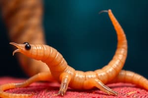
Toxocara canis Overview
- Commonly known as the dog roundworm.
- Adult females reside in the small intestine of infected dogs.
- Produce thick-shelled, unembryonated eggs.
Egg Development and Transmission
- Eggs are excreted through the feces of infected dogs.
- Development of Toxocara canis eggs is influenced by temperature and humidity.
- Eggs develop into infective third-stage larvae (L3) within a few weeks depending on environmental conditions.
Infection Routes for Dogs
- Primary infection routes: ingestion of infective eggs, consumption of paratenic hosts, transplacental transmission, transmammary transmission.
- Ingested eggs hatch into L3 larvae in the small intestine, which then penetrate the intestinal wall and enter the bloodstream.
Life Cycle Migration
- L3 larvae migrate to the liver and lungs via the bloodstream after penetrating the intestinal wall.
- Larvae undergo further development in the lungs and can be coughed up and swallowed.
- Somatic migration occurs in older dogs, with larvae migrating to tissues (muscles, organs) where they remain dormant as second-stage somatic larvae.
Adult Worm Maturation and Symptoms
- Larvae mature into adult worms in the small intestine after migrating through the lungs.
- Young puppies with heavy infections may exhibit coughing, noisy breathing, vomiting, diarrhea, stunted growth, distended abdomen, and abdominal discomfort.
- Adult dogs often show asymptomatic infections.
Diagnosis and Treatment
- Diagnosis of Toxocara canis is made by detecting eggs in feces.
- Difficulty in diagnosing older dogs is due to dormant somatic stage larvae.
- Common treatments include deworming medications such as piperazine, fenbendazole, or mebendazole.
Human Infection and Larval Migrans
- Humans can become infected by ingesting eggs of Toxocara canis.
- Two types of larval migrans: Visceral Larva Migrans (VLM) and Ocular Larva Migrans (OLM).
- Children under age four are most at risk for VLM.
Clinical Manifestations in Humans
- VLM signs include fever, elevated white blood cell count, weight loss, wheezing, and coughing.
- Toxocara canis larvae migrate to various organs, such as the liver and lungs, during VLM.
- OLM is characterized by larvae migrating to the eye, potentially causing severe complications.
- OLM clinical signs include formation of a granulomatous mass in the eye and potential vision problems or blindness.
Morphological Differences
- Cervical alae of Toxocara canis have clear cuticular flanges running along the anterior lateral margins.
- Distinct from Toxocara cati, which has broad alae ending in an "arrow-head" shape.
- Differentiated from Toxascaris leonina, which has a "lance-like" appearance with gradually terminating cervical alae.
Overview of Toxocara cati
- Toxocara cati is commonly known as the cat roundworm.
Infection Routes
- Cats can become infected by:
- Ingesting infective eggs from the environment (L3 stage).
- Consuming paratenic hosts, such as rodents.
- Transmammary transmission via mother's milk.
Paratenic Hosts
- Paratenic hosts are animals that carry Toxocara cati larvae within their tissues and can infect cats when ingested.
Transmammary Transmission
- Kittens can acquire Toxocara cati infection through their mother's milk if the mother is infected.
Lifecycle in Cats
- Ingested Toxocara cati eggs hatch in the small intestine, releasing L3 larvae that penetrate the intestinal wall.
- Larvae migrate through tissues to the lungs, ascend through the trachea to the pharynx, and are then swallowed back into the small intestine.
- Once in the intestines, larvae mature into adult worms.
Adult Worms
- Adult Toxocara cati worms feed on intestinal contents in the cat's small intestine.
- The eggs produced are excreted in the cat's feces.
Egg Infectivity
- It takes approximately 2-4 weeks for Toxocara cati eggs to become embryonated and infective.
Public Health Implications
- Toxocara cati poses a zoonotic risk, particularly to children, who might accidentally ingest infective eggs.
- Potential conditions resulting from infection include visceral larva migrans (VLM) and ocular larva migrans (OLM).
Clinical Signs in Kittens
- Young kittens infected with Toxocara cati may exhibit:
- Noisy breathing
- Coughing
- Vomiting
- Diarrhea
- Abdominal discomfort
- Distended abdomen
- Stunted growth
- Fever
- Lethargy
Severe Infections
- Heavy infections can lead to life-threatening complications such as intestinal obstruction or perforation.
Diagnosis
- Toxocara cati infection is diagnosed by identifying eggs in the cat's feces.
Deworming Protocol
- Kittens should be routinely dewormed:
- At 2 weeks of age
- Again two weeks later
- Another dose at two months
Importance of Regular Deworming
- Regular deworming is crucial, particularly for cats that hunt, to reduce the risk of infection from ingested larvae.
Hygiene Practices
- Prevent spread by:
- Regularly cleaning litter boxes.
- Avoiding soil contamination where cats might defecate.
Monitoring Hunting Behavior
- Limiting a cat's hunting activity reduces the risk of them ingesting paratenic hosts that carry Toxocara cati larvae.
Importance of Public Education
- Raising awareness about Toxocara cati helps cat owners understand risks and encourage protective measures for both pets and humans.
Overview of Toxascaris leonina
- Toxascaris leonina is a roundworm affecting both dogs and cats.
- Primary infection route is through ingestion of infective eggs containing L3 larvae or paratenic hosts.
Anatomical Characteristics
- Cervical alae appear lance-like due to a gradual termination merging into the cuticle.
- Differentiation from Toxocara canis: Toxocara canis has clear-cut, pronounced cervical alae along the anterior lateral margins.
- Differentiation from Toxocara cati: Toxocara cati features broad cervical alae which end abruptly, creating an arrow-head appearance.
Life Cycle and Size
- Eggs measure approximately 80 x 65 µm, are thick-shelled and smooth.
- Adult worms reside in the small intestine of definitive hosts (dogs or cats).
- Larvae hatch in the small intestine and develop directly into adults without significant migration.
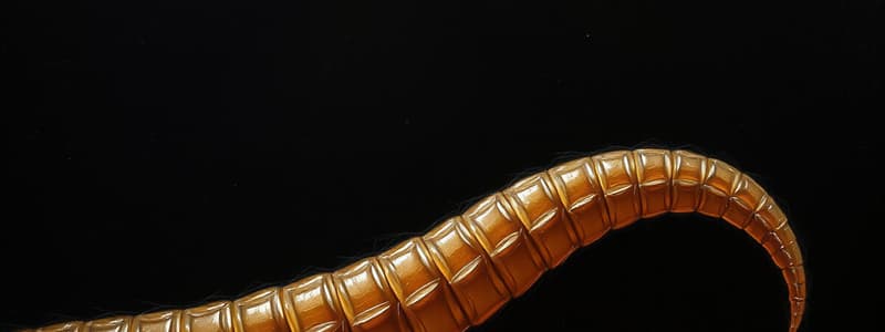
- Females can reach lengths up to 10 cm; males can grow up to 7 cm.
Feeding and Excretion
- Toxascaris leonina feeds on the host's gut contents within the small intestine.
- Adult females lay eggs in the intestines, which are then excreted in feces.
Transmission and Infection in Young Animals
- Toxascaris leonina does not transmit via transplacental or transmammary routes.
- Puppies and kittens typically become infected through environmental exposure to infective eggs or by consuming infected paratenic hosts.
Clinical Signs and Diagnosis
- Common clinical signs include diarrhea, dehydration, poor health, and rare cases of death.
- Infections are usually not severe but can lead to distress to pets.
- Diagnosis is made by identifying Toxascaris leonina eggs during fecal examinations.
Deworming Recommendations
- Regular deworming is crucial for pets at risk, particularly those that hunt or have access to environments where they might ingest infective eggs or paratenic hosts.
Overview of Baylisascaris procyonis
- Commonly known as the raccoon roundworm.
- Adults reside in the small intestine of raccoons.
- Eggs are excreted in the feces of raccoons.
Egg Characteristics and Infectivity
- Eggs become infective within 2 to 4 weeks under optimal environmental conditions.
- Feature a thick, mammillated outer shell for resilience against environmental factors.
Infection Pathways
- Raccoons ingest infective eggs from contaminated soil, water, or food, or through incidental hosts with encysted larvae.
- Incidental hosts include over 90 species of mammals and birds, including humans.
Effects on Incidental Hosts
- Upon ingestion, larvae hatch and migrate through host tissues, particularly affecting the ocular and central nervous system.
- Can lead to severe conditions: visceral larval migrans (VLM), ocular larval migrans (OLM), and neural larval migrans (NLM), which can be fatal.
Public Health Concerns
- Baylisascaris procyonis poses a significant public health risk due to the potential for accidental human ingestion of infective eggs.
- NLM is the most clinically significant condition resulting from infection.
Diagnostic Methods
- Primary diagnosis involves identifying eggs in feces using fecal flotation techniques.
- Additional methods include serological tests and molecular techniques (e.g., PCR) to confirm the presence of the parasite.
Treatment and Prevention
- No effective treatment exists for incidental hosts like humans.
- Preventive measures include decontaminating environments through methods like flaming with portable propane torches or turning and flaming surface soil.
Larvae Characteristics
- Baylisascaris procyonis larvae can grow up to 60 mm in width.
- Diagnostic features include prominent lateral alae and excretory columns.
Pathogenicity in Raccoons
- Generally nonpathogenic in raccoons, their natural host, thus causing minimal harm.
Nanophyetus salmincola Overview
- Commonly known as the salmon poisoning fluke.
- Approximately 1 mm in length.
- Transmits the pathogenic bacterium Neorickettsia helminthoeca.
Habitat and Hosts
- Primarily found in the Pacific Northwest of the United States.
- Infects species that consume raw or undercooked salmon, trout, or Pacific Giant Salamander.
Life Cycle Stages
- Egg Stage: Eggs laid in the intestines of definitive hosts, typically canids (e.g., dogs), and excreted in feces.
- Miracidium Stage: Free-swimming larvae called miracidia hatch from the eggs.
- Intermediate Host: Freshwater snail, where miracidia develop into sporocysts, then rediae, followed by cercariae.
- Cercariae: Free-swimming larvae released into water, actively seeking and penetrating the fish's skin or fins.
Infection Process
- Inside Fish: Cercariae transform into metacercariae, the infective stage.
- Definitive host (e.g., dogs) gets infected by consuming raw or undercooked fish containing metacercariae.
Adult Fluke and Disease
- Adult flukes reside in the small intestine of the definitive host, maturing and laying eggs.
- Causes Salmon Poisoning Disease (SPD) associated with Neorickettsia helminthoeca.
Clinical Signs of Infection
- Symptoms include high fever, weight loss, vomiting, diarrhea.
- Without treatment, can lead to death within 7 to 10 days.
Diagnosis and Testing
- Diagnosis involves identifying eggs in feces, history of raw fish ingestion, and clinical signs.
- Confirmation of Neorickettsia helminthoeca can be achieved through serology or polymerase chain reaction (PCR) tests.
Parasite Overview
- Ancylostoma spp. and Uncinaria spp. are hookworms affecting dogs and cats.
- Adult female hookworms live in the small intestine of their hosts.
Egg Excretion and Development
- Hookworm eggs are excreted in the feces of infected hosts.
- Development from egg to infective larvae (L3) takes 5-7 days in the environment.
Infection Routes
- Primary infection routes include:
- Skin penetration by infective larvae in the environment.
- Ingestion of infected paratenic hosts.
- Transmammary transmission to puppies from arrested larvae in maternal tissues.
Larvae Migration
- When infective hookworm larvae are ingested through paratenic hosts, they penetrate the oral mucosa and migrate as if entering through the skin.
- After skin penetration, larvae enter the bloodstream, migrate to the lungs, ascend the trachea, and are swallowed to reach the small intestine.
Prepatent Period and Infection Effects
- The prepatent period for hookworms is 15-18 days before eggs are passed in feces.
- Main pathological effects include:
- Blood sucking leading to anemia.
- Gastrointestinal symptoms, e.g., black tarry feces.
- Dermatitis at sites of skin penetration by larvae.
Clinical Signs of Severe Infection
- Severe infections may present with:
- Pale mucous membranes due to anemia.
- Symptoms of lethargy and weakness.
- Pruritic skin lesions, notably interdigital lesions.
Diagnosis
- Diagnosis of Ancylostoma or Uncinaria spp. infection is performed through fecal examination to detect eggs.
Cutaneous Larval Migrans (CLM)
- A skin condition caused by the migration of hookworm larvae through human skin.
Causative Agents
- Primarily caused by L3 larvae of hookworms, specifically Ancylostoma braziliense and Ancylostoma caninum.
Mode of Infection
- Infection occurs upon contact with contaminated soil or sand harboring hookworm larvae.
Environmental Context
- Commonly contracted in warm, moist settings such as beaches, playgrounds, and areas where dogs or cats defecate.
Larvae Penetration
- The L3 larvae typically penetrate human skin through exposed areas, often the feet.
Life Cycle in Humans
- Hookworm larvae cannot complete their life cycle in humans, as they do not reach the intestines to mature into adult worms.
Symptoms
- Characterized by an intensely pruritic rash, displaying serpiginous (snake-like) tracks on the skin.
Duration of Condition
- CLM is self-limiting, with larvae dying within 5-6 weeks; however, itching may persist beyond this period.
Diagnosis
- Diagnosed based on the distinctive appearance of skin lesions along with the patient's exposure history to contaminated environments.
Treatments
- Treatment may include topical anti-itch medications like corticosteroids or antihistamines, and in some cases, oral antiparasitic medications like ivermectin or albendazole.
Preventive Measures
- Reduce risk by avoiding barefoot walking in pet-defecated areas, covering sandboxes, and ensuring pets are regularly dewormed.
Public Health Significance
- CLM underscores the zoonotic potential of certain parasites, emphasizing the importance of pet hygiene and environmental sanitation awareness among the public.
Host and Habitat
- Primary hosts for Strongyloides stercoralis include dogs and primates.
- Adult female worms inhabit the mucosa of the anterior half of the small intestine.
Reproductive Cycle
- Reproduction occurs through mitotic parthenogenesis, resulting in eggs passed as L1 larvae (both male and female) in feces.
- In the environment, larvae can develop into free-living males and females that reproduce, leading to infective L3 larvae.
- Parasitic females can also develop directly into infective L3 larvae which infect hosts through skin penetration.
Auto-infection Mechanism
- L1 larvae in the intestine can transform into L3 and penetrate the intestinal mucosa or perineal skin.
- The larvae migrate through the bloodstream to the lungs, then to the small intestine, where they mature into adult females that lay eggs.
Free-Living Stage
- L1 larvae develop into free-living adult males and females in the environment.
- These adults mate and produce more larvae, developing them into infective L3 larvae.
Infection Process
- Infective L3 larvae penetrate the skin of a new host and migrate through the bloodstream to the lungs.
- Upon reaching the lungs, they invade the alveoli, travel up the trachea, and are swallowed to reach the small intestine.
Developmental Pathways
- Homogonic development entails direct development into infective L3 larvae.
- Heterogonic development involves free-living adults that reproduce before developing into infective L3 larvae.
Associated Pathology
- Infection can cause mucosal damage, diarrhea (potentially bloody), abdominal pain, and respiratory issues such as coughing and wheezing.
Clinical Signs in Puppies
- Common symptoms in puppies include severe diarrhea (occasionally bloody), weight loss, and respiratory signs like coughing and wheezing.
Severe Conditions
- Pneumonia can occur, especially due to the migration of larvae through the lungs.
Diagnosis
- Diagnosis involves fecal examination (via direct smear or Baermann technique), assessment of clinical signs, serological tests, and understanding the environmental history.
Taenia spp. Overview
- Taenia spp. are tapeworms that primarily infect dogs and cats.
- Adult tapeworms can grow between 1 to 9 feet long in the small intestine of definitive hosts.
Hosts
- Definitive Hosts: Dogs and cats serve as the primary definitive hosts for Taenia spp.
- Intermediate Hosts: Grazing animals such as cattle, pigs, and sheep act as intermediate hosts depending on the specific Taenia species.
Life Cycle
- Egg Ingestion: Intermediate hosts ingest Taenia eggs, which are excreted in the feces of definitive hosts.
- Larval Development: Upon ingestion, the eggs hatch in the intermediate host's intestine, releasing larvae that penetrate the intestinal wall and migrate to specific tissues, where they develop into larval forms.
Larval Forms
- Cysticerci: Contain a single protoscolex, representing one larval stage of Taenia spp.
- Coenuri: Contain multiple protoscolices, another larval form of Taenia spp.
Infection in Definitive Hosts
- Definitive hosts become infected by consuming undercooked or raw meat from intermediate hosts containing larval forms (cysticerci or coenuri).
- Taenia spp. tapeworms mature in the small intestine of the definitive host, where they can establish infections.
Pathology
- Asymptomatic Infections: Infections in definitive hosts are usually asymptomatic, though mild gastrointestinal discomfort can occur.
- Effects on Intermediate Hosts: The development of cysticerci may cause organ damage in intermediate hosts and potentially lead to meat condemnation at slaughter.
Clinical Signs and Diagnosis
- Common clinical signs in infected dogs and cats are often absent; however, occasional gastrointestinal upset or weight loss may occur.
- Diagnosis is primarily through fecal examination for Taenia eggs or visualization of proglottids in feces or around the perianal area.
Egg and Proglottid Characteristics
- Under microscopic examination, Taenia eggs appear round and radially striated, which aids in distinguishing them from other parasites.
- Proglottids: Segments of the tapeworm that can be seen in feces or around the perianal area are crucial for diagnosis of Taenia spp. infections.
Echinococcus spp. Overview
- Echinococcus spp. are a type of tapeworm.
- Two primary species affecting dogs and cats are Echinococcus granulosus and Echinococcus multilocularis.
Definitive Hosts
- Echinococcus granulosus: definitive hosts include dogs and other canids.
- Echinococcus multilocularis: definitive hosts are red foxes, with occasional infections in dogs and cats.
Size of Adult Tapeworms
- Adult Echinococcus granulosus tapeworms can grow up to approximately 7 mm long.
- Adult Echinococcus multilocularis tapeworms can grow up to about 3.5 mm long.
Intermediate Hosts
- Echinococcus granulosus intermediate hosts include herbivores, pigs, humans, and other mammals.
- Echinococcus multilocularis primarily infects microtine rodents, such as voles, and can also infect humans.
Cyst Formation
- Echinococcus granulosus larvae form hydatid cysts in intermediate hosts.
- Echinococcus multilocularis larvae form alveolar hydatid cysts in intermediate hosts.
Transmission Route
- Infection occurs when intermediate hosts ingest eggs shed in the feces of definitive hosts.
- Ingested eggs hatch in the intestine, releasing oncospheres that penetrate the intestinal wall and migrate to organs.
Lifecycle in Definitive Hosts
- When definitive hosts consume cysts found in intermediate hosts, protoscolices emerge and mature into adult tapeworms, shedding eggs via feces.
Pathology in Intermediate Hosts
- Echinococcus granulosus larvae develop into large hydatid cysts, causing pressure on surrounding organs.
- Echinococcus multilocularis larvae form invasive alveolar hydatid cysts that proliferate and infiltrate tissues, resembling malignant growths.
Clinical Signs
- Echinococcus spp. rarely cause immediate problems in definitive hosts, typically presenting as cosmetic concerns.
- Echinococcus granulosus may cause abdominal pain, nausea, and symptoms due to organ pressure, particularly affecting the liver.
- Echinococcus multilocularis can lead to severe, often fatal complications within a few years if untreated, due to organ invasion.
Diagnosis
- Diagnosis is made through fecal examination, imaging techniques like ultrasound, CT, MRI, and serological tests.
- Diagnostic challenges arise due to the morphologically similar eggs of Echinococcus spp. and Taenia spp., making distinction difficult with fecal flotation alone.
Key Differences Between Species
- Echinococcus granulosus primarily affects dogs and forms hydatid cysts.
- Echinococcus multilocularis mainly affects foxes, forming more aggressive alveolar hydatid cysts that can be fatal without treatment.
Dipylidium caninum Overview
- Commonly known as the flea tapeworm, double-pored tapeworm, or cucumber seed tapeworm.
- Primarily affects dogs, cats, and humans.
Size and Growth
- Adult tapeworms can grow approximately 10 to 70 cm in length.
Hosts
- Definitive hosts include dogs, cats, and humans.
- Intermediate hosts are fleas and lice.
Transmission
- Transmitted to definitive hosts by ingestion of an infected flea or louse containing the larval form of the tapeworm.
Life Cycle
- Begins with adult tapeworms residing in the small intestine of the host.
- Eggs are shed through proglottids (segments) in feces.
- Flea larvae ingest the eggs, which develop into infectious cysticercoids within fleas.
- Definitive hosts become infected by ingesting fleas.
Location within Host
- Adult Dipylidium caninum tapeworms reside in the small intestine.
Pathological Impact
- Generally does not cause significant medical issues; mainly considered a cosmetic concern.
Clinical Signs
- Most adult animals show no symptoms (asymptomatic).
- Young animals may experience mild gastrointestinal upset or discomfort, but these signs are not specific to this parasite.
Notable Characteristics
- Characteristic sign noticed by pet owners includes proglottids in feces or around the pet's anus, resembling grains of rice or cucumber seeds.
Diagnosis
- Diagnosed through fecal examination, visualization of proglottids, and identifying eggs under a microscope.
- Eggs are oval and have a distinct appearance when observed microscopically.
Flea Connection
- Life cycle closely tied to flea populations; fleas act as the intermediate host.
- Controlling fleas is critical to prevent Dipylidium caninum infections.
Preventive Measures
- Regular flea control and deworming of pets.
- Maintaining good hygiene practices to reduce infection risk.
Trichuris Species in Canines and Felines
- Trichuris vulpis is the species of most concern in dogs.
- Trichuris campanula is a species of concern in cats, albeit rare.
Common Name and Morphology
- Common name for Trichuris spp. is whipworms.
- Adult Trichuris vulpis worms typically measure 4 to 8 cm in length.
- Distinctive morphological feature: long, slender anterior end making up about two-thirds of the body length, resembling a whip.
Life Cycle and Egg Characteristics
- Trichuris spp. have a direct life cycle.
- Eggs are excreted in the host's feces.
- Eggs demonstrate significant environmental resilience, surviving for extended periods.
- Infective stage: Trichuris eggs typically take 9-12 days to develop.
Infection and Pathophysiology
- Ingested infective Trichuris eggs hatch in the intestine, releasing larvae that penetrate the intestinal wall and migrate to the cecum.
- Larvae mature into adult worms in the cecum and colon of the host.
- Typhlitis refers to the inflammation of the cecum due to heavy worm burdens of Trichuris spp.
- Adult worms cause damage by embedding into the intestinal mucosa, leading to inflammation and tissue damage.
Clinical Signs of Infection
- Adult dogs often remain asymptomatic but may show mild signs.
- Puppies tend to be more severely affected, exhibiting symptoms like mucoid diarrhea and rare occurrences of blood in stool.
Diagnosis
- Diagnosis of Trichuris spp. infection is performed through fecal flotation, which identifies the characteristic bipolar eggs.
- Under microscopic examination, Trichuris spp. eggs feature a distinctive bipolar appearance.
Zoonotic Helminths in Cats and Dogs
- Several helminths, including hookworms like Ancylostoma braziliense, can infect humans and cause Cutaneous Larva Migrans (CLM).
- Cutaneous Larva Migrans occurs when hookworm larvae penetrate human skin, creating pruritic, serpiginous lesions.
Infection with Dipylidium caninum
- Humans can become infected with Dipylidium caninum by ingesting infected fleas, highlighting the need for effective flea control.
Public Health Concerns of Tapeworms
- Echinococcus spp. tapeworms pose a significant public health risk, potentially causing hydatid disease in humans.
- Hydatid disease results from ingesting eggs shed in the feces of infected animals, leading to cyst formation in organs such as the liver and lungs.
Diagnostic Methods for Helminths
- The primary identification method for helminths in pets is fecal flotation, where feces are mixed with a solution, allowing eggs to float for microscopic examination.
- Strongyloides stercoralis is typically identified through a direct smear of feces for larvae or the Baermann technique.
Proglottids and Tapeworm Diagnosis
- A proglottid is a segment of a tapeworm, often found in feces or around the peri-rectal area, crucial for diagnosing tapeworm infections like Dipylidium caninum.
Serological Testing
- Serological tests are employed to detect specific antibodies or antigens associated with helminth infections.
Preventive Measures Against Helminths
- Regular deworming is essential for pets, best done using approved anthelminthic medications.
- Puppies should be dewormed at 2, 4, 6, and 8 weeks of age, followed by monthly treatments when possible.
Feces Management
- Promptly picking up dog feces minimizes the risk of parasite eggs or larvae contaminating the environment, thus reducing reinfection potential.
Sanitation Practices
- Maintaining good sanitation in pet areas is vital; this includes washing surfaces to eliminate free-living parasite stages.
Importance of Flea Control
- Flea control is critical for preventing Dipylidium caninum infections since fleas act as intermediate hosts.
Sandbox Safety Measures
- Covering sandboxes can prevent contamination from pet feces, thereby reducing the risk of helminth exposure.
Public Health Awareness
- Educating pet owners about the zoonotic potential of parasites like Echinococcus spp., along with hygiene and preventive measures, is essential for public health.
Role of Routine Veterinary Check-Ups
- Routine veterinary visits, including fecal examinations, aid in the early detection and treatment of parasites, preventing serious health issues in pets.
Studying That Suits You
Use AI to generate personalized quizzes and flashcards to suit your learning preferences.