Podcast
Questions and Answers
Which of the following cell types is LEAST likely to regenerate after significant damage?
Which of the following cell types is LEAST likely to regenerate after significant damage?
- Fibroblasts
- Neurons (correct)
- Epidermis
- Hepatocytes
In liver damage, which outcome is most likely when the supporting framework is severely damaged?
In liver damage, which outcome is most likely when the supporting framework is severely damaged?
- Normal tissue growth
- Cirrhosis with disorganized regeneration and fibrosis (correct)
- Complete regeneration of liver tissue
- Development of labile cells
What is the primary function of osteoclasts during the remodeling phase of bone fracture healing?
What is the primary function of osteoclasts during the remodeling phase of bone fracture healing?
- Forming the bone matrix
- Removing unnecessary callus (correct)
- Providing oxygen and nutrients to cells
- Secreting alkaline phosphatase
What is the first critical step in tissue healing?
What is the first critical step in tissue healing?
If a fracture is poorly immobilized, which of the following is most likely to occur?
If a fracture is poorly immobilized, which of the following is most likely to occur?
Which event characterizes the cleaning phase of tissue healing?
Which event characterizes the cleaning phase of tissue healing?
In the healing of a simple bone fracture, what role do capillaries play during the formation of provisional callus?
In the healing of a simple bone fracture, what role do capillaries play during the formation of provisional callus?
Why is the proliferation phase important during tissue repair?
Why is the proliferation phase important during tissue repair?
Following damage to the epidermis, which type of repair is most likely to occur?
Following damage to the epidermis, which type of repair is most likely to occur?
What is the primary distinction between labile and stable cells in terms of tissue repair?
What is the primary distinction between labile and stable cells in terms of tissue repair?
Flashcards
Tissue Repair
Tissue Repair
Replacement of damaged tissue by new, healthy tissue.
Regeneration
Regeneration
Replacement of damaged tissue with the same tissue type.
Fibrosis
Fibrosis
Replacement of damaged tissue with fibrous connective tissue, especially in the CNS (called Gliosis).
Labile Cells
Labile Cells
Signup and view all the flashcards
Permanent Cells
Permanent Cells
Signup and view all the flashcards
Stable Cells
Stable Cells
Signup and view all the flashcards
Cleaning Phase
Cleaning Phase
Signup and view all the flashcards
Proliferation Phase
Proliferation Phase
Signup and view all the flashcards
Remodeling Phase
Remodeling Phase
Signup and view all the flashcards
Osteoid (Soft Callus)
Osteoid (Soft Callus)
Signup and view all the flashcards
Study Notes
- Tissue repair is the replacement of damaged tissue by new, healthy tissue.
Types of Repair
- Classical types of repair include regeneration and fibrosis (gliosis in the CNS).
- Organization is another type of repair.
Factors Determining the Type of Repair
- Type of damaged cells
- The state of the supporting framework
Types of Damaged Cells
Labile Cells
- Continuously dividing cells that heal by regeneration if damaged.
- Examples include epidermis, mucous membranes (epithelial cells), and blood and lymphoid tissue.
Permanent Cells
- Cells that do not proliferate.
- Muscle cells heal by fibrosis if damaged.
- Nerve cells heal by gliosis if damaged.
Stable Cells
- Cells that can proliferate with limited capacity.
- They can heal by mixed regeneration and/or fibrosis if damaged.
- Parenchymal cells (hepatocytes, renal tubules, endocrine & exocrine glands), and mesenchymal cells (fibroblasts, osteoblasts, & chondroblasts) are examples.
Supporting Framework
- Intact framework leads to healing by regeneration.
- Destroyed framework results in disorganized cell growth and improper regeneration mixed with fibrosis, such as in liver cirrhosis.
Phases of Healing
- Cleaning phase involves the removal of necrotic debris by phagocytic cells.
- Proliferation phase involves cells responsible for repair proliferating to replace the damaged ones.
- Re-modeling phase involves maturation of the new tissue to simulate the original tissue.
Healing by Regeneration
- Regeneration is the replacement of damaged tissue by healthy tissue of the same kind.
- Labile cells and stable cells (with intact framework) heal by regeneration.
Examples of Healing by Regeneration
Healing in the Skin
- Damage to the epidermis or mucosa only leads to regeneration.
- Damage to the epithelium and sub-epithelium results in fibrosis with the loss of hair follicles, and sweat and sebaceous glands.
Healing of Liver Damage
- Intact supporting framework (mild damage) leads to regeneration.
- Destroyed framework (severe damage) results in cirrhosis, disorganized regeneration mixed with fibrosis, and loss of architecture.
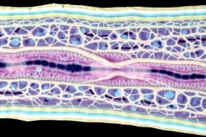
Healing of Simple Bone Fracture
Cleaning Phase
- Macrophages and osteoclasts clean blood and necrotic debris between the fracture ends.
Proliferation Phase
- A modified type of granulation tissue develops between the fracture ends, formed of capillaries, fibroblasts, and osteoblasts.
- Capillaries provide O2 and nutrients to the active cells.
- Fibroblasts begin to form collagen of bone.
- Osteoblasts form the bone matrix.
- Early formation of osteoid (soft callus) unites the fracture ends, formed of internal, external, and intermediate callus.
- Late formation of hard callus (osseous) occurs as osteoblasts secrete alkaline phosphatase with mineral deposition in the soft callus, leading to complete union of the fracture.
Remodeling Phase
- Osteoclasts remove unnecessary internal and external callus.
- The remaining intermediate callus matures into lamellar bone, and bone marrow regeneration occurs.
Causes of Failure of Bony Union
- Poor immobilization of the fractured ends leads to fibrous union.
- Interposition of soft tissue (muscle or fascia) can cause non-union.
- Poor apposition results in mal-union.
- Other factors include delayed union, pathological fracture caused by bone disease, poor blood supply, infection, old age, and malnutrition.
Studying That Suits You
Use AI to generate personalized quizzes and flashcards to suit your learning preferences.