Podcast
Questions and Answers
Metastases to the thyroid gland, though uncommon, can occur with which combination of cancers?
Metastases to the thyroid gland, though uncommon, can occur with which combination of cancers?
- Prostate, kidney, and bladder cancer
- Lung, breast, and colon cancer (correct)
- Sarcoma, lymphoma, and melanoma
- Ovarian, uterine, and cervical cancer
Which sonographic feature is least likely to suggest a malignant thyroid nodule?
Which sonographic feature is least likely to suggest a malignant thyroid nodule?
- Punctate calcifications
- Hypoechoicity
- Poorly defined margin
- Well-defined thin halo (correct)
What percentage of the general population is estimated to have sonographically demonstrable thyroid nodules?
What percentage of the general population is estimated to have sonographically demonstrable thyroid nodules?
- 30%
- 50%
- 15% (correct)
- 5%
True cystic lesions of the thyroid are most often what type of cyst, and how do they typically appear on ultrasound?
True cystic lesions of the thyroid are most often what type of cyst, and how do they typically appear on ultrasound?
What is a benign inflammatory condition of the thyroid known as?
What is a benign inflammatory condition of the thyroid known as?
In chronic cases of Hashimoto's thyroiditis, which of the following is least likely to be encountered?
In chronic cases of Hashimoto's thyroiditis, which of the following is least likely to be encountered?
Which statement regarding markedly hypoechoic and solid nodules is most accurate?
Which statement regarding markedly hypoechoic and solid nodules is most accurate?
Which of the following is NOT a way of grouping the provided information?
Which of the following is NOT a way of grouping the provided information?
Approximately what percentage of the general population exhibits sonographically detectable thyroid nodules?
Approximately what percentage of the general population exhibits sonographically detectable thyroid nodules?
Which finding is LEAST suggestive of an increased risk of thyroid cancer within a thyroid nodule?
Which finding is LEAST suggestive of an increased risk of thyroid cancer within a thyroid nodule?
What is generally the initial imaging modality of choice for evaluating a palpable thyroid nodule?
What is generally the initial imaging modality of choice for evaluating a palpable thyroid nodule?
Which radiographic finding is LEAST likely to be associated with cretinism?
Which radiographic finding is LEAST likely to be associated with cretinism?
Which factor is LEAST likely to cause nodular thyroid disease?
Which factor is LEAST likely to cause nodular thyroid disease?
A parathyroid nodule becomes increasingly suspicious for pathology when its size exceeds what threshold?
A parathyroid nodule becomes increasingly suspicious for pathology when its size exceeds what threshold?
According to the TIRADS classification, what is the risk of malignancy for a thyroid nodule classified as TIRADS 2?
According to the TIRADS classification, what is the risk of malignancy for a thyroid nodule classified as TIRADS 2?
What sonographic feature is LEAST characteristic of most benign thyroid nodules?
What sonographic feature is LEAST characteristic of most benign thyroid nodules?
Which of the following is NOT a typical sonographic feature of leiomyomas?
Which of the following is NOT a typical sonographic feature of leiomyomas?
A patient presents with pelvic pain and a suspected pelvic mass. Which imaging modality is typically the MOST cost-effective initial diagnostic tool?
A patient presents with pelvic pain and a suspected pelvic mass. Which imaging modality is typically the MOST cost-effective initial diagnostic tool?
What is a limitation of using transvaginal sonography (TVS) compared to other imaging modalities?
What is a limitation of using transvaginal sonography (TVS) compared to other imaging modalities?
Which diagnostic imaging procedure is MOST appropriate for evaluating tubal occlusion as a potential cause of infertility related to uterine anomalies?
Which diagnostic imaging procedure is MOST appropriate for evaluating tubal occlusion as a potential cause of infertility related to uterine anomalies?
Which of the following is NOT a typical indication for performing a hysterosalpingogram (HSG)?
Which of the following is NOT a typical indication for performing a hysterosalpingogram (HSG)?
Which of the following is NOT generally considered a contraindication for hysterosalpingogram (HSG)?
Which of the following is NOT generally considered a contraindication for hysterosalpingogram (HSG)?
Which of the following is NOT a recognized complication of hysterosalpingogram (HSG)?
Which of the following is NOT a recognized complication of hysterosalpingogram (HSG)?
During what phase of the menstrual cycle does the trophoblast start producing Human Chorionic Gonadotropin (HCG)?
During what phase of the menstrual cycle does the trophoblast start producing Human Chorionic Gonadotropin (HCG)?
Pes anserine bursitis of the knee involves the insertion of what three muscles?
Pes anserine bursitis of the knee involves the insertion of what three muscles?
Which set of bones correctly comprise the ankle joint?
Which set of bones correctly comprise the ankle joint?
What imaging modality offers an efficient and inexpensive way to assess tendons and tendon sheaths around the ankle?
What imaging modality offers an efficient and inexpensive way to assess tendons and tendon sheaths around the ankle?
The 'Tom, Dick, and Harry' tendons are located posterior to the medial malleolus. What are the corresponding muscles?
The 'Tom, Dick, and Harry' tendons are located posterior to the medial malleolus. What are the corresponding muscles?
Which muscle group is located at the lateral malleolus of the ankle?
Which muscle group is located at the lateral malleolus of the ankle?
Which of the following is NOT a joint that contributes to the overall ankle joint complex?
Which of the following is NOT a joint that contributes to the overall ankle joint complex?
What is the approximate ratio of cervix to corpus size in a postmenopausal uterus?
What is the approximate ratio of cervix to corpus size in a postmenopausal uterus?
How does the endometrium typically appear on ultrasound during menstruation (days 1-4)?
How does the endometrium typically appear on ultrasound during menstruation (days 1-4)?
In a parous adult, what is the typical volume range of the uterus?
In a parous adult, what is the typical volume range of the uterus?
What is the approximate cervix/corpus ratio observed in a nulliparous adult?
What is the approximate cervix/corpus ratio observed in a nulliparous adult?
Which of the following is a limitation associated with using Transvaginal Sonography (TVS)?
Which of the following is a limitation associated with using Transvaginal Sonography (TVS)?
Which of the following is NOT typically an indication for performing transvaginal sonography (TVS)?
Which of the following is NOT typically an indication for performing transvaginal sonography (TVS)?
What is generally considered the most practical imaging modality for assessing ovarian tumors?
What is generally considered the most practical imaging modality for assessing ovarian tumors?
During the luteal phase of menstruation, if fertilization occurs, what hormone does the developing trophoblast produce?
During the luteal phase of menstruation, if fertilization occurs, what hormone does the developing trophoblast produce?
Which uterine structure is most readily identifiable on MRI, crucial in uterine pathology assessment, yet less distinct on ultrasound and CT?
Which uterine structure is most readily identifiable on MRI, crucial in uterine pathology assessment, yet less distinct on ultrasound and CT?
Which statement is NOT true regarding the junctional zone of the uterus?
Which statement is NOT true regarding the junctional zone of the uterus?
Which imaging technique is typically preferred for evaluating congenital uterine anomalies?
Which imaging technique is typically preferred for evaluating congenital uterine anomalies?
The main ligamentous support for the uterus and cervix is provided by which of the following?
The main ligamentous support for the uterus and cervix is provided by which of the following?
Which ligament supporting the pelvic organs is described as a double reflection of the peritoneum?
Which ligament supporting the pelvic organs is described as a double reflection of the peritoneum?
Which uterine ligament is considered analogous to the male gubernaculum?
Which uterine ligament is considered analogous to the male gubernaculum?
Which of the following accurately describes the endometrium?
Which of the following accurately describes the endometrium?
Which of the following structures does NOT form a border of the retroperitoneum?
Which of the following structures does NOT form a border of the retroperitoneum?
The kidneys are located within which space of the retroperitoneum?
The kidneys are located within which space of the retroperitoneum?
Which of the following retroperitoneal spaces typically does NOT contain any major organs?
Which of the following retroperitoneal spaces typically does NOT contain any major organs?
Flashcards
Thyroid Nodules Prevalence
Thyroid Nodules Prevalence
Sonographically demonstrable thyroid nodules are present in nearly 25-40% of the population.
Risk Factors for Thyroid Cancer
Risk Factors for Thyroid Cancer
Prior neck radiation, MEN-II Syndrome, known prior thyroid cancer, and a positive neck node all increase the risk for thyroid cancer.
Suggests Lower Risk for Thyroid Cancer
Suggests Lower Risk for Thyroid Cancer
Eggshell calcifications in thyroid nodules.
Initial Imaging for Thyroid Nodule
Initial Imaging for Thyroid Nodule
Signup and view all the flashcards
Parathyroid Nodule Size
Parathyroid Nodule Size
Signup and view all the flashcards
Thyroid Ultrasound Frequency
Thyroid Ultrasound Frequency
Signup and view all the flashcards
Thyroid Nodule Echogenicity
Thyroid Nodule Echogenicity
Signup and view all the flashcards
TIRADS 4 Classification
TIRADS 4 Classification
Signup and view all the flashcards
Hyperplastic/Adenomatous Nodules
Hyperplastic/Adenomatous Nodules
Signup and view all the flashcards
Thyroid Metastasis
Thyroid Metastasis
Signup and view all the flashcards
Common Primary Cancers that Metastasize to Thyroid
Common Primary Cancers that Metastasize to Thyroid
Signup and view all the flashcards
Malignant Thyroid Nodule Ultrasound Signs
Malignant Thyroid Nodule Ultrasound Signs
Signup and view all the flashcards
Hypoechoic Nodule
Hypoechoic Nodule
Signup and view all the flashcards
Punctate Calcifications
Punctate Calcifications
Signup and view all the flashcards
True Cystic Lesions
True Cystic Lesions
Signup and view all the flashcards
Hashimoto's Thyroiditis
Hashimoto's Thyroiditis
Signup and view all the flashcards
What is Hysterosalpingogram (HSG)?
What is Hysterosalpingogram (HSG)?
Signup and view all the flashcards
Pes Anserine Bursitis
Pes Anserine Bursitis
Signup and view all the flashcards
HSG Indications: Ectopic Pregnancy?
HSG Indications: Ectopic Pregnancy?
Signup and view all the flashcards
HSG: Ligated Abortion Okay?
HSG: Ligated Abortion Okay?
Signup and view all the flashcards
Ankle Joint Bones
Ankle Joint Bones
Signup and view all the flashcards
HSG Complication: Irregular Menses?
HSG Complication: Irregular Menses?
Signup and view all the flashcards
Ankle Ultrasound
Ankle Ultrasound
Signup and view all the flashcards
Ovarian Tumor Evaluation?
Ovarian Tumor Evaluation?
Signup and view all the flashcards
"Tom, Dick, and Harry" Tendons
"Tom, Dick, and Harry" Tendons
Signup and view all the flashcards
Ankle Joint Components
Ankle Joint Components
Signup and view all the flashcards
Luteal Phase Trophoblast Produces?
Luteal Phase Trophoblast Produces?
Signup and view all the flashcards
Technique of choice in the evaluation of uterine congenital anomalies
Technique of choice in the evaluation of uterine congenital anomalies
Signup and view all the flashcards
Primary Support of the Uterus
Primary Support of the Uterus
Signup and view all the flashcards
Hematometrium/colpos: cause?
Hematometrium/colpos: cause?
Signup and view all the flashcards
Endometrium During Menstruation
Endometrium During Menstruation
Signup and view all the flashcards
Normal uterine volume (parous)
Normal uterine volume (parous)
Signup and view all the flashcards
Cervix/Corpus ratio (nulliparous)
Cervix/Corpus ratio (nulliparous)
Signup and view all the flashcards
Trophoblast hormone after fertilization
Trophoblast hormone after fertilization
Signup and view all the flashcards
Uterine Junctional Zone
Uterine Junctional Zone
Signup and view all the flashcards
TVS Indications
TVS Indications
Signup and view all the flashcards
Best modality for ovarian tumors
Best modality for ovarian tumors
Signup and view all the flashcards
TVS Disadvantage
TVS Disadvantage
Signup and view all the flashcards
CT Scan
CT Scan
Signup and view all the flashcards
Endometrium
Endometrium
Signup and view all the flashcards
Cardinal ligament
Cardinal ligament
Signup and view all the flashcards
MRI (for uterine anomalies)
MRI (for uterine anomalies)
Signup and view all the flashcards
Round ligament
Round ligament
Signup and view all the flashcards
Broad Ligament
Broad Ligament
Signup and view all the flashcards
Perirenal Space
Perirenal Space
Signup and view all the flashcards
Anterior Pararenal Space
Anterior Pararenal Space
Signup and view all the flashcards
Study Notes
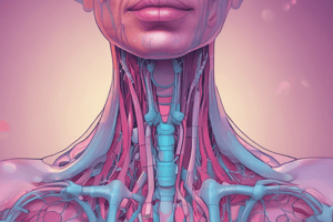
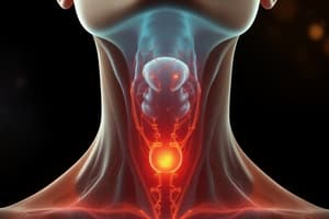
Thyroid Imaging and Disease
- Thyroid hormones T4 and T3 are produced by the cells of the thyroid follicles.
- Medullary carcinoma results from malignant transformation of the thyroid follicle cells.
- Calcitonin is produced by the thyroid gland's A cells, which are the second cell type.
- Papillary, follicular, and aplastic carcinomas are formed by malignant changes in A cells.
- Due to its superficial location, thyroid scanning preferably employs frequency linear array transducers.
- It is most suited for 10 MHz and above.
- Ovulation occurs on day 14 and is stimulated by LH.
- Determining the echogenicity of nodules can be of use.
- Hypoechoic lesions are more likely to be malignant than other lesions.
- When a nodule has cystic changes, it is more likely to be benign.
- Malignant calcifications, which are linked to psammoma bodies, frequently manifest sonographically as fine punctate regions of calcification that appear as high amplitude echoes without acoustic shadowing.
- Color Doppler is not highly specific in differentiating benign from malignant thyroid nodules
- Prominent internal vascularity is vascularity seen in thyroid nodules
- Colloid cysts are benign thyroid lesions.
- Hyperplastic and adenomatous nodules are benign lesions of the thyroid.
- Benign lesions often manifest as isoechoic or echogenic compared to the thyroid's normal background echogenicity.
- Incomplete or irregular haloes are common in hyperplastic and adenomatous nodules.
- Color Doppler detects blood flow in degenerated hemorrhagic nodules.
- Curvilinear and peripheral calcification, also referred to as "eggshell" calcification, are common in benign nodules.
- True thyroid cystic lesions are frequently colloid cysts.
- Comet-tail artifacts known as high amplitude reverberation artifacts are present in predominately anechoic lesions.
- Curvilinear calcifications known as eggshell calcifications are present in anechoic lesions
- Hashimoto's Thyroiditis is a benign inflammatory condition which can include an enlarged gland exhibiting hypoechoic areas and a heterogeneous pattern.
- Vascularity may be seen with Hashimoto's Thyroiditis due to chronic inflammation
- Papillary carcinoma is the most common malignancy of the thyroid.
- Uncommon metastases to the thyroid gland may occur with lung cancer, breast cancer, and colon cancer.
- Color Doppler can show increased internal flow is often seen and but is not specific for malignant nodules.
- A poorly defined margin, hypoechoicity, and punctate calcifications suggest a malignant thyroid nodule.
- Well-defined, thin halos seen on the periphery do not suggest a malignant thyroid nodule but rather a benign one
- Approximately 40% of the population has thyroid nodules that can be sonographically shown.
- An increased risk of thyroid cancer may be indicated by prior neck radiation, MEN-II Syndrome, prior thyroid cancer and a positive neck node.
- Parathyroid nodules that are greater than 5mm are highly suspicious for pathology.
- 10 megahertz or higher is the right, high frequency for the superficial location of the thyroid.
- Unless there is a high risk of thyroid cancer or suspicious characteristics, patients with nodules smaller than 1 cm should not be considered for biopsy.
- Because hypoechoic lesions have a higher clinical probability of being malignant, the echogenicity of nodules must be characterized.
- Isoechoic solid ovoid lesions do not commonly tend to be malignant.
- Lymphoma is not benign.
- Markedly hypoechoic and solid nodules are not classically considered to be benign lesions
- Predominantly anechoic may be considered as true for thyroid cystic lesions that may show tiny amplitude echoes demonstrating known comet tail reverberation factors
- Hashimoto's Thyroiditis is a recognized benign inflammatory thyroid state.
- Papillary carcinoma is not seen in cases of Hashimotos Thyroiditis
- Lung, breast, and colon cancer metastasize .
- Well-defined thin halos seen on the periphery does not suggest a malignant thyroid nodule.
- 40% of individuals have the ability to sonographically display thyroid nodules.
- Eggshell calcifications in thyroid nodules do not suggest an increased risk for thyroid cancer.
- Ultrasound is usually the imaging choice modality for seeing palpable thyroid nodules.
- Premature skull suture closure is not a sign of cretinism.
- Poor vitamin D absorption does not always cause thyroid nodular disease.
- Back of the tongue and foramen cecum is where ectopic thyroids most commonly reside.
- A normal thyroid gland by ultrasound is classified as TIRADS 1
- TIRADS 2 are thyroid nodules that have 0% risk of malignancy.
- TIRADS 4 are thyroid nodules that are classified with irregular micro calcification
- Microcalcification is not a characteristic of benign thyroid nodules.
- Eggshell calcification is frequently seen in malignant nodules.
- Ultrasound is the preferred modality for viewing the Thyroid Gland.
- Solid nodules that were taller rather than wide were signs of thyroid Nodule malignancy.
- A thyroid cyst is a predominately benign nodule trait.
- TIRAD, or Thyroid Imaging Reporting and Data System, is the process.
- The hyoid bone is bone cartilage that's U shaped, inverted, and marks of the hypopharynx onset.
- The retropharyngeal region has the possibility to elevate disease spreading malignancy of the neck.
- The cricoid cartilage differentiates the 3 and 4 degree of the cervical lymph location levels.
- A thyroid nodule is marked at the spongiform echopattern for recognizing benignity.
- A inverting papilloma in the paranasals presents a potential malignant mass within ethmoid sinuses .
- The pharyngeal mucosal is invested with a pharyngeal Mucosal Space.
- The Parapharyngeal space can also be an answer .
- When the patient may be receiving radioactive iodine the contrast CT result scan is then not advised.
- A: A and C are typical sono markers demonstrating vascularity and anecho pattern and comet
- Predominately papillary is what the most thyroid common cancer is
Reproductive Imaging
- The following are anticipated normal uterus ultrasound appearances except the separation of the cervix and body by the exterior os
- Ultrasound relies on the bladder in discerning and highlighting uterus form and position
- Ultrasound ranges vary in the uterus , however, non pregnant and Nulliparous is in range of 30 to 40 mL
- The cervix:corpus ratio in the nulliparous adult is 1:2
- During menstruation the following can appear *Hypoechoic lining, by endometrial with surrounding membrane *
- The evaluation of potential mass formation in the pelvis is not likely done with or by Transvaginal sonography.
- Sonography is usually the procedure for highlighting the ovaries or potential adnexal masses and is usually a high and sensitive modality
- A TVS is a transvaginal ultrasound . However, the field and angles can be limited
- Hysterosalpingogram is what diagnostic imaging is used for when checking uterine anomalies as a potential diagnosis when ruling out a tubal occlusion"
- Ectopic anomaly is not expected nor likely when following Transvaginal procedures.
- Ligated abortion .Is a potential answer for the next line" Following or expecting to follow TVS for abnormalities to the uterus"
- Irregular menstruation is the main side effect and happens in most cases when following TVS or potential Uterine issues
- transvaginal .is still the primary imaging modality for ovarian mass or growth in the Uterus.
- junctional zone .Is the more highlight recognizable zone for knowing or defining uterus.
- The Basal layer of the endometrium is not the true about the junctional zone of the uterus.
- perirenal is the space you see the kidney in. Anterior pararenal is space where pancreas can reside.
- Predominately cystic appearance is not the *Typical sono feature of Leiomyomata * which may show pelvic implications and lacerations. Ultrasound :is one of the most reliable sources for patients with potential PID and needing to determine and see PID within.
- limited field angle by transvaginal angle with potential probes.
- hysterosalpingogram. Diagnostic procedure that is best for highlighting any uterine anomalies." "Pregnancy is likely least side effective reason for hysterosalpingram" Repeat abortions is the *side effects we don't likely need to expect from TVS
- *Endovaginal ultrasound is the key to evaluation of ovarian tumor
- The placenta or trophoblast usually builds HCG in the luteal phases of menses"
- MRI- This is best tech when highlighting the uterus" The above statement is likely key to expect to see in future questions.
- Hematrometrium and hematocolpos" This cause severe Dysmenorrhea. "Hematocolops AND metrium causes high rates of single kidneys" Increased size with thickness is false claim about Adenomyosis.
- *Normal Ovaries * are usually the point to follow the polycycstic or potential abnormalies polycystic causes "
- Heated endometriums is never or not very true *Torsion in cysts or ovaries "
- Cysts that are found to be more that *7cm likely need further MRI analysis or further analysis due to its volume." Two bodies and cervix is likely a side effect or sign known as uterus Didelphys" The uterus is covered by peritoneum Broad support if key or primary for highlighting issues with uterus. "It would be expected that you can see the body connected to the cervix because of its external make up"
- Uterine abnormalities highlight or show potential complications with unicornate version of that diagnosis" "When the or or if potential uterus didelphys what would be true is by or more caused from partial or bad m stress ducts" If the M ducts are fully complete with fusions that is called "Septate uterus MRI is the best for highlihting j zones of the abdominal cavity. fibrous is most associated to highlihigts to posteno pauses women. If there is more than 4mil of fluid from ano pause that should be expected or recognized. Hypoechhoic id side to expect myometrium . Highlihtnsedy is is usually for is what you'ds see for endimetruims Subserosal in likely expect if you can see fibroids on inside of the utering with high cavity submucous : what expect to find of utering for cavity with fibroid inside out "Intramural- for cavit, we also need to take In what in myometrium is seen with" junction all zone measures in the uterus with <8 in cavit* y" Menopauses al endimetrum in the uterus by or often can be is *Endometrial carcinoma " 14, disruption for knowing if myometrial and or tumor, the key is "disruited junction all Thick and large not related ovarian side effects.
Stroke Imaging
- Subtle low T1 signal and no enhancement with contrast is not an MRI finding of acute stroke
- T2-weighted MRI shows hypointense area of predominantly intracellular methemoglobin
- T2-weighted MRI shows hyperacute hours old hemorrhage as a hypointense area surrounded by margin.
- MRI shows chronic hemorrhage hypointense slitor and or surrounding fluid cavity
- MRI is superior to CT for imaging vessels in the posterior fossa and may identify absent flow, compromised circulation of basilar and vertebral arteries.
- Magnetic Resonance Angiography (MRA) is not indicated in cases of otic or cochlear implants instead is more useful when highlighting intraorbital fragments"
- CT scan signs can include loss of insular ribbon""obstruction in lentiform nucleus"
- MRI studies use water for showing brain schematics in the correct or true light for brain ischemia . Sulcal obliteration is of hyper acute infarcts and helps high spot different types of what is or what shows early signs of infraction with those times Epidural hematoma b . Biconvex lentiform is key to follow hematoma CT-scan best helps for subarrinoids when the scan shows hematoma due to its potential to show if there is another type of aneurysm Anterior artery is 2 the anterior side Gliablastoma ( mult multiforme :is known to best connect it to the intra cranial for knowing if issues will follow" Hydranencephaly " is the best congenital abnormailtiy " A-R is the Middle distribution 8,, Not a side effect of the signs acute infections Subdrual for know the trauma A->is the scan/imaging to best side effect of the brain injury " Demylenarion is a intravitreall side effect """"
Neck Spaces & Cervical Lymph nodes
- The hard palate separates the nasopharynx and the oropharynx.
- The parapharyngeal space has no significant structure, except fat, is used as landmark to determine the location of masses in the neck.
- Nasal polyp is the major or most key factor here
- Level II is the correct level for areas close or at the skul base when knowing if there is cervical lymph nodes
- Level 5 is the likely sign of bounders for knowing Cervial lymph node is the key when side effects might occur
- Ultrasound is used in visualizing thyroid glands.
- Solid with a bit height is the known side effects if one is to be malignancy
- C ;by knowing what is below
- For knowing what sepoihhyd does Hyoid" is always key /
- infer or belly to what is to "what anatomic to this triagnel "o
- Enclose is " Cervical vertal" which is deep vertibral that's is vert deep Carotoid sheet is "highway " is fasica "Deep, to lay for investng : Retrophyrangral. Side effect" is one of the most common statements and likely ones being quizzing To be sure it has been that is vert deep, not superficial Danger space: *what in Suirapoid and located " Para space Is it the is at what or close to ( Supryhoid "location "" Parotoid GLANDS is not side effect "the visceral for visceral location. Pyra for like is parphangeal """" Vegula, internal - in what the pa" "carotoid space"" is and in what the medial or posterios" " Lower spread: that that it is danger"" "" Carotin Lateral"""" " Hard Palate to Oro :This Struct to nasopphrany or oral cavity" A; ton" ""Carotiod" is a sheet that connects or is like Lincolin"" is that correct then for the layers" Parahpyrangrak "" in which"
Lung Anatomy via CT
- That that this ( relatively space mall is not in the neck is Paranhoyngeal"
- The visceral space that it is trachra and infra hyoid "An elevator : that at it that spread its likely in the para space. Pops are chronic with nasals cavity to high light" Lever 2 where skull to hyoid "lymb Levelv,,where is interior and bounders"" Under for "Thyroid" Wider or for malig"
Thytoid, this, "That :systic "for benights "
Paeds : "retnoblastia" Phtiths bulbs. For to "end stage" High resolution or scan of the "temporary for bone " MRI """"
""Carotids is what and what""
Meaning Tirad." Only space or fasca: Cart is key for what separate levels" Echod of be" Potential Parenynfial"" Ratiio active y" Signet of the ring"
Studying That Suits You
Use AI to generate personalized quizzes and flashcards to suit your learning preferences.