Podcast
Questions and Answers
In the context of imaging for lower back pain, what is a primary consideration when deciding whether to order radiographic studies?
In the context of imaging for lower back pain, what is a primary consideration when deciding whether to order radiographic studies?
- Not all patients with low back pain require imaging. (correct)
- All patients with low back pain should undergo immediate imaging to rule out serious pathology.
- The patient's age is the sole determinant for ordering imaging.
- Imaging is only necessary if the patient reports pain lasting longer than six weeks.
Which of the following is an indication for imaging in patients with back pain?
Which of the following is an indication for imaging in patients with back pain?
- Patients with a history of well-managed chronic back pain.
- Back pain that resolves within a week with rest.
- Mild muscle soreness after physical activity.
- Patients who have a neurologic deficit. (correct)
A younger patient presents with morning stiffness that improves with exercise, alternating buttock pain, and night awakenings. Which condition should be suspected, potentially warranting imaging?
A younger patient presents with morning stiffness that improves with exercise, alternating buttock pain, and night awakenings. Which condition should be suspected, potentially warranting imaging?
- Acute muscle strain.
- Osteoporosis.
- Vertebral fracture due to trauma.
- Ankylosing Spondylitis. (correct)
A patient presents with unexplained weight loss and multiple risk factors for cancer, alongside new-onset back pain. What is the most appropriate next step?
A patient presents with unexplained weight loss and multiple risk factors for cancer, alongside new-onset back pain. What is the most appropriate next step?
A patient presents with urinary retention, motor deficits in the lower extremities, and saddle anesthesia. Which condition is most likely?
A patient presents with urinary retention, motor deficits in the lower extremities, and saddle anesthesia. Which condition is most likely?
Which of the following is the MOST appropriate imaging modality to evaluate for possible spinal infection?
Which of the following is the MOST appropriate imaging modality to evaluate for possible spinal infection?
What is the primary advantage of using MRI over plain radiography for evaluating back pain?
What is the primary advantage of using MRI over plain radiography for evaluating back pain?
On a plain radiograph, what specific anatomical feature is evaluated in an oblique view of the lumbar spine to assess for spondylolysis?
On a plain radiograph, what specific anatomical feature is evaluated in an oblique view of the lumbar spine to assess for spondylolysis?
When evaluating a lateral radiograph of the spine, which of the following findings is MOST indicative of a vertebral compression fracture?
When evaluating a lateral radiograph of the spine, which of the following findings is MOST indicative of a vertebral compression fracture?
What anatomical landmark is typically aligned with the L4 vertebral level?
What anatomical landmark is typically aligned with the L4 vertebral level?
Which of the following best describes a kyphotic curve?
Which of the following best describes a kyphotic curve?
What is the primary characteristic of Scheuermann's disease on radiographic imaging?
What is the primary characteristic of Scheuermann's disease on radiographic imaging?
Which of the following statements is MOST accurate regarding kyphosis?
Which of the following statements is MOST accurate regarding kyphosis?
A patient with exaggerated thoracic kyphosis is MOST likely to develop which of the following compensatory changes?
A patient with exaggerated thoracic kyphosis is MOST likely to develop which of the following compensatory changes?
What intervention is typically considered when a patient's kyphosis exceeds 60 degrees or when pain is persistent despite conservative management?
What intervention is typically considered when a patient's kyphosis exceeds 60 degrees or when pain is persistent despite conservative management?
Which of the following distinguishes spondylolysis from spondylosis?
Which of the following distinguishes spondylolysis from spondylosis?
Which of the following is the MOST accurate definition of spondylolisthesis?
Which of the following is the MOST accurate definition of spondylolisthesis?
A patient is diagnosed with spondylolysis. Which anatomical structure is MOST likely affected?
A patient is diagnosed with spondylolysis. Which anatomical structure is MOST likely affected?
Which of the following MOST accurately describes 'retrolisthesis'?
Which of the following MOST accurately describes 'retrolisthesis'?
An adolescent patient presents with lower back pain and tight hamstrings. Which condition should be considered in the differential diagnosis?
An adolescent patient presents with lower back pain and tight hamstrings. Which condition should be considered in the differential diagnosis?
Upon physical examination, an anteriorly located spinous process is noted in the lumbosacral region. What condition does this finding MOST strongly suggest?
Upon physical examination, an anteriorly located spinous process is noted in the lumbosacral region. What condition does this finding MOST strongly suggest?
In the context of spondylolisthesis, what does the term 'drop-off sign' refer to during a physical examination?
In the context of spondylolisthesis, what does the term 'drop-off sign' refer to during a physical examination?
What radiographic finding is associated with spondylolysis?
What radiographic finding is associated with spondylolysis?
A patient's radiograph reveals a 'Scotty dog' appearance with a visible fracture line. What does this suggest?
A patient's radiograph reveals a 'Scotty dog' appearance with a visible fracture line. What does this suggest?
When managing lumbar lordosis, which of the following OMT techniques should be avoided in the lumbosacral area?
When managing lumbar lordosis, which of the following OMT techniques should be avoided in the lumbosacral area?
What is the MOST appropriate recommendation regarding lifting techniques for a patient with excessive lumbar lordosis?
What is the MOST appropriate recommendation regarding lifting techniques for a patient with excessive lumbar lordosis?
Which of the following is a major goal of OMT in the treatment of excessive lumbar lordosis?
Which of the following is a major goal of OMT in the treatment of excessive lumbar lordosis?
What percentage of patients demonstrate the hallmark symptom of tight hamstrings in cases of spondylolysis?
What percentage of patients demonstrate the hallmark symptom of tight hamstrings in cases of spondylolysis?
What is a common early symptom of ankylosing spondylitis that may prompt an individual to seek treatment?
What is a common early symptom of ankylosing spondylitis that may prompt an individual to seek treatment?
What radiographic finding is MOST indicative of advanced ankylosing spondylitis?
What radiographic finding is MOST indicative of advanced ankylosing spondylitis?
What is the PRIMARY focus of osteopathic manipulation in the initial treatment of ankylosing spondylitis?
What is the PRIMARY focus of osteopathic manipulation in the initial treatment of ankylosing spondylitis?
What is a key characteristic of the pain associated with Psoas Syndrome?
What is a key characteristic of the pain associated with Psoas Syndrome?
A patient with suspected psoas syndrome presents with the trunk flexed forward, rotated and listing to one side. What does this postural adaptation indicate?
A patient with suspected psoas syndrome presents with the trunk flexed forward, rotated and listing to one side. What does this postural adaptation indicate?
Which spinal level is MOST commonly associated with somatic dysfunction in Psoas Syndrome?
Which spinal level is MOST commonly associated with somatic dysfunction in Psoas Syndrome?
What is the MOST important consideration when using OMT to manage Psoas Syndrome?
What is the MOST important consideration when using OMT to manage Psoas Syndrome?
What is the typical pain referral pattern associated with Quadratus Lumborum Syndrome?
What is the typical pain referral pattern associated with Quadratus Lumborum Syndrome?
Which of the following is MOST indicative of Quadratus Lumborum Syndrome during a physical examination?
Which of the following is MOST indicative of Quadratus Lumborum Syndrome during a physical examination?
When evaluating range of motion in a patient with suspected quadratus lumborum syndrome, which movement is MOST likely to be limited and painful?
When evaluating range of motion in a patient with suspected quadratus lumborum syndrome, which movement is MOST likely to be limited and painful?
Which of the following is a common finding upon palpation of the quadratus lumborum muscle in a patient with Quadratus Lumborum Syndrome?
Which of the following is a common finding upon palpation of the quadratus lumborum muscle in a patient with Quadratus Lumborum Syndrome?
What is the key consideration when interpreting neurodiagnostic imaging studies for lower back pain?
What is the key consideration when interpreting neurodiagnostic imaging studies for lower back pain?
Which of the following patient demographics would warrant imaging of the lumbar spine?
Which of the following patient demographics would warrant imaging of the lumbar spine?
A patient presents with lower back pain, a history of osteoporosis, and is currently taking corticosteroids. Which condition is MOST likely suspected?
A patient presents with lower back pain, a history of osteoporosis, and is currently taking corticosteroids. Which condition is MOST likely suspected?
What condition is MOST likely when a patient presents with urinary retention, motor deficits in the lower extremities, and saddle anesthesia, warranting immediate MRI?
What condition is MOST likely when a patient presents with urinary retention, motor deficits in the lower extremities, and saddle anesthesia, warranting immediate MRI?
What view on plain radiograph is MOST useful for assessing transverse processes, the psoas muscle, and scoliosis?
What view on plain radiograph is MOST useful for assessing transverse processes, the psoas muscle, and scoliosis?
What anatomical structure is assessed for defects in the 'Scotty dog' appearance on an oblique view radiograph?
What anatomical structure is assessed for defects in the 'Scotty dog' appearance on an oblique view radiograph?
Which radiographic finding is MOST indicative of Scheuermann's disease?
Which radiographic finding is MOST indicative of Scheuermann's disease?
What best describes the nature of kyphosis in patients with Scheuermann’s disease upon physical exam?
What best describes the nature of kyphosis in patients with Scheuermann’s disease upon physical exam?
A patient is diagnosed with Scheuermann's kyphosis. What is the MOST likely age range for this patient?
A patient is diagnosed with Scheuermann's kyphosis. What is the MOST likely age range for this patient?
What percentage of cases of back pain in specialty clinics for children and adolescents is accounted for by Scheuermann's kyphosis?
What percentage of cases of back pain in specialty clinics for children and adolescents is accounted for by Scheuermann's kyphosis?
A tall male adolescent is diagnosed with Scheuermann's disease. What is a potential risk factor associated with his gender and body type?
A tall male adolescent is diagnosed with Scheuermann's disease. What is a potential risk factor associated with his gender and body type?
A patient with thoracic kyphosis is experiencing compromised respiratory function. How does this curvature affect the intercostal spaces?
A patient with thoracic kyphosis is experiencing compromised respiratory function. How does this curvature affect the intercostal spaces?
A patient has been diagnosed with osteoporosis. What is the MOST appropriate OMT approach when providing pain relief?
A patient has been diagnosed with osteoporosis. What is the MOST appropriate OMT approach when providing pain relief?
What is the primary focus of conservative management for spondylolisthesis?
What is the primary focus of conservative management for spondylolisthesis?
What is the BEST advice regarding lifting techniques for a patient with lumbar hyperlordosis?
What is the BEST advice regarding lifting techniques for a patient with lumbar hyperlordosis?
Spondylolysis is defined as which of the following?
Spondylolysis is defined as which of the following?
Which of the following is the BEST definition of spondylolisthesis?
Which of the following is the BEST definition of spondylolisthesis?
In children and adolescents with spondylolysis, what clinical finding is MOST commonly observed?
In children and adolescents with spondylolysis, what clinical finding is MOST commonly observed?
During a physical exam for suspected spondylolysis, what palpatory finding MOST strongly suggests the condition?
During a physical exam for suspected spondylolysis, what palpatory finding MOST strongly suggests the condition?
What radiographic finding is MOST suggestive of spondylolysis?
What radiographic finding is MOST suggestive of spondylolysis?
What is a primary goal of OMT in the treatment of excessive lumbar lordosis?
What is a primary goal of OMT in the treatment of excessive lumbar lordosis?
Ankylosing spondylitis primarily affects what anatomical region?
Ankylosing spondylitis primarily affects what anatomical region?
What early symptom is MOST commonly associated with the onset of ankylosing spondylitis, prompting individuals to seek medical care?
What early symptom is MOST commonly associated with the onset of ankylosing spondylitis, prompting individuals to seek medical care?
What is the MOST appropriate focus of osteopathic manipulation in the initial treatment for a patient with ankylosing spondylitis?
What is the MOST appropriate focus of osteopathic manipulation in the initial treatment for a patient with ankylosing spondylitis?
What spinal level is MOST commonly associated with somatic dysfunction in Psoas Syndrome, potentially leading to compensatory changes?
What spinal level is MOST commonly associated with somatic dysfunction in Psoas Syndrome, potentially leading to compensatory changes?
A patient exhibiting a forward flexed posture and listing to one side MOST likely indicates:
A patient exhibiting a forward flexed posture and listing to one side MOST likely indicates:
What is a key consideration when using OMT to manage Psoas Syndrome?
What is a key consideration when using OMT to manage Psoas Syndrome?
In a patient with Quadratus Lumborum Syndrome, what is the anticipated direction and posture of deflection in the lumbar spine?
In a patient with Quadratus Lumborum Syndrome, what is the anticipated direction and posture of deflection in the lumbar spine?
During a physical exam for suspected Quadratus Lumborum Syndrome, which of the following ROM findings is MOST expected?
During a physical exam for suspected Quadratus Lumborum Syndrome, which of the following ROM findings is MOST expected?
What finding is anticipated during palpation of the quadratus lumborum in a patient with Quadratus Lumborum Syndrome?
What finding is anticipated during palpation of the quadratus lumborum in a patient with Quadratus Lumborum Syndrome?
In Quadratus Lumborum Syndrome, where would a patient MOST likely experience pain referral?
In Quadratus Lumborum Syndrome, where would a patient MOST likely experience pain referral?
Regarding osteoporosis, what is included in the definition?
Regarding osteoporosis, what is included in the definition?
The T-score in bone densitometry compares a patient's bone mineral density (BMD) to that of:
The T-score in bone densitometry compares a patient's bone mineral density (BMD) to that of:
According to FRAX, which of the following is a clinical risk factor for fracture, independent of bone mineral density?
According to FRAX, which of the following is a clinical risk factor for fracture, independent of bone mineral density?
Flashcards
Vertebral Column Regions
Vertebral Column Regions
The vertebral column consists of Atlas (C1), Axis (C2), 7 cervical, 12 thoracic, 5 lumbar, Sacrum (5 segments), and Coccyx (4 segments)
Imaging Indications for Back Pain
Imaging Indications for Back Pain
Imaging is indicated for young patients (<18 years old), patients greater than 50 years old, trauma cases, and patients who have a neurologic deficit
X-Ray Indications
X-Ray Indications
X-rays are indicated for vertebral fractures, cancer, and Ankylosing Spondylitis
MRI Indications
MRI Indications
Signup and view all the flashcards
AP View Radiograph
AP View Radiograph
Signup and view all the flashcards
Lateral View Radiograph
Lateral View Radiograph
Signup and view all the flashcards
Oblique View Radiograph
Oblique View Radiograph
Signup and view all the flashcards
Kyphosis
Kyphosis
Signup and view all the flashcards
Lordosis
Lordosis
Signup and view all the flashcards
Scoliosis
Scoliosis
Signup and view all the flashcards
Thoracic Kyphosis
Thoracic Kyphosis
Signup and view all the flashcards
Scheuermann's Disease
Scheuermann's Disease
Signup and view all the flashcards
Rigid Kyphosis Characteristics
Rigid Kyphosis Characteristics
Signup and view all the flashcards
Osteoporosis
Osteoporosis
Signup and view all the flashcards
Causes of hyperlordosis
Causes of hyperlordosis
Signup and view all the flashcards
Spondylolisthesis
Spondylolisthesis
Signup and view all the flashcards
Spondylolysis
Spondylolysis
Signup and view all the flashcards
Spondylosis
Spondylosis
Signup and view all the flashcards
Dysplastic Spondylolisthesis
Dysplastic Spondylolisthesis
Signup and view all the flashcards
Isthmic Spondylolisthesis
Isthmic Spondylolisthesis
Signup and view all the flashcards
Degenerative Spondylolisthesis
Degenerative Spondylolisthesis
Signup and view all the flashcards
Scotty Dog Collar
Scotty Dog Collar
Signup and view all the flashcards
Spondylolisthesis - Palpation
Spondylolisthesis - Palpation
Signup and view all the flashcards
Spondylolisthesis - ROM
Spondylolisthesis - ROM
Signup and view all the flashcards
Ankylosing Spondylitis
Ankylosing Spondylitis
Signup and view all the flashcards
Symptoms of Ankylosing Spondylitis
Symptoms of Ankylosing Spondylitis
Signup and view all the flashcards
Treatment for Ankylosing Spondylitis
Treatment for Ankylosing Spondylitis
Signup and view all the flashcards
Psoas Syndrome
Psoas Syndrome
Signup and view all the flashcards
Causes of Psoas Syndrome
Causes of Psoas Syndrome
Signup and view all the flashcards
Symptoms of Psoas Syndrome
Symptoms of Psoas Syndrome
Signup and view all the flashcards
Biomechanical Impacts of Psoas Syndrome
Biomechanical Impacts of Psoas Syndrome
Signup and view all the flashcards
Diagnosing Psoas Syndrome
Diagnosing Psoas Syndrome
Signup and view all the flashcards
OMT for Psoas Syndrome
OMT for Psoas Syndrome
Signup and view all the flashcards
Quadratus Lumborum Syndrome
Quadratus Lumborum Syndrome
Signup and view all the flashcards
QL Syndrome symptoms
QL Syndrome symptoms
Signup and view all the flashcards
QL Findings
QL Findings
Signup and view all the flashcards
Study Notes
- Thoracolumbar Clinical Syndromes is the topic
Objectives
- Recognize common thoracolumbar conditions
- Interpret radiographic studies for patients with back pain
- Arrive at a diagnosis for patients with back pain
- Formulate a treatment plan for future patients with back pain
- Identify common clinical scenarios related to back pain and how to address them
Imaging for Back Pain
- Imaging is not required for all patients with low back pain
- Neurodiagnostic imaging studies have high sensitivity but low specificity
Imaging Indications
- Young patients (under 18 years old)
- Patients older than 50 years old
- Trauma cases
- Patients who have a neurologic deficit
Low Back Pain Imaging Guidelines
- Vertebral Fracture:
- Osteoporosis combined with corticosteroid use and advanced age
- Trauma
- Cancer: unexplained weight loss, multiple risk factors for cancer
- Ankylosing Spondylitis:
- Younger patients present with morning stiffness improving with exercise
- Alternating buttock pain and night awakening
MRI Indications
- Neurologic Deficits: Central or peripheral nervous system compression or disease.
- Infection: Fever, recent infection, or IV drug abuse history.
- Cancer: History of cancer with new onset low back pain.
- Cauda Equina Syndrome: Urinary retention, motor deficits, fecal incontinence, or saddle anesthesia.
Plain Radiograph Views
- AP View shows transverse processes, psoas muscle, and scoliosis
- Lateral View shows disk space narrowing, compression fractures, facet joints, vertebral endplate changes, and kyphosis/lordosis
- Spot View provides information about L5-S1 intervertebral disk space and alignment
- Oblique View demonstrates facet joints and pars interarticularis, may reveal "Scotty dog" shape in the lumbar spine.
Thoracic Spine Radiograph View
- Bony Landmarks:
- Rib
- Pedicle
- Intervertebral disc
- Costotransverse joint
- Transverse process
- Vertebral body
- Costovertebral joint
- Spinous process
Lumbar Spine
- Anatomical Features:
- Pedicle
- Vertebral foramen
- Spinous process
- Transverse process
- Intervertebral disc
- Inferior vertebral notch of L2 vertebra
- Intervertebral disc space
- Superior vertebral notch of L3 vertebra
- Pedicle of L3 vertebra
- Intervertebral foramen
- Inferior articular process of L3 vertebra
- Superior articular process of L4 vertebra
- Spinous process of L4 vertebra
- Vertebral body
Spinal Curves
- Cervical lordosis
- Thoracic kyphosis
- Lumbar lordosis
- Sacral kyphosis
- C2-3 corresponds to the mandible
- C3 corresponds to the hyoid bone
- C4-5 corresponds to the thyroid cartilage
- C6 corresponds to the cricoid cartilage
- C7 corresponds to vertebra prominens
- T3 corresponds to the spine of scapula
- T8 corresponds to the level that inferior vena cava pierces respiratory diaphragm
- T10 corresponds to:
- Xiphisternal junction
- Level that esophagus pierces respiratory diaphragm
- T12 corresponds to:
- Level that aorta pierces respiratory diaphragm
- L1 corresponds to the end of spinal cord (conus medullaris)
- L3 corresponds to the subcostal plane
- L3-4 corresponds to the umbilicus
- L4 corresponds to bifurcation of abdominal aorta
- L4 corresponds to the iliac crests
- S2 corresponds to the end of dural sac
Posture
- Kypholordotic posture includes:
- Exaggerated lumbar curve, forward head, and rounded shoulders
- Lordosis posture includes:
- Increased lumbar curve, anterior pelvic tilt, and hyperextended knees
- Flat back posture includes:
- Decreased lumbar curve, posterior pelvic tilt, and hips flexed forward
- Military posture includes:
- Increased lumbar curve, anterior pelvic tilt, and extended thoracic spine
- Anterior postural deviation posture includes:
- Forward head, rounded shoulders, and flexed hips and knees
- Posterior postural deviation posture includes:
- Extended hips with knees slightly flexed
- Rotary posture includes:
- Rotated trunk with one shoulder or hip forward
Spinal Curvature
- Kyphotic curves refer to the outward curve of the thoracic spine at the level of the ribs
- Lordotic curves refer to the inward curve of the lumbar spine, just above the buttocks
- Scoliotic curving is a sideways curvature of the spine and is always abnormal
- A small degree of both kyphotic and lordotic curvature is normal
- Too much kyphotic curving causes round shoulders or hunched shoulders (Scheuermann's disease)
- Too much lordotic curving is called swayback (lordosis)
- Lordosis tends to make the buttocks appear more prominent
Exaggerated Thoracic Kyphosis
- Long-term poor posture can lead to exaggerated thoracic kyphosis
- Imbalances in muscle strength can contribute to abnormal spinal curvature
- Conditions like Scheuerman's disease, osteoporosis, and Paget disease can affect spinal structure
- Somatic dysfunction of flexed articulation
Impact on Respiratory Function
- Kyphotic Curvature of the spine moves ribs downward, diminishing or eliminating intercostal spaces
- Intercostal Muscle Loss prevents upward and outward movement of ribs on inhalation
- Compromised Respiration reduces the depth of respiratory excursion and efficacy of respiratory movements
Scheuermann's Disease
- Juvenile kyphosis
- Developmental disorder causing increased kyphosis in the thoracic spine
- Presents in adolescence with back pain and postural changes
- Radiographs show anterior wedging of 3 adjacent vertebral bodies
Epidemiology and Clinical Features of Scheuermann's Disease
- Accounts for up to 20% of cases in specialty clinics for children and adolescents with back pain
- The estimated prevalence ranges from 4 to 8%
- More common in males with tall males at increased risk for severe disease
- Wedging may reflect compression fractures associated with osteoporosis or stress on the spine due to a short sternum
- May be associated with spondylolysis and, rarely, myelopathy
- Patients have a rigid kyphosis with a sharp angulation
- Curvature does not flatten with forward bending/extension/lying supine
- Compensatory increases in lumbar lordosis and hamstring tightening
Scheuermann's Disease Treatment
- Surgical Correction is considered if pain is persistent or kyphosis exceeds 60°
- Orthopedic Interventions may use bracing in severe cases
- Conservative Management uses strengthening and stretching exercises, osteopathic manipulation, and analgesics for pain control
Osteoporosis
- Bones become weak and brittle, increasing the risk of fractures
- Bone resorption outpaces bone formation, leading to decreased bone density
- Sarcopenia, or loss of muscle mass, can significantly affect a person's mobility and quality of life
Osteoporosis and the Downward Spiral
- Compromised Respiration causes hypoxia and reduced motion
- Increased Bone Loss due to restricted movements
- Progressive Kyphosis causes loss of thoracic motion
Axial Osteoporosis
- Vertebral compression fractures cause continuous (acute) or intermittent (chronic) back pain from the midthoracic to the midlumbar region, and occasionally to the lower lumbar region
Characteristics of Osteoporosis
- Etiology involves postmenopausal women, genetics, vitamin D synthesis deficiency, and idiopathic factors
- Risk factors involves family history, white female, increasing age, estrogen deficiency, vitamin D deficiency, low calcium intake, smoking, excessive alcohol use, and an inactive lifestyle
- Complications include vertebral compression fractures, fracture of proximal femur or humerus, ribs, and distal radius (Colles' fracture)
Diagnosis of Osteoporosis
- T-score calculates standard deviations from the average bone mineral density (BMD) of healthy young women
- Osteoporosis is diagnosed when T-score < -2.5 SD
- Z-score calculates standard deviations from the average BMD of women of the same age as the patient
Osteopenia vs Osteoporosis
- Osteoporosis = T-score < -2.5 SD (2.5 SD or more below the average BMD mean value)
- Osteopenia = T-score between -1 and -2.5 SD
Clinical Risk Factors for Fracture
- Advancing age
- Female gender
- Low body weight (less than 58 kg [127 lbs])
- Previous fracture
- Parental history of hip fracture
- Glucocorticoid therapy
- Rheumatoid arthritis
- Secondary osteoporosis
- Hypogonadism or premature menopause/malabsorption/chronic liver disease or inflammatory bowel disease
- Current cigarette smoking
- Excessive alcohol consumption (3 or more units/day)
FRAX® (Fracture Risk Assessment Tool)
- Used to assess the risk of fracture
- Consider bisphosphonates, elemental calcium 1200 mg, and vitamin D 800 international units
- Smoking cessation and avoidance of heavy alcohol use
- Weight-bearing and muscle-strengthening physical activity are beneficial
Osteoporosis - OMT
- Use of osteopathic manipulative treatment for pain relief should involve mainly indirect techniques
- NEVER HVLA (high-velocity, low-amplitude thrust)
Excessive Lumbar Lordosis
- Pregnancy/obesity can lead to excessive lumbar lordosis due to changes in body weight distribution
- Extended somatic dysfunction in the lumbar spine or compensatory dysfunctions in pelvic/thoracic regions
- Spinal Diseases: Spondylolysis and Spondylolisthesis
- Muscle Weakness: Abdominal wall muscle weakness can contribute
Lordosis, Spondylolysis, Spondylolisthesis
- Spondylosis is age-related degeneration of the spine affecting discs and facet joints causing arthritis of the spine
- Spondylolysis is a defect or stress fracture in the pars interarticularis of the vertebra
- Spondylolisthesis is forward slippage of a vertebra over the one below, often due to spondylolysis
Anterolisthesis, Spondylolisthesis, Retrolisthesis
- Anterolisthesis is anterior displacement of a vertebral body relative to the one below.
- Spondylolisthesis is anterolisthesis secondary to spondylolysis used to denote anterolisthesis from any cause
- Retrolisthesis is posterior displacement of a vertebral body relative to the one below
Symptoms and Presentation of Spondylolisthesis
- Children and Adolescents are often asymptomatic or with mild symptoms
- May present with changes in gait or posture, dull ache in buttock/posterior thigh, tight hamstrings (80% of cases), or difficulty touching toes
- Adults are more likely to experience pain, typically:
- Low back pain, with/without radiculopathy
- Pain aggravated by activity/prolonged standing and relieved with rest/limited activity.
- Possible neurological symptoms in severe cases
- Laxity of the pelvicotrochanteric group and hamstrings able to touch toes without bending knees or obliterating lumbar lordosis
Physical Examination Findings of Spondylolisthesis
- Palpation reveals anteriorly located spinous process or drop-off sign and increased intersegmental motion in the lumbosacral region
- Paraspinal tissues are boggy/congested
- Range of Motion: Decreased forward flexion due to tight hamstrings
- Increased flexibility in older patients with degenerative spondylolisthesis
- Only 2% of young patients show objective neurologic changes
- Postural Assessment reveals flared ilia, forward-thrusted abdomen, short waist, and flattened heart-shaped buttocks in severe cases
Types of Spondylolisthesis
- Type I (Dysplastic): Congenital deficiency of neural arch of L5 or upper sacrum and insufficiency of superior sacral facets
- Type IIA (Isthmic): Pars interarticularis defect due to lytic-fatigue fracture of pars
- Type III (Degenerative): Degenerative changes at apophyseal joints due to long-standing intersegmental instability
- Type IV (Traumatic): Due to fractures in other areas of the bony hook other than the pars
- Type V (Pathologic): Generalized or localized bone disease like neoplasm/Paget disease
Grades of Spondylolisthesis
- Grade I: 1 to 25% slip
- Grade II: 26 to 50% slip
- Grade III: 51 to 75% slip
- Grade IV: 76 to 100% slip
- Grade III and Grade IV slips and some milder grade slips may benefit from surgery if persistent and disabling symptoms are present
Scotty Dog
- Term used for visualization of the pars interarticularis
- Spondylolysis: "Scotty dog with collar"
- Spondylolisthesis: "Scotty dog decapitated"
Spondylolisthesis Examination & OMT
- Reduce lumbar lordosis, address somatic dysfunction, and promote maximal functional weight-bearing posture
- Starting with Pelvic and Sacral Balance
- Focus on balancing the pelvis horizontally, unilateral sacral shear, and leveling the sacral base
- Consider All Regions
- Treat hamstrings, thoracic spine, thoracolumbar junction, diaphragm, and quadratus lumborum
- Address lumbars, use indirect techniques
- Avoid HVLA in the lumbosacral area
- OMT: Soft tissue, counterstrain, myofascial release, and fascial unwinding techniques
Conservative Spondylolisthesis Management
- Back School provides 36 hours of instruction on back mechanics and optimal back care
- Emphasize flexion-type exercises and include pelvic narrow/coil exercises
- Proper Lifting Techniques: Lift correctly and avoid improper lifting to reduce lumbar lordosis
- Posture Improvement: Advise on correct sleeping, sitting, and standing postures and wearing proper footwear
- Weight Management: Counsel on weight loss if necessary to reduce stress on the spine
Outcomes for Conservative Spondylolisthesis Management
- 85-90% Conservative Success Rate for degenerative spondylolisthesis in adults
- 76% Improved Outcomes when combining manipulation, exercise, and functional orthosis
- 50% Pediatric Success Rate for symptomatic children managed conservatively, severe cases require potential surgical intervention
- Ongoing monitoring and adjustments to the treatment plan are essential for optimal outcomes
Loss of Lumbar Lordosis
- Muscular Etiology: Paravertebral muscle spasm
- Disease Etiology: Ankylosing spondylitis
- Somatic Etiology: Flexed or neutral somatic dysfunction in the lumbar spine and compensatory dysfunctions in pelvic or lowerextremity regions
Seronegative Spondyloarthropathies
- Psoriatic Arthritis
- Ankylosing Spondylitis
- Bamboo Spine, Flat Back
- Inflammatory Bowel Disease-associated Arthritis
- Crohn's or Ulcerative Colitis
- Reactive Arthritis (Reiter's Syndrome)
- Follows to enteric infection by Yersinia, Shigella, Salmonella, or Campylobacter
Ankylosing Spondylitis
- Seronegative spondyloarthropathy lacking typical markers (ANA, Anti-CCP)
- Inflammatory arthritis affecting the spine and sacroiliac joints.
- Affects 1 in 2000 persons, Men > Women (3:1 ratio), manifests in 20's to 30's
- Genetic association with HLA-B27 gene (90% of AS cases), family history increases risk
Early Symptoms of Ankylosing Spondylitis
- Gelling phenomenon (prominent stiffness and pain in the morning) AND back pain that forces the individual out of bed at night
- Sacroiliitis: gluteal pain that radiates down the posterior thigh is misdiagnosed as hip disease or sciatica
- Disease Progression:calcification of the anterior spinal ligament leads to fusion of the spine, "bamboo spine."
Management and Treatment of Ankylosing Spondylitis
- Medications such as NSAIDs, DMARDs and Biologics
- Prescribe Osteopathic Manipulation to helps maintain joint function if started early
- Promote Physical Therapy, emphasize exercises to maintain flexibility and posture
- In severe cases surgery and joint replacement may be performed
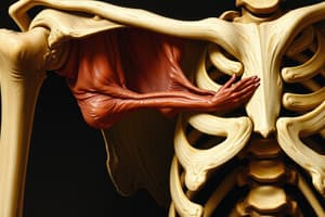
Psoas Syndrome
- Complex condition affecting the psoas muscle, which connects the lower back to the hips
- A chronic psoas spasm can create a persistent strain across the lumbosacral junction
- Impedes resolution of lumbosacral somatic dysfunction in spite of osteopathic manipulative therapy and exercises
Attachments of Psoas
- Psoas muscles attach to the vertebral bodies and anterior surface of the lumbar vertebrae's transverse processes and pass along the pelvis
- Joins the iliacus muscles, and insert on the lesser trochanter of each femur
Biomechanical Function of Psoas
- Primary function is hip flexion
- As a long restrictor muscle spanning multiple joints, it affects the hip joint, SI joint, & lumbar spine
Causes of Psoas Syndrome
- Prolonged Shortened Positions: sitting in soft chairs or bending over for extended periods/working at a desk.
- Overuse: Activities like doing sit-ups with fully extended lower extremities.
- Sudden Movements: Getting up quickly after prolonged sitting or bending can trigger the syndrome.
- Underlying Conditions: Disk herniations/organic issues like arthritis, diverticulitis, or prostatitis.
Signs and Symptoms of Psoas Syndrome
- Lower Back Pain: Central lower back pain accompanied by sciatic-type pain down the opposite leg
- Postural Changes: Patients may walk flexed forward/listing to one side, severe cases may present with hip flexion contracture
- Lower Extremity: Shortening and external rotation of the ipsilateral lower extremity, with possible Trendelenburg gait
Lumbar Dysfunction with Psoas Syndrome
- L1 or L2 vertebrae is flexed, rotated, and sidebent ipsilateral to hypertonic psoas
Pelvic Shift with Psoas Syndrome
- Pelvic side shift occurs contralateral to hypertonic psoas
Sacroiliac Joint with Psoas Syndrome
- Restrictions caused by psoas hypertonicity compressing SI joints and Backwards torsion around ipsilateral hypertonic axis
Compensatory Changes with Psoas Syndrome
- Contralateral piriformis trigger point and sciatic nerve pain above the knee
- Ipsilateral has externally rotated short left leg
- Long term dysfunction can lead to compensatory changes in the thoracolumbar junction/lumbar spine
Diagnosis of Psoas Syndrome
- Type II (flexed) L1 or L2 rotated and side bent to the left where Sacral torsion dysfunction (right torsion on a left oblique axis) with Pelvic side shift to the right
- Right piriformis trigger point and sciatic nerve pain above the knee along with Left externally rotated short left leg
- Prone and Supine Tests compare tension/end feel during Hip Extension & positive Thomas test on left Hip Flexion
- Palpatation indicates tenderness with in Psoas and Iliacus
- Rule Out Organic Causes: Like arthritis, diverticulitis, or prostatitis
Osteopathic Manipulative Treatment Approach for Psoas Syndrome
- Address Key Dysfunction via L1 or L2 somatic dysfunction
- Resolve Psoas Tension using techniques to relieve psoas hypertonicity
- Treat Associated Areas address lumbosacral, sacral, and pelvic dysfunctions
- Patient Education Provide home exercises and preventive strategies
Management of Psoas Syndrome
- OMT (Counterstrain, Myofascial Release, Still Technique, Muscle Energy)
- Home Exercises (prescribe specific stretches and exercises for the psoas muscle)
- Lifestyle Modifications (Advise on posture, ergonomics, plus activity modifications)
- Additional Therapies (Consider ice, medications, or treatment of underlying conditions if necessary)
Quadratus Lumborum Syndrome
- Myofascial pain syndrome caused by spasm and hypertonic quadratus lumborum muscle in the flank region, along the iliac crest, and around the front of the upper groin with referral into the sacroiliac joint
- Caused by poor posture and sitting in a slumped position
Anatomy and Function of the Quadratus Lumborum
- 12rh rib, transverse processes of L1-L4 vertebrae insertion and iliolumbar ligament connection
- Stabilizes/depresses -12th rib
- Ipsilateral sidebending Extends lumbar spine via bilateral contraction
Symptoms and Presentation of Quadratus Lumborum Syndrome
- Postural Changes resulting in extended and side-bent affected side
- Limited Range of Motion Contralateral side-bending and flexion becomes limited and painful
- Muscular Weakness indicates Opposite quadratus lumborum muscle becomes weak and pain
- Develop Trigger Points produce aching/sharp pain primarily worse standing
Physical Exam Findings for Quadratus Lumborum Syndrome
- Side-bent toward the contracted side
- Spinal Curve: Neutral group curve type 1 somatic dysfunction (side-bending toward and internal rotation away)
- Pelvic Alignment: Elevated pelvis on contracted quadratus lumborum side
- Leg Length: Apparent short leg on affected side because the muscle "pulls up" on the leg
Palpation and Range of Motion Evaluation for Quadratus Lumborum Syndrome
- Palpation will reveal hypertonic and tender quadratus lumborum muscles and transverse processes.
- There is Decreased flexion with pain to bending side
Special Tests for Quadratus Lumborum Syndrome
- Hip Drop Test will produce a restriction to Hip Motion
- There is Leg Length Shortening
- Quadratus Luborum Strength Tests show restriction
Treatment of Quadratus Lumborum Syndrome
- OMT techniques like Muscle Energy, Myofascial Release, & Counterstrain
- Follow up with Trigger Point Therapy and specific exercises
Viscerosomatics
- Viscerosomatic reflexes provide diagnostic clues on the somatic system (muscles, joints, blood, & lymph)
- Caused by a visceral disorder or disease which similar TART findings, that are in fact a reflex from the viscera
- Sympathetic nervous system and Parasympathetic Nervous System involvement
Studying That Suits You
Use AI to generate personalized quizzes and flashcards to suit your learning preferences.