Podcast
Questions and Answers
What is the main function of the corticospinal tracts?
What is the main function of the corticospinal tracts?
- Regulating autonomic functions
- Processing sensory information
- Facilitating reflex actions
- Controlling voluntary motor activity (correct)
How do motor neurons in the spinal cord transfer signals?
How do motor neurons in the spinal cord transfer signals?
- They project their axons contralaterally
- They use chemical signals exclusively
- They have no direct connections to muscles
- They project their axons ipsilaterally (correct)
Which structure is responsible for transmitting signals from the cerebral cortex to the brainstem or spinal cord?
Which structure is responsible for transmitting signals from the cerebral cortex to the brainstem or spinal cord?
- Upper motor neuron (correct)
- Corticobulbar tract
- Anterior horn cell
- Lower motor neuron
What characterizes a monosynaptic reflex?
What characterizes a monosynaptic reflex?
What is the main function of the lower motor neuron?
What is the main function of the lower motor neuron?
Which area is primarily associated with sensory information processing?
Which area is primarily associated with sensory information processing?
What distinguishes upper motor neuron lesions from lower motor neuron lesions?
What distinguishes upper motor neuron lesions from lower motor neuron lesions?
What percentage of fibers decussate at the level of the medulla?
What percentage of fibers decussate at the level of the medulla?
In the context of motor pathways, how is the term 'extrapyramidal' best described?
In the context of motor pathways, how is the term 'extrapyramidal' best described?
Which part of the internal capsule contains the corticobulbar tracts?
Which part of the internal capsule contains the corticobulbar tracts?
What is the primary role of the internal capsule?
What is the primary role of the internal capsule?
How is the body represented on the motor cortex?
How is the body represented on the motor cortex?
What is the primary function of the primary sensory cortex?
What is the primary function of the primary sensory cortex?
What structure is primarily involved in coordinating voluntary movements?
What structure is primarily involved in coordinating voluntary movements?
Where is the primary motor cortex located in relation to the central sulcus?
Where is the primary motor cortex located in relation to the central sulcus?
What does the term 'motor homunculus' refer to?
What does the term 'motor homunculus' refer to?
What is the primary role of the medial reticulospinal tracts in the spinal cord?
What is the primary role of the medial reticulospinal tracts in the spinal cord?
Which condition is likely to show hyper-reflexia?
Which condition is likely to show hyper-reflexia?
What is the result of a diminished patellar reflex response?
What is the result of a diminished patellar reflex response?
What does the term 'paresis' refer to in a clinical context?
What does the term 'paresis' refer to in a clinical context?
Multiple Sclerosis primarily affects which part of the neuronal structure?
Multiple Sclerosis primarily affects which part of the neuronal structure?
Which of the following best describes hemiplegia?
Which of the following best describes hemiplegia?
The patellar tendon reflex tests which aspect of the nervous system?
The patellar tendon reflex tests which aspect of the nervous system?
In motor neuron disease, which motor neurons can be affected?
In motor neuron disease, which motor neurons can be affected?
What does a positive Babinski sign indicate?
What does a positive Babinski sign indicate?
In which age group is the Babinski sign commonly present as a normal reflex?
In which age group is the Babinski sign commonly present as a normal reflex?
What characteristic is NOT associated with lower motor neuron lesions?
What characteristic is NOT associated with lower motor neuron lesions?
Which cranial nerve has mixed function among all motor cranial nerves?
Which cranial nerve has mixed function among all motor cranial nerves?
Which statement about the innervation of cranial motor neurons is true?
Which statement about the innervation of cranial motor neurons is true?
What is the consequence of an upper motor neuron injury to the facial nerve?
What is the consequence of an upper motor neuron injury to the facial nerve?
Which of the following conditions is typically related to lower motor neuron lesions?
Which of the following conditions is typically related to lower motor neuron lesions?
What clinical sign indicates reduced resistance to passive stretching?
What clinical sign indicates reduced resistance to passive stretching?
What type of lesion results in paralysis of the lower right side of the face?
What type of lesion results in paralysis of the lower right side of the face?
Which part of the brain is primarily involved in the pyramidal system for upper motor neurons?
Which part of the brain is primarily involved in the pyramidal system for upper motor neurons?
Which type of movement is the extrapyramidal system primarily responsible for?
Which type of movement is the extrapyramidal system primarily responsible for?
What is the primary role of the rubrospinal pathway?
What is the primary role of the rubrospinal pathway?
Which structure is NOT part of the basal ganglia?
Which structure is NOT part of the basal ganglia?
What is the primary function of the tectospinal tract?
What is the primary function of the tectospinal tract?
Which of the following statements correctly describes the extrapyramidal system?
Which of the following statements correctly describes the extrapyramidal system?
What characterizes a right-sided lower motor neuron lesion of the facial nerve?
What characterizes a right-sided lower motor neuron lesion of the facial nerve?
What is the main pathway used by the motor neurons for sending signals to the same side of the body?
What is the main pathway used by the motor neurons for sending signals to the same side of the body?
Which type of motor neuron lesion typically results in muscle atrophy and flaccid paralysis?
Which type of motor neuron lesion typically results in muscle atrophy and flaccid paralysis?
In the context of reflex actions, how many neurons are involved in the efferent pathway?
In the context of reflex actions, how many neurons are involved in the efferent pathway?
Which structure is the initial point for the upper motor neurons of the pyramidal system?
Which structure is the initial point for the upper motor neurons of the pyramidal system?
What is the role of the internal capsule in the motor system?
What is the role of the internal capsule in the motor system?
What is the primary distinction between corticobulbar and corticospinal tracts?
What is the primary distinction between corticobulbar and corticospinal tracts?
Which area of the brain is primarily involved in generating voluntary motor commands?
Which area of the brain is primarily involved in generating voluntary motor commands?
Which reflex is exemplified by monosynaptic neurons responding directly to a stimulus?
Which reflex is exemplified by monosynaptic neurons responding directly to a stimulus?
What role does the lower motor neuron play in the motor pathway?
What role does the lower motor neuron play in the motor pathway?
Which structure is located posterior to the central sulcus and primarily involved in sensory processing?
Which structure is located posterior to the central sulcus and primarily involved in sensory processing?
At what anatomical location do the upper motor neuron fibers decussate in the pyramidal system?
At what anatomical location do the upper motor neuron fibers decussate in the pyramidal system?
Which motor pathway is primarily associated with involuntary or automatic movements?
Which motor pathway is primarily associated with involuntary or automatic movements?
What is meant by the term 'bulb' in the context of the pyramidal system?
What is meant by the term 'bulb' in the context of the pyramidal system?
Which structures are considered components of the basal nuclei?
Which structures are considered components of the basal nuclei?
What is the primary function of the superior colliculus?
What is the primary function of the superior colliculus?
Where do the upper motor neurons of the pyramidal system originate?
Where do the upper motor neurons of the pyramidal system originate?
Which of the following statements is true regarding the extrapyramidal system?
Which of the following statements is true regarding the extrapyramidal system?
Which area of the brainstem is involved in both homeostatic and reflex functions?
Which area of the brainstem is involved in both homeostatic and reflex functions?
What defines the primary pathway of the upper motor neurons in the pyramidal system?
What defines the primary pathway of the upper motor neurons in the pyramidal system?
Which nucleus is NOT included in the lentiform nucleus?
Which nucleus is NOT included in the lentiform nucleus?
Which of the following is primarily associated with reflexive movements in response to auditory input?
Which of the following is primarily associated with reflexive movements in response to auditory input?
What is supplied by the anterior perforating arteries in the brain?
What is supplied by the anterior perforating arteries in the brain?
What do the anterior perforating arteries originate from?
What do the anterior perforating arteries originate from?
In which part of the pyramidal motor pathway does the corticobulbar tract primarily reside?
In which part of the pyramidal motor pathway does the corticobulbar tract primarily reside?
What is the primary effect of a unilateral upper motor neuron lesion in the cerebral hemisphere?
What is the primary effect of a unilateral upper motor neuron lesion in the cerebral hemisphere?
What percentage of fibers decussate in the lateral corticospinal tracts?
What percentage of fibers decussate in the lateral corticospinal tracts?
What condition is characterized by spasticity and increased resistance to muscle stretching?
What condition is characterized by spasticity and increased resistance to muscle stretching?
What is described as a rhythmic series of muscle contractions induced by stretching the tendon?
What is described as a rhythmic series of muscle contractions induced by stretching the tendon?
What aspect of the pyramidal motor pathway corresponds to the decussation near termination for anterior corticospinal fibers?
What aspect of the pyramidal motor pathway corresponds to the decussation near termination for anterior corticospinal fibers?
What is the main role of the superior colliculus?
What is the main role of the superior colliculus?
Which of the following correctly defines the tegmentum?
Which of the following correctly defines the tegmentum?
What anatomical feature distinguishes tectum from tegmentum?
What anatomical feature distinguishes tectum from tegmentum?
Which brain structure is responsible for reflex movements in response to auditory stimuli?
Which brain structure is responsible for reflex movements in response to auditory stimuli?
Which statement about the tegmentum is correct?
Which statement about the tegmentum is correct?
What type of tracts does the tegmentum contain?
What type of tracts does the tegmentum contain?
What is the primary function of the reticulospinal tract mentioned in relation to the tegmentum?
What is the primary function of the reticulospinal tract mentioned in relation to the tegmentum?
In which part of the brainstem is the tegmentum located?
In which part of the brainstem is the tegmentum located?
Flashcards are hidden until you start studying
Study Notes
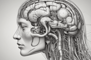
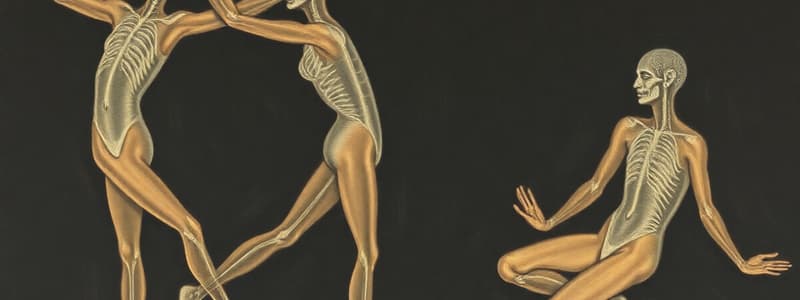
The Motor System
- The motor system is responsible for movement and is controlled by both the central nervous system (CNS) and peripheral nervous system (PNS).
- The simplest motor response is a monosynaptic reflex which is triggered by a stimulus and causes a muscle to contract without conscious control (e.g., knee jerk, biceps reflex).
- Motor neurons in the spinal cord and cranial nerves are ipsilateral, meaning they project their axons to the periphery on the same side of the body.
- Controlled responses to stimuli require a more complex pathway involving sensory (afferent) and motor (efferent) neurons.
- Afferent neurons carry information from the periphery to the CNS.
- Efferent neurons carry commands from the CNS to the periphery.
Motor Cortex and Pathways
- The pre-central gyrus is the primary motor cortex where voluntary movements are initiated.
- The motor homunculus is a distorted representation of the body on the motor cortex, reflecting the relative amount of cortical area dedicated to controlling different body parts.
- The pyramidal motor pathway is responsible for conscious voluntary movements.
- Upper motor neurons (UMN) originate in the cerebral cortex and project to the brainstem or spinal cord.
- Lower motor neurons (LMN) originate in the brainstem or spinal cord and project to muscles.
- Most (85%) of the corticospinal tract fibers cross over (decussate) in the medulla forming the lateral corticospinal tracts.
- The internal capsule is a white matter structure that carries axons from the cerebral cortex to the brainstem and spinal cord.
- The genu of the internal capsule carries the corticobulbar tract, which controls cranial nerve nuclei.
- The posterior limb carries the corticospinal tract, which controls spinal motor neurons.
- Damage to the pyramidal tract can result in weakness, paralysis, wasting of muscles, fasciculations, and hypotonia.
Motor Cranial Nerves
- Cranial nerves III, IV, VI, XI, and XII are primarily involved in motor control, while V, VII, IX, and X are mixed, containing both motor and sensory components.
- The facial nerve (VII) has bilateral innervation for the upper face, meaning it receives input from both hemispheres.
- Injury to the upper motor neuron (UMN) of the facial nerve will result in paralysis of the lower face on the contralateral side of the body, but the forehead will be spared.
- Injury to the lower motor neuron (LMN) of the facial nerve will result in paralysis of the entire face on the ipsilateral side of the body.
Extrapyramidal System
- The extrapyramidal system is responsible for fine-tuning movements, regulating muscle tone, and controlling posture.
- The basal ganglia are a group of subcortical nuclei that play a key role in the extrapyramidal system.
- The rubrospinal tract originates in the red nucleus of the midbrain and helps control the tone of flexor muscles.
- The tectospinal tract originates in the superior colliculi and controls reflex movements in response to visual stimuli.
- The vestibulospinal tract originates in the vestibular nuclei and controls the tone of extensor muscles, maintaining posture and balance.
Clinical Considerations
- UMN lesions can result in hyperreflexia, spasticity, clonus, and a positive Babinski sign.
- LMN lesions can result in hyporeflexia, flaccid paralysis, muscle atrophy, and fasciculations.
- Neurological exams can help assess the integrity of the motor system by testing reflexes, muscle tone, and strength.
Additional Information
- Stroke is a common cause of UMN lesions, leading to weakness or paralysis on the contralateral side of the body.
- Poliomyelitis (polio) is a viral infection that damages anterior horn cells, leading to LMN lesions.
- Multiple sclerosis is an autoimmune disease that damages the myelin sheath of neurons, affecting both UMNs and LMNs.
- Motor neuron disease is a group of disorders that affect motor neurons, leading to progressive muscle weakness and atrophy.
Key Terms
- Hyporeflexia (hypo) Reduced reflex response.
- Hyperreflexia (hyper) Increased or exaggerated reflex response.
- Fasciculations: Small, localized, involuntary twitches in muscle fibers.
- Spasticity: Increased muscle tone and resistance to stretching.
- Clonus: Rhythmic, involuntary muscle contractions often seen in response to sudden stretching.
- Babinski sign: Dorsal extension of the big toe in response to stroking the bottom of the foot.
- Palsy: Paralysis.
- Paralysis: Complete or partial loss of motor function.
- Paresis: Weakness or partial loss of motor function.
- Paraplegia: Paralysis of the lower limbs.
- Hemiplegia: Paralysis of one side of the body.
- Monoplegia: Paralysis restricted to one limb.
Motor System Function
- The corticospinal tracts are a group of upper motor neurons that start in the cortex and end in the spinal cord.
- The corticobulbar tracts are a group of upper cranial motor nerves that start in the cortex and end in the brainstem.
- Upper motor neurons begin in the cerebral cortex.
- Lower motor neurons begin in the brainstem or spinal cord.
- Monosynaptic neurons are the simplest motor neurons, they respond to stimuli directly without any control.
- The internal capsule is a part of the brain that is supplied by the anterior perforating arteries.
- The anterior perforating arteries enter the brain through the anterior perforated substance and are branches of the middle cerebral artery.
Motor Pathway Anatomy
- Cerebral peduncles are the pathways that connect the cerebrum to the brainstem.
- Pyramids are the bulges on the ventral surface of the medulla.
- The pyramidal decussation occurs when 85% of the fibers cross to the opposite side in the medulla
- Fibers in the anterior corticospinal tract decussate near the termination of the spinal cord.
- The substantia nigra and red nuclei are located in the tegmentum of the midbrain.
- The tectum is located in the midbrain posterior to the cerebral aqueduct.
- The superior colliculus controls reflex movements from visual input.
- The inferior colliculus controls reflex movements from auditory input.
- The tegmentum is located in the brainstem and connects the thalamus to the brainstem, it is involved in homeostatic and reflex pathways.
Upper and Lower Motor Neuron Lesions
- An upper motor neuron lesion results in contralateral weakness or paralysis.
- If the lesion occurs in the spinal cord, then the weakness or paralysis is ipsilateral.
- Lower motor neuron lesions result in ipsilateral weakness or paralysis, atrophy, fasciculations, and hyporeflexia.
Comparing Pyramidal and Extrapyramidal Systems
- The pyramidal system consists of upper motor neurons that travel through the pyramids of the medulla and are responsible for conscious movement.
- The extrapyramidal system consists of neurons that start in other brain regions and fine-tune movement.
Other Extrapyramidal Tracts
- The reticulospinal tract originates from the pons and medulla.
Clinical Presentation
- Spasticity is increased resistance to stretch of muscles, with a clasp-knife component.
- Clonus is a rhythmic series of muscle contractions that occurs when stretching the tendon.
- Flaccidity is a loss of muscle tone, can be caused by a lower motor neuron lesion.
- Fasciculations are spontaneous muscle twitching, can also be caused by a lower motor neuron lesion.
- Hyporeflexia is a decrease in reflexes, can also be caused by a lower motor neuron lesion.
Key Structures
- Basal nuclei include the lentiform nucleus (putamen + globus pallidus), caudate nucleus, and substantia nigra.
- The lentiform nucleus is a large nucleus located in the forebrain that is involved in motor control.
- The caudate nucleus is a curved structure that is involved in planning and executing movement.
Motor Neuron Pathways
- Motor areas are located in the pre-central gyrus and are responsible for controlling voluntary movement.
- The internal capsule contains the corticospinal and corticobulbar tracts.
- The genu of the internal capsule contains the corticobulbar tracts, which innervate the muscles of the head and face.
- The posterior limb of the internal capsule contains the corticospinal tracts, which innervate the muscles of the trunk and limbs.
- The crus cerebri is the part of the cerebral peduncles that is located in the midbrain.
- The spinal cord conveys motor signals to the muscles.
Remember
- "Bulb" is an outdated word for the medulla.
Studying That Suits You
Use AI to generate personalized quizzes and flashcards to suit your learning preferences.