Podcast
Questions and Answers
What is the cellular mechanism of cardiac contraction?
What is the cellular mechanism of cardiac contraction?
- Ca2+ is released from the sarcoplasmic reticulum (SR)
- ATP binds to myosin head and detaches head from actin
- Ca2+ binds to actin and forms cross-bridges with myosin (correct)
- Myosin pulls on actin to shorten the sarcomere and generate force
How does the force of cardiac contraction increase?
How does the force of cardiac contraction increase?
- By decreasing the cytosolic Ca2+ concentration
- By decreasing the number of cross-bridges formed
- By increasing the cytosolic Ca2+ concentration (correct)
- By increasing the pumping of Ca2+ into the sarcoplasmic reticulum (SR)
What is the purpose of the valves in the heart?
What is the purpose of the valves in the heart?
- To generate force during contraction
- To pump blood to the body
- To regulate the flow of blood (correct)
- To relax the cardiomyocytes
How does cardiac relaxation occur?
How does cardiac relaxation occur?
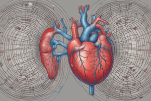
What is the composition of the mammalian heart?
What is the composition of the mammalian heart?
How do the pulmonary and systemic circuits work in the heart?
How do the pulmonary and systemic circuits work in the heart?
What is the purpose of the two pumps in the heart?
What is the purpose of the two pumps in the heart?
How are cardiomyocytes activated during each heartbeat?
How are cardiomyocytes activated during each heartbeat?
What happens when ATP binds to myosin head during cardiac relaxation?
What happens when ATP binds to myosin head during cardiac relaxation?
Which phase of the cardiac cycle is characterized by the relaxation and falling pressure of the ventricular chambers?
Which phase of the cardiac cycle is characterized by the relaxation and falling pressure of the ventricular chambers?
What is the main function of the overlapping diastole of the atria and ventricles during the cardiac cycle?
What is the main function of the overlapping diastole of the atria and ventricles during the cardiac cycle?
What is the difference between systole and diastole in terms of their durations?
What is the difference between systole and diastole in terms of their durations?
What is the highest point on the blood pressure trace?
What is the highest point on the blood pressure trace?
What is the lowest point on the blood pressure trace?
What is the lowest point on the blood pressure trace?
What is the difference between systolic and diastolic pressure called?
What is the difference between systolic and diastolic pressure called?
What is the average pressure across the full cardiac cycle called?
What is the average pressure across the full cardiac cycle called?
What is hypertension?
What is hypertension?
What is hypotension?
What is hypotension?
During the cardiac cycle, the heart is in diastole for two-thirds of the time.
During the cardiac cycle, the heart is in diastole for two-thirds of the time.
The diastole of the atria and ventricles overlaps, which helps maintain forward, unidirectional flow.
The diastole of the atria and ventricles overlaps, which helps maintain forward, unidirectional flow.
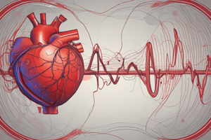
The blood pressure trace can be used to determine the systolic and diastolic pressure in arteries.
The blood pressure trace can be used to determine the systolic and diastolic pressure in arteries.
Diastole is typically longer (60%) than systole (40%) in the cardiac cycle.
Diastole is typically longer (60%) than systole (40%) in the cardiac cycle.
The systemic arterial pressure is higher than the pulmonary arterial pressure.
The systemic arterial pressure is higher than the pulmonary arterial pressure.
The highest point on the blood pressure trace is the systolic pressure.
The highest point on the blood pressure trace is the systolic pressure.
The lowest point on the blood pressure trace is the diastolic pressure.
The lowest point on the blood pressure trace is the diastolic pressure.
The pulse pressure is the difference between the systolic and diastolic pressure.
The pulse pressure is the difference between the systolic and diastolic pressure.
The mean pressure is slightly shifted towards the diastolic pressure because more time is spent in diastole than systole.
The mean pressure is slightly shifted towards the diastolic pressure because more time is spent in diastole than systole.
Hypertension refers to high blood pressure, while hypotension refers to low blood pressure.
Hypertension refers to high blood pressure, while hypotension refers to low blood pressure.
True or false: The mammalian cardiovascular system consists of a four-chambered heart with 2 atria and 2 ventricles.
True or false: The mammalian cardiovascular system consists of a four-chambered heart with 2 atria and 2 ventricles.
True or false: Arterial blood flows away from the heart.
True or false: Arterial blood flows away from the heart.
True or false: The pulmonary and systemic circuits in the heart work simultaneously, one after the other in series.
True or false: The pulmonary and systemic circuits in the heart work simultaneously, one after the other in series.
True or false: Valves in the heart open and close to direct blood in a single direction, thus ensuring unidirectional flow
True or false: Valves in the heart open and close to direct blood in a single direction, thus ensuring unidirectional flow
True or false: Ca2+ is released from the sarcoplasmic reticulum (SR) and binds to actin to generate force during cardiac contraction.
True or false: Ca2+ is released from the sarcoplasmic reticulum (SR) and binds to actin to generate force during cardiac contraction.
True or false: Every cardiomyocyte is activated during each heartbeat.
True or false: Every cardiomyocyte is activated during each heartbeat.
True or false: Decreases in cytosolic Ca2+ concentration lead to cardiac relaxation.
True or false: Decreases in cytosolic Ca2+ concentration lead to cardiac relaxation.
True or false: ATP binds to myosin head during cardiac relaxation to detach it from actin.
True or false: ATP binds to myosin head during cardiac relaxation to detach it from actin.
True or false: The force of contraction can be altered by changing the Ca2+ released by the SR.
True or false: The force of contraction can be altered by changing the Ca2+ released by the SR.
True or false: The purpose of the valves in the heart is to ensure unidirectional flow of blood.
True or false: The purpose of the valves in the heart is to ensure unidirectional flow of blood.
During cardiac contraction, Ca2+ is released from the sarcoplasmic reticulum (SR) and binds to actin to form ______ with myosin.
During cardiac contraction, Ca2+ is released from the sarcoplasmic reticulum (SR) and binds to actin to form ______ with myosin.
Every ______ is activated during each heartbeat, unlike skeletal muscles which activate muscle fibers via recruitment.
Every ______ is activated during each heartbeat, unlike skeletal muscles which activate muscle fibers via recruitment.
The ______ cardiovascular system consists of a four-chambered heart with 2 atria and 2 ventricles.
The ______ cardiovascular system consists of a four-chambered heart with 2 atria and 2 ventricles.
Which node acts as the 'pacemaker' of the heart?
Which node acts as the 'pacemaker' of the heart?
What is the function of the AV node in the conduction pathway?
What is the function of the AV node in the conduction pathway?
Which structure distributes the contractile signal to the muscle cells of the left atrium?
Which structure distributes the contractile signal to the muscle cells of the left atrium?
What is the function of the AV bundle and bundle branches in the conduction pathway?
What is the function of the AV bundle and bundle branches in the conduction pathway?
Which cells rapidly propagate electrical conduction to the contractile cells of the ventricles?
Which cells rapidly propagate electrical conduction to the contractile cells of the ventricles?
What does an electrocardiogram (ECG) detect?
What does an electrocardiogram (ECG) detect?
Which wave in an ECG represents atrial depolarization?
Which wave in an ECG represents atrial depolarization?
During which phase of the cardiac cycle does ventricular depolarization occur?
During which phase of the cardiac cycle does ventricular depolarization occur?
What happens during ventricular repolarization?
What happens during ventricular repolarization?
When does ventricular repolarization complete?
When does ventricular repolarization complete?
Which type of cells in the heart are responsible for triggering and spreading electrical impulses to all parts of the myocardium?
Which type of cells in the heart are responsible for triggering and spreading electrical impulses to all parts of the myocardium?
What is the main difference between electrical conduction cells and contractile cells in the heart?
What is the main difference between electrical conduction cells and contractile cells in the heart?
What is the role of gap junctions in the intercalated disks of cardiac cells?
What is the role of gap junctions in the intercalated disks of cardiac cells?
What is NOT the composition of conduction cells in the heart?
What is NOT the composition of conduction cells in the heart?
How do electrical impulses spread between electrical and contractile cells in the heart?
How do electrical impulses spread between electrical and contractile cells in the heart?
What is the role of contractile cells in the heart?
What is the role of contractile cells in the heart?
What is the main difference between electrical and contractile cells in terms of appearance?
What is the main difference between electrical and contractile cells in terms of appearance?
What is the function of conduction cells in the heart?
What is the function of conduction cells in the heart?
Electrical conduction cells make up 1% of cardiac cells.
Electrical conduction cells make up 1% of cardiac cells.
Electrical conduction cells are modified cardiomyocytes.
Electrical conduction cells are modified cardiomyocytes.
Electrical conduction cells have a high content of actin and myosin.
Electrical conduction cells have a high content of actin and myosin.
Electrical conduction cells are involved in intercellular conduction within the heart.
Electrical conduction cells are involved in intercellular conduction within the heart.
Electrical conduction cells have a pale striated appearance.
Electrical conduction cells have a pale striated appearance.
Electrical conduction cells are mainly composed of nervous tissue.
Electrical conduction cells are mainly composed of nervous tissue.
Contractile cells make up 99% of cardiac cells.
Contractile cells make up 99% of cardiac cells.
Contractile cells have a high content of actin and myosin.
Contractile cells have a high content of actin and myosin.
Contractile cells are not involved in electrical conduction.
Contractile cells are not involved in electrical conduction.
Contractile cells are responsible for the contraction of the myocardium.
Contractile cells are responsible for the contraction of the myocardium.
True or false: The SA node is responsible for initiating electrical depolarization in the right atrium and spreading it to the left atrium and AV node.
True or false: The SA node is responsible for initiating electrical depolarization in the right atrium and spreading it to the left atrium and AV node.
True or false: The internodal bundle distributes the contractile signal to the right atrium muscle contractile cells.
True or false: The internodal bundle distributes the contractile signal to the right atrium muscle contractile cells.
True or false: The AV node is a relatively slow conductor and creates a pause in the conduction pathway to allow for the completion of atrial systole and ventricular depolarisation preparation.
True or false: The AV node is a relatively slow conductor and creates a pause in the conduction pathway to allow for the completion of atrial systole and ventricular depolarisation preparation.
True or false: The AV bundle and bundle branches provide electrical connection between the atria and ventricles along the interventricular septum.
True or false: The AV bundle and bundle branches provide electrical connection between the atria and ventricles along the interventricular septum.
True or false: The Purkinje fibers rapidly propagate electrical conduction to the contractile cells of the ventricles, causing a ventricular contraction from the apex upwards and outwards.
True or false: The Purkinje fibers rapidly propagate electrical conduction to the contractile cells of the ventricles, causing a ventricular contraction from the apex upwards and outwards.
True or false: Leads in an ECG detect the difference or change in electrical activity between two surface electrodes.
True or false: Leads in an ECG detect the difference or change in electrical activity between two surface electrodes.
True or false: Atrial depolarization is initiated by the AV node and causes the P wave in an ECG.
True or false: Atrial depolarization is initiated by the AV node and causes the P wave in an ECG.
True or false: Ventricular depolarization and atrial repolarization cause the QRS complex in an ECG.
True or false: Ventricular depolarization and atrial repolarization cause the QRS complex in an ECG.
True or false: Ventricular repolarization starts from the base of the ventricles and causes the T wave in an ECG.
True or false: Ventricular repolarization starts from the base of the ventricles and causes the T wave in an ECG.
True or false: Ventricular repolarization is complete when the ventricles are fully repolarized and relaxed.
True or false: Ventricular repolarization is complete when the ventricles are fully repolarized and relaxed.
Flashcards are hidden until you start studying