Podcast
Questions and Answers
Which of the following scientists is credited with the observation of cells leading to the term 'cell'?
Which of the following scientists is credited with the observation of cells leading to the term 'cell'?
- Rudolf Virchow
- Walther Flemming
- Antony van Leeuwenhoek
- Robert Hooke (correct)
Cytogenetics is best defined as the branch of cytology focused on the study of cell movement.
Cytogenetics is best defined as the branch of cytology focused on the study of cell movement.
False (B)
Which type of microscope is most suitable for observing the surface properties of living cells without staining?
Which type of microscope is most suitable for observing the surface properties of living cells without staining?
- Transmission Electron Microscope (TEM)
- Polarizing microscope
- Phase contrast microscope (correct)
- Fluorescence microscope
What fixative is commonly used in cytological and histological examination to inactivate enzymes and prevent tissue breakdown?
What fixative is commonly used in cytological and histological examination to inactivate enzymes and prevent tissue breakdown?
The PAS reaction is used to demonstrate which class of biomolecules in tissues?
The PAS reaction is used to demonstrate which class of biomolecules in tissues?
Enzyme histochemistry requires the use of fixed tissue sections to preserve enzyme activity.
Enzyme histochemistry requires the use of fixed tissue sections to preserve enzyme activity.
Which method is used to identify specific mRNA sequences within a tissue sample?
Which method is used to identify specific mRNA sequences within a tissue sample?
In cell culture, cells are bathed in a fluid similar to ______ which provides the molecules required for survival and growth.
In cell culture, cells are bathed in a fluid similar to ______ which provides the molecules required for survival and growth.
Which technique allows the identification of cells replicating DNA within a tissue?
Which technique allows the identification of cells replicating DNA within a tissue?
What is the primary difference between Transmission Electron Microscopy (TEM) and Scanning Electron Microscopy (SEM) in terms of how the electron beam interacts with the specimen?
What is the primary difference between Transmission Electron Microscopy (TEM) and Scanning Electron Microscopy (SEM) in terms of how the electron beam interacts with the specimen?
Prokaryotic cells contain long linear DNA molecules within a nucleus.
Prokaryotic cells contain long linear DNA molecules within a nucleus.
Which structure is primarily responsible for the synthesis and processing of RNA in eukaryotic cells?
Which structure is primarily responsible for the synthesis and processing of RNA in eukaryotic cells?
In eukaryotic cells, the cytoplasm is composed of organelles and the ______ matrix.
In eukaryotic cells, the cytoplasm is composed of organelles and the ______ matrix.
Which of the following elements are considered macroelements, comprising 98-99% of cell mass?
Which of the following elements are considered macroelements, comprising 98-99% of cell mass?
Lipids play main role in blood group antigenicity.
Lipids play main role in blood group antigenicity.
Which level of protein structure is primarily stabilized by hydrogen bonds?
Which level of protein structure is primarily stabilized by hydrogen bonds?
Match the following types of RNA with their respective functions:
Match the following types of RNA with their respective functions:
Which type of molecule inserts at varying densities among phospholipid fatty acids, modulating the fluidity of the plasma membrane?
Which type of molecule inserts at varying densities among phospholipid fatty acids, modulating the fluidity of the plasma membrane?
The glycocalyx, a carbohydrate-rich coating on the cell surface, is ______ positive due to its chemical composition.
The glycocalyx, a carbohydrate-rich coating on the cell surface, is ______ positive due to its chemical composition.
Peripheral proteins are integrated directly within the lipid bilayer of a cell membrane.
Peripheral proteins are integrated directly within the lipid bilayer of a cell membrane.
Which of the following is a function of membrane proteins?
Which of the following is a function of membrane proteins?
What is the name of the process where molecules are expelled from cells by vesicle fusion with the plasma membrane?
What is the name of the process where molecules are expelled from cells by vesicle fusion with the plasma membrane?
Regulated secretion is characterized by the immediate and continuous release of cellular products.
Regulated secretion is characterized by the immediate and continuous release of cellular products.
Which of the following specialized cell membrane structures contain bundles of actin filaments?
Which of the following specialized cell membrane structures contain bundles of actin filaments?
Cell adhesion and communication are primarily executed through which structures?
Cell adhesion and communication are primarily executed through which structures?
Gap junctions contain proteins called ______, which form a hydrophilic pore for communication between cells.
Gap junctions contain proteins called ______, which form a hydrophilic pore for communication between cells.
Smooth endoplasmic reticulum is involved in ribosome production.
Smooth endoplasmic reticulum is involved in ribosome production.
What is the role of the signal-recognition particle (SRP) in protein synthesis?
What is the role of the signal-recognition particle (SRP) in protein synthesis?
What enzyme removes the signal sequence from most proteins once they are inside the lumen of the rough endoplasmic reticulum (RER)?
What enzyme removes the signal sequence from most proteins once they are inside the lumen of the rough endoplasmic reticulum (RER)?
The major role of enzymes in the smooth ER is protein degradation.
The major role of enzymes in the smooth ER is protein degradation.
What coat protein is primarily involved in the forward movement of vesicles in the golgi?
What coat protein is primarily involved in the forward movement of vesicles in the golgi?
What term describes granules with digestive enzymes that are produced by some cells?
What term describes granules with digestive enzymes that are produced by some cells?
Heterolysosomes function to remove nonfunctional organelles by autophagy
Heterolysosomes function to remove nonfunctional organelles by autophagy
Which specific enzyme is found in peroxisomes and acts to break down hydrogen peroxide, preventing cellular damage?
Which specific enzyme is found in peroxisomes and acts to break down hydrogen peroxide, preventing cellular damage?
Which specialized transmembrane proteins allow small molecules to pass from the cytoplasm through the intermembrane space in mitochondria?
Which specialized transmembrane proteins allow small molecules to pass from the cytoplasm through the intermembrane space in mitochondria?
Cytochrome C is released from the mitochondrial membrane space to trigger______
Cytochrome C is released from the mitochondrial membrane space to trigger______
Clathrin-coated vesicles are responsible for retrograde transport from the endoplasmic reticulum to the Golgi.
Clathrin-coated vesicles are responsible for retrograde transport from the endoplasmic reticulum to the Golgi.
What is the role of nucleoporins in the nuclear pore complexes?
What is the role of nucleoporins in the nuclear pore complexes?
Which type of chromatin is more transcriptionally active?
Which type of chromatin is more transcriptionally active?
What is the name for the structural unit of DNA and histones that comprises a core of 8 small histones wrapped with DNA?
What is the name for the structural unit of DNA and histones that comprises a core of 8 small histones wrapped with DNA?
Flashcards
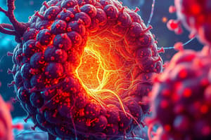
Cytology
Cytology
The study of cells, including cell structure, function, and life processes.
Histology
Histology
Study of tissues
Embryology
Embryology
The study of the development of an embryo
Light (optical) microscope
Light (optical) microscope
Signup and view all the flashcards
Phase contrast microscope
Phase contrast microscope
Signup and view all the flashcards
Interference microscope
Interference microscope
Signup and view all the flashcards
Fluorescence microscope
Fluorescence microscope
Signup and view all the flashcards
Polarizing microscope
Polarizing microscope
Signup and view all the flashcards
Electron microscope (EM)
Electron microscope (EM)
Signup and view all the flashcards
Transmission EM (TEM)
Transmission EM (TEM)
Signup and view all the flashcards
Cell fractionation
Cell fractionation
Signup and view all the flashcards
Autoradiography
Autoradiography
Signup and view all the flashcards
X-ray crystallography
X-ray crystallography
Signup and view all the flashcards
The Cell
The Cell
Signup and view all the flashcards
Prokaryotic
Prokaryotic
Signup and view all the flashcards
Eukaryotic
Eukaryotic
Signup and view all the flashcards
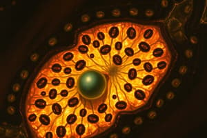
Nucleus
Nucleus
Signup and view all the flashcards
Cytoplasm
Cytoplasm
Signup and view all the flashcards
Macroelements
Macroelements
Signup and view all the flashcards
Microelements
Microelements
Signup and view all the flashcards
Simple lipids
Simple lipids
Signup and view all the flashcards
Compound lipids
Compound lipids
Signup and view all the flashcards
Proteins
Proteins
Signup and view all the flashcards
DNA
DNA
Signup and view all the flashcards
Carbohydrates
Carbohydrates
Signup and view all the flashcards
Plasma membrane
Plasma membrane
Signup and view all the flashcards
Glycocalyx
Glycocalyx
Signup and view all the flashcards
Enzymes
Enzymes
Signup and view all the flashcards
Receptors
Receptors
Signup and view all the flashcards
Vesicular Transport
Vesicular Transport
Signup and view all the flashcards
Endocytosis
Endocytosis
Signup and view all the flashcards
Exocytosis
Exocytosis
Signup and view all the flashcards
Microvilli
Microvilli
Signup and view all the flashcards
Stereocilia
Stereocilia
Signup and view all the flashcards
Cilia
Cilia
Signup and view all the flashcards
Mitosis.
Mitosis.
Signup and view all the flashcards
Cilia
Cilia
Signup and view all the flashcards
Intercellular junctions
Intercellular junctions
Signup and view all the flashcards
Endoplasmic reticulum (rough)
Endoplasmic reticulum (rough)
Signup and view all the flashcards
SER
SER
Signup and view all the flashcards
Study Notes
Historical Development
- Robert Hooke observed cells in 1665
- Antony van Leeuwenhoek observed cells in 1678
- Cell theory was developed by Schleiden and Schwann (1838-1839)
- "Omnis cellula e cellula" (all cells come from cells) was postulated by Rudolf Virchow in 1852
- "Omnis nucleus e nucleo" (all nuclei come from nuclei) was postulated by Walther Flemming in 1860
- 20th century saw focus expand to biochemistry, cytogenetics, cytophysiology, molecular biology, and cellular ecology
Microscopy Types
- Light microscope uses a mechanical part (stand, holder, tube) and an optical part (ocular, objective, condenser)
- Phase contrast does not need staining, cells can be studied while alive
- Interference microscope assesses surface properties of cells and biological objects
- Fluorescence microscope visualises naturally occurring fluorescent molecules like neurotransmitters and vitamin A
- Polarizing microscope is designed for specimens visible by their anisotropic qualities, recognising structures with oriented molecules, uses 2 filters (polarizer and analyzer)
- Electron microscope (EM) uses electron beams, high resolution due to short wavelength
- Transmission EM (TEM) has 3nm resolution, up to 400,000x magnifications
- Scanning EM (SEM) similar to TEM but the electron beam is focused and doesn't pass through the specimen
Cytological exam principles
- Formalin fixes the tissue (inactivates enzymes)
- Alcohol dehydrates sample
- Paraffin/wax for embedding
- Microtome sectioning
- Hematoxylin and eosin (H and E) staining (basic and acidic dye)
Histochemistry
- Used to demonstrate carbohydrates, lipids, and proteins
- PAS reaction demonstrates glycogen, glycoproteins, & glycosaminoglycans
- Lipid soluble dyes demonstrate lipids like Sudan 3 (orange), Sudan B (black), Sudan 4 (red)
- Protein demonstration uses elastin, orcein, amino acids, immunocytochemistry, chemical groups, paraldehyde-fuschin, solubility, and isoelectric point.
- Nucleic acid demonstration is based on basophilia, with DNA using Feulgen reaction and RNA using method of brachet (methyl green pyronin)
Enzyme Histochemistry
- Reactivity of enzymes produces visible products
- Fresh, unfixed cryostat tissue is used
- Optimum pH of enzyme is used
- Useful enzymes are phosphatases and peroxidases
- Gomori's method is an example
Immunohistochemistry
- Based on antigen-antibody reaction
- Direct: fluorescent/gold binds to antigen on tissue
- Indirect: secondary antibody is how mRNA sequences are found w/ in situ hybridization
Cell Culture
- Living cells/tissues maintained/studied outside the body
- Cells in organism are bathed in fluid for survival/growth
- Medical applications include metabolism study, developing new drugs, studying intracellular parasites, vaccine creation, cytogenetic studies, human genetic disorders, and gene/cell engineering.
- Paraffin technique steps: remove/biopsy, fix, rinse, dehydrate, clear, infiltrate, embed, section, stain, and mount.
Cell Fractionation
- Separates cell components while preserving function
- Isolation of cell constituents by differential centrifugation
Autoradiography
- Localizes radioactive tissues using radiation effects on photographic emulsions
- A radioactive precursor in tissue shows preparation to divide and replicating DNA
- Indicates radioactive protein migration
- X-ray crystallography determines arrangement of atoms in a crystal
- It solves crystal structures of proteins, cholesterol, vitamin B12, haemoglobin, and myoglobin.
Cell Makeup
- Prokaryotes and Eukaryotes are two types of cells
- Cell is life's basic structural and functional unit
- Cell is the human body's building block
- Major cell abilities are reproduction, retaining contents, adaptation, and energy use
Prokaryotes
- Prokaryotes contains bacteria and archaea
- Prokaryotes are 1–10 μm in size
- Circular DNA, no cytoskeleton
- RNA/Protein share compartment
- Chromosomes attach to plasma membrane
- Prokaryotes are mainly unicellular
Eukaryotes
- Eukaryotes are multi-cellular
- Eukaryotes are 5-100 μm in size
- Linear DNA that is non-coding
- RNA processed in nucleus
- A Cytoskeleton of protein filaments
- Chromosomes use spindle apparatus
Eukaryotic cell
- Size varies from 5-200µm
- May be colourless or pigmented
- Cells are spherical, cuboidal or spindle shape with specialized organelles and cytoplasm
- Diameter size varies
- Small cells up to 10µm
- Medium cells 10–20 µm
- Large cells greater than 20μm
- Its parts are the nucleus, cytosol, macromolecules, organelles consisting of
- Proteins
- Transfers RNA
- Messenger RNA
- Ribosomal RNA
- Carbohydrates all in 70% volume
Cell Chemistry
- Essential elements are macroelements (98–99%), microelements and Water 70–80%
- Organic and inorganic molecules include ions and bonded molecules
- Carbohydrates include
- Polysaccharides
- Monosaccharides
- Disaccharides
- Pentoses
- Hexoses
- Lipids, proteins and nucleic acids.
- Functions as blood group antigenicit, adhesion, and recognition
- They are receptors for hormones ions and ligand
Chemical elements
- Lipids contain fatty acids and alcohol groups
- Proteins determine the shape and structure of the cell
- The functional classification are for enzymes, structural proteins and hormones
- Functional classifications include structural proteins, transport proteins, storage proteins, defense proteins, and contractile proteins
- Protein structures include a sequence called amino acids
- Nucleic acids and DNA are ribosomal RNA
- RNA: Messenger, ribosomal and transfer.
Cell membranes
- Cell membranes thickness size is 7-10nm only visible with a microscope and is asymmetrical
- The function of the cell membrane serves as a connection and barrier used for transferring information
- Permeability regulates waste and ions
- Composition is cholesterol insertion at varying densities that modulate fluidity components
- Proteins also maintain with hydrophobic molecules contributing to delicate glycoalyx
- Molecules of glycoproteins provide protein protection, the external surface that gives a lot of moeties that restricts diffusion
- Glycocalyx: carbohydrate moieties/glycoproteins project from external surface of plasma
- Peripheral proteins: membrane surface protein
Glycocalyx
- Glycocalyx is PAS positive up to 100 nm thick
- Composition is of the glycoprotein and glycolipids
- The membrane are proteins that contain transmebrane proteins
- Protruding membranes contain proteins and fluids
- Freeze fracture is for proteins adjacent to cytoplasm
- Function is defense, absorption, and cell adhesion and recognition,
- Lipid layers contain trans-membrane and firmly embedded proteins
Cell memebrane proteins
- Cell membrane proteins synthesized in the endoplasmic reticulumn
- Golgi apparatus completes their transfter into the vesticles in the cell surface
- Enzymes catalyze extra/intracelluar reactions
- Alkaline phosphatase + digestive enzymes + nucleoditase 5
- Junction between calls have proteins families + superimmnoglobulin
- Receptors reconginze ligands + ions and antibodies
- Channels transfer watet and ions
- Vescular transport goes into and out of the cells.
- Endocytosis transports though vesicles
- 3 types are the active transport such as endocytosis folding for enclosed material
- phogocytosis
- Pinocytosis
- fluid phase endocytosis -RME
Cell signal
- Exocytosis then removes the extra- inter material through cyctosolic Calcium
- The signal releases constitutive recreation
- Regulated recreation releases digestive enzymes through pancreas in response stimuli
- Cells co ordinate through receptors
- Signals have different routes
Structure of the cell
- Signal molecules in Synaptic signalling is a type of paracrine interaction that controls adjacent synapses
- Autocrine cell are signal receptors on the same molecules.
- Endocrine cell receptors are cell surfaces
- Hydrophilic signalling Molecules are small in surface
- Types are; -G - Linked receptors. -Ion channelled receptors. -Receptors enzymes
4 Stages of cell cycle control
- Overall Regulation = Cyclins,
- Cell present affect different cell phases by activating -Kinases
- Activated CDK.
- Specific protein CDK
Inhibitors
- Mitosis takes < 60 minutes.
- G1 gap takes 5 to 10 hours
- S gap takes 6 to 8 hours
- G2 gap takes 2 t0 5 hours
- Cell division is for Mitosis M phase = Amitosis
- Parent cells divides that produce new daughter cells during a process controlled at several checkpoints.
-Prophase in 10 to 60 min.
- nucleolus disappears -metaphase is a spindle with kineticore 10 to 20 min -Anaphase has 5- 8 min
- Telophase is 2 sets chromosomes to build new state. 20/ 30MINS
- Cytokenisis as a belt like ring that produces clevage.
- Endomitosis is chromosome replica
Melosis features
- Prophase in chromosomes
- Metaphase I /1 Homoglobins Separate.
Cell Reactivity
- Cell reactance id irritibailitt and excitaition
- cell movement is to go to chemicals types;
- chemo taxi
- proto taxi
- reao Taxi
- amboid dialation -Mytoliia
- Muscle cantricion is done by actin differentiation is proliferation of cells. Cell differentiation or specialization have specific function
Cell Death of Apotosis:
- Necrosis id external of the cell
- Apotosis ia a controlled cellular function
Histology classification of tissues
- Tissues are 4.
- epethial. 2 connective
- Mascular four are regeneration as physiological
Epithelial Tissue:
- Tissue and the hollow region of organs;
- Protection and secreation for function
- Shapes; is columnar squamous or epticuboial
- Is also in blood vessels. Avascular
- Polar in region and has a lumen.
Covering cell in Epathilia
-Squamous for vessels
- Ephithila for lining –Visible nuecli cuboidal Resembles spherical nuclear and has protection for exocrine and secretion function
- found as glands
- Columner
- elongated nuclei, secretion
- pseudostratified as one layer with basement. Transaltioal eolithilia to form liner.
- Urotheiloum lines unitary tra
- Shape rounds.
Ephathilia and Layers
- Squamous Keratin layer epidermis —Non K surface lines, and lines what
- Columnnar +Rare
- columnnAR+ conjunctiva
Layers of skin
- Basale strata –Stratatum
- grandsa statrum
- Lucid and core
3 layer is exocrome (cell secreetion)
-
Reticanular
-
They connect
-
Epithelium
-
Structure is of cells by branches that are tubular are rounded by multi lubular.
-
secretion is the 3 group is meocrine secretion.
 -Apocrine products.
 -Horcne
Accumulate and release is product as debris: Histology contd
Exocrine glands layers
- Exocrine glands: have surface connection
- Conmrection is to connect glands w/ a epithelium
- Classified into shapes; simple acinar tubular and composed
- secretion is seruous that make matris
- accinuces is to create centers -Mucosus is Spherical Mixed glands/acinar, and Myoethliluc
- Gland cells most are slibvary types
- Merociian are most common is secretion with the apocrien
Glanduler cell properties:
- Conneaction sureface
- Secrete from direct hormes
- Types; -cell cord -pituitary -parathryoiud -Adrenal type/thymous -cell vessel thyriod.
Connectives tissue features
- Contraction of extraxcellulat matris and necer found
- Adatiive ad regenerayice
- Func is for bond and for tissue and protection+ and immulogucal properties
- classifcation
Types;
- Connect and lose
- Bundle Coligen
- Dense and resist the stress+ connect.
- Special tissue bone +cardlidge Emeryonic tissue +
Cellularity + function (conective portion)
- Retular and fibre calloagen 3 with cell adhesion
- Multi adhesion- glyco protein- and fibres.
- Alllouch to cell . The matrix in trans parent and the main
- Compoent is GAG and it binds to water-
- Proteogllucins. And hualauric acid + helps with regulaltion and filbrote.
Main Cellularity:
- Fibrous to build.
- syntheisese extretulcat
- Defenxe cells Macrophage –phagtic
- oval kidney shape =lyzomes = mast -Anti COAGulants + histamine + Protrase Plasma cells + Blood cell
- Nutrive
Fibres collagens
Most is for 3 body types -Low and easy
- Forms
3 collagen fibre is is type I AND Fibsils-+ for shaths and sheet type IV and VII
Histogeins
Is cell division . And for forming for the cell membrane.
Cartage
Is to help is found in the following
Types is found in the following Founcd in the 3 cartlidge / Elastic fibre or hylernic
Tissue of Body
Types;
-
Bone
-
Support organs.
-
Bone is a connective tissue type 5 -Osteoblast +Oestites -OSTOBLASTS - multicie OSTROCLASS
-
Bone matrix types
-
Collagen type 1
-
Cartilage + bone .
-
Cellular of Oganiac cells Hydrophite /phosophis
-
Calcium , carbonate *Fluonide
Bones by tissue
- primary is woven
- secundary iscompact 80/ Cansillusous
Blood supply
Is harvested with vessels and cells that cannalclui and the outer membrane
Cartlagae of the 3 regions . . *Red rest 11/Prolifilc 21Hypretrothic. 13calcifed.
Bone
-Uses cells to singal during re pairing the bone with hemorrgae and callous formations
Blood
Cell are suspeemded in special tyssue and connec tissue for Types ( ERLThio. Lukoes + thrombo. Function trasport remove toix and balance . And Clott and temp Plasma the water,proteins. elates and gasses
PROTEINS + albumin , fibrogen + Compliemt +
Antibiotics
- Glycrproetine of G family Producess and bponds to 0Lasm and makes chains
5 Antibodies
_G
_A
_M
_E
- D +Erythotes are Terminallly differenatice Lack and fill cells Shale is bi concave make a space surface
- 472
- Lipis Carb and Protiend
- Types:
- Combined for oxygen and for CO . LIfespsn Is 190 .
The 3 types the
1.Granolytces ( types of cells the Neuthills. Eishills+ basophills has multi and the cytoplasm / and types: 3
- Lymph ( small media to large = -cells t . a and nature of cells
Devlpment of monocytes
Cells: is 1111 + function as lyzyms The cells are agriotes and make the proteasomes
Thromboytes
Are platelet types .
- And have all the funciotn for platelet and clots: -Membrane with adhesion
- Alpha Granloles
- Function + —Primaary and Clot Is to make and attarcjt
The orin go to cell.
Himoipeies + cells
- Erythrocytes —Theomcytes 4 is liner cells
100/85 Hemopoetics
-
Hiempoetic and cell the is . liner
-
Bone cart + cells help grow
-
Cell and • -Mytolliner
- The cells to from liner, Lmphoyd
Reagulles Hiesmepeitosys
Types .Granulo + types of macrophages
- Erythoiding of 0o And the gnes
Muscle
-
The tissure has proetienm
-
Cell all . the desmalgion from + to type , and function. And contraction + or mylin and mylin And in smooth ,.
Histology
- 81
- Skel tissue the
- Is abundant. Contraction -
- The 4 point alls. Has cell of hyertrophy that function by.
• And .
Organizaitions:
Skelatal muse
Muscles in details.
- Filiment of and actins / bands
42 the conttaciot.
- Polarizarotn • Fast with muscle is contrioty
-
- cardiac muscle and cardiac
Types to look too.
- Mythotes -Contariont for heart . They help pump blood. -Serves hormones
- The tissue of caridacs
Nerve tissuse charrterristiics
And has netwrok of celles as support: has a fuincon to setmilar and control Prperties
- Irratibilty -Coduccuticy Divisition
- CNS spinal or brain -preropelth and nerves on duse 3rd
- Types Neurons types are ,Axon the cells . the body, dendrites
TYPES_ BI POP . MULTI /UNoPOLOR+ Ananoiac Has cell to support Axons for brain and has axon as mylin to supproet + Axolema and synapse to help transfeer cells
Cell Properties
Axilal and collatreal are for for generating to create cells /sinal. Organellers cell and the retro and help
- Myllin in Fibre, to provide in all nerve to cell functipons the shwarm to the
Studying That Suits You
Use AI to generate personalized quizzes and flashcards to suit your learning preferences.