Podcast
Questions and Answers
What primarily causes synaptic fatigue during rapid and intense stimulation?
What primarily causes synaptic fatigue during rapid and intense stimulation?
- Depletion of neurotransmitter stores (correct)
- Overproduction of neurotransmitters
- Increased receptor sensitivity
- Excessive calcium ion influx
Which condition is a protective mechanism against excessive neuronal activity?
Which condition is a protective mechanism against excessive neuronal activity?
- Alkalosis
- Hypoxia
- Acidosis
- Synaptic fatigue (correct)
What happens to neuron excitability during marked hypoxia?
What happens to neuron excitability during marked hypoxia?
- Neuronal excitability is unaffected
- Neurons maintain normal function
- Excitability is lost after a few seconds (correct)
- Excitability increases immediately
How does acidosis affect neuronal excitability?
How does acidosis affect neuronal excitability?
What effect does caffeine have on neuronal excitability?
What effect does caffeine have on neuronal excitability?
What is the primary role of the presynaptic neuron in synaptic transmission?
What is the primary role of the presynaptic neuron in synaptic transmission?
Which component of the synapse directly opposes the synaptic knob?
Which component of the synapse directly opposes the synaptic knob?
What occurs first in the sequence of events during synaptic transmission?
What occurs first in the sequence of events during synaptic transmission?
What distinguishes excitatory postsynaptic potentials (EPSPs) from inhibitory postsynaptic potentials (IPSPs)?
What distinguishes excitatory postsynaptic potentials (EPSPs) from inhibitory postsynaptic potentials (IPSPs)?
What can affect synaptic transmission through changes in pH, specifically in terms of acidosis and alkalosis?
What can affect synaptic transmission through changes in pH, specifically in terms of acidosis and alkalosis?
What does the influx of Ca++ trigger during synaptic transmission?
What does the influx of Ca++ trigger during synaptic transmission?
Which of the following is NOT a property of chemical synapses?
Which of the following is NOT a property of chemical synapses?
What describes the nature of an Excitatory Postsynaptic Potential (EPSP)?
What describes the nature of an Excitatory Postsynaptic Potential (EPSP)?
Which ion influx is primarily responsible for the depolarization during an EPSP?
Which ion influx is primarily responsible for the depolarization during an EPSP?
What happens when the summated EPSPs reach the firing level?
What happens when the summated EPSPs reach the firing level?
What is the primary mechanism of an Inhibitory Postsynaptic Potential (IPSP)?
What is the primary mechanism of an Inhibitory Postsynaptic Potential (IPSP)?
Which statement correctly describes synaptic transmission?
Which statement correctly describes synaptic transmission?
What is the primary cause of synaptic delay?
What is the primary cause of synaptic delay?
What occurs during the termination of synaptic transmission?
What occurs during the termination of synaptic transmission?
How can inhibitory postsynaptic potentials (IPSPs) affect the generation of action potentials?
How can inhibitory postsynaptic potentials (IPSPs) affect the generation of action potentials?
In which way can neurotransmitters be removed from the synaptic cleft?
In which way can neurotransmitters be removed from the synaptic cleft?
What is the typical duration of synaptic delay?
What is the typical duration of synaptic delay?
Flashcards
Synapse
Synapse
Specialized connection between neurons or between a neuron and another cell type (like muscle or gland) where communication occurs.
Presynaptic Neuron
Presynaptic Neuron
The neuron that sends the signal.
Postsynaptic Neuron
Postsynaptic Neuron
The cell that receives the signal.
Synaptic Cleft
Synaptic Cleft
Signup and view all the flashcards
Neurotransmitters
Neurotransmitters
Signup and view all the flashcards
Excitatory Postsynaptic Potential (EPSP)
Excitatory Postsynaptic Potential (EPSP)
Signup and view all the flashcards
Inhibitory Postsynaptic Potential (IPSP)
Inhibitory Postsynaptic Potential (IPSP)
Signup and view all the flashcards
Synaptic Fatigue
Synaptic Fatigue
Signup and view all the flashcards
Rate of release vs. Rate of reuptake
Rate of release vs. Rate of reuptake
Signup and view all the flashcards
What is an Excitatory Postsynaptic Potential (EPSP)?
What is an Excitatory Postsynaptic Potential (EPSP)?
Signup and view all the flashcards
How does an EPSP work?
How does an EPSP work?
Signup and view all the flashcards
Hypoxia's impact on synapses
Hypoxia's impact on synapses
Signup and view all the flashcards
pH's effect on neural activity
pH's effect on neural activity
Signup and view all the flashcards
What happens when EPSPs summate?
What happens when EPSPs summate?
Signup and view all the flashcards
Drugs and their effects on synaptic activity
Drugs and their effects on synaptic activity
Signup and view all the flashcards
What is an Inhibitory Postsynaptic Potential (IPSP)?
What is an Inhibitory Postsynaptic Potential (IPSP)?
Signup and view all the flashcards
How does an IPSP work?
How does an IPSP work?
Signup and view all the flashcards
What is synaptic transmission termination?
What is synaptic transmission termination?
Signup and view all the flashcards
What is one way to terminate synaptic transmission?
What is one way to terminate synaptic transmission?
Signup and view all the flashcards
What is another way to terminate synaptic transmission?
What is another way to terminate synaptic transmission?
Signup and view all the flashcards
What is a third way to terminate synaptic transmission?
What is a third way to terminate synaptic transmission?
Signup and view all the flashcards
What is one-way conduction in synaptic transmission?
What is one-way conduction in synaptic transmission?
Signup and view all the flashcards
Study Notes
Synapse Structure and Function
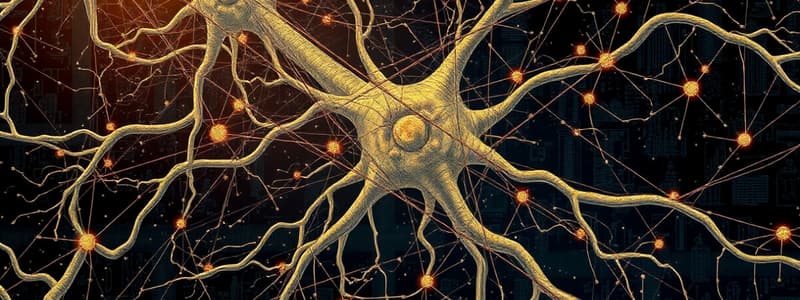
- A synapse is a specialized junction where one neuron communicates with another neuron or another cell type, such as a muscle or gland. Information typically flows from a neuron to its target.
- The presynaptic neuron is the first part of the connection to communicate, while the target is considered postsynaptic.
- The synaptic knob, a small round or oval shaped structure, contains vesicles and mitochondria.
- The postsynaptic region contains receptors for neurotransmitters.
- The synaptic cleft is a tiny space between the two neurons. (200-300 angstroms)
- Neurotransmitters are molecules released from the presynaptic neuron into the cleft; then they bind to receptors on the postsynaptic membrane.
Synaptic Transmission
-
Neurotransmitter release occurs in three steps:
- Release of neurotransmitters into the synaptic cleft.
- Action of the neurotransmitters on the postsynaptic membrane.
- Termination of synaptic transmission.
-
Neurotransmitter release is triggered by an action potential arriving at the synaptic knob. This triggers an influx of calcium ions
-
This causes vesicles containing neurotransmitters to fuse with the membrane and release neurotransmitters via exocytosis into the synaptic cleft.
-
Neurotransmitters are removed from the cleft via uptake and degrading enzymes.
Postsynaptic Potentials
- Postsynaptic potentials (PSPs) are two major types of signals in a neuron:
- Excitatory Postsynaptic Potentials (EPSPs)
- These cause partial depolarization of the postsynaptic membrane, bringing it closer to threshold.
- Inhibitory Postsynaptic Potentials (IPSPs)
- These cause partial hyperpolarization of the postsynaptic membrane, moving it further away from threshold.
- Excitatory Postsynaptic Potentials (EPSPs)
- EPSPs are triggered by excitatory neurotransmitters binding to receptors, usually allowing sodium ions to enter the cell.
- IPSPs are triggered by inhibitory neurotransmitters binding to receptors, usually allowing chloride ions to enter the cell or potassium ions to leave the cell.
- Summation of EPSPs and IPSPs determines whether or not an action potential occurs in the postsynaptic neuron.
Termination of Synaptic Transmission
- Synaptic transmission ends when the neurotransmitter is removed from the synaptic cleft. This happens through various mechanisms:
- Active reuptake of the neurotransmitter into the presynaptic terminal.
- Enzymatic degradation (breaking down the neurotransmitter).
- Diffusion of the neurotransmitter out of the synaptic cleft.
General Properties of Synaptic Transmission
- One-way conduction: impulses move in one direction (from presynaptic to postsynaptic). This is because the neurotransmitter is located only in the presynaptic neuron.
- Synaptic delay (approximately 0.5 milliseconds): the time it takes for the neurotransmitter to be released, diffuse across the synapse, and bind to receptors. This delay is a result of several steps in the process.
- Synaptic fatigue: the synapse's ability to transmit signals progressively diminishes if stimulated continuously for a long period. A depletion of neurotransmitters could be the cause of the progressive decline.
Effects of Environmental Factors
- Hypoxia: Reduced oxygen supply to neurons leads to loss of excitability and cessation of synaptic transmission, which can result in coma quickly (within less than 7 seconds).
- pH: Changes in pH (acidosis or alkalosis) alter neuronal excitability; alkalosis generally increases excitability, while acidosis typically reduces it.
Effects of Drugs
- Some drugs affect synaptic transmission by altering neuronal excitability.
- Caffeine and theophylline decrease the threshold for excitation, thereby increasing neuronal excitability.
- Anesthetics and hypnotics increase the threshold, thus decreasing neuronal excitability and synaptic transmission.
Studying That Suits You
Use AI to generate personalized quizzes and flashcards to suit your learning preferences.


